



SCR7 pyrazine
Based on 9 publication(s) in Google Scholar
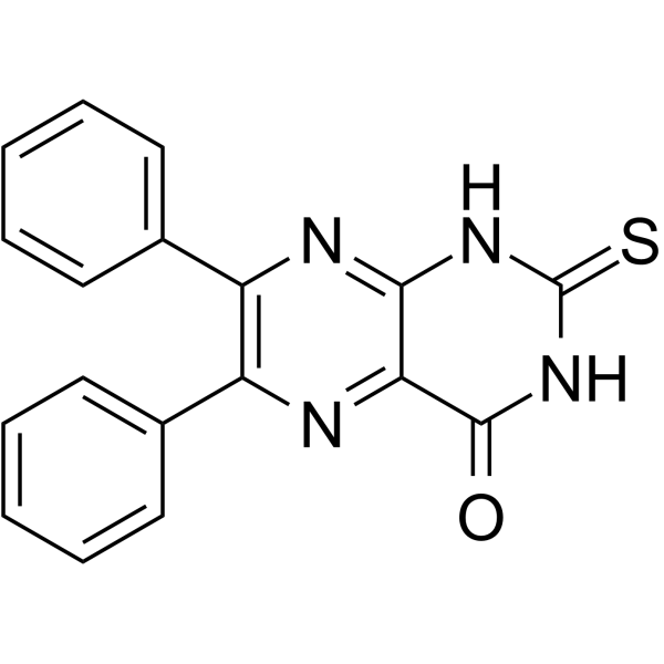
SCR7 pyrazine is a DNA ligase IV inhibitor that blocks nonhomologous end-joining (NHEJ) in a ligase IV-dependent manner. SCR7 pyrazine increases the efficiency of Cas9-mediated homology-directed repair (HDR). SCR7 pyrazine induces cell apoptosis and has anticancer activity.
商品は「研究用試薬」です。人や動物の医療用・臨床診断用・食品用の製品ではありません。
研究用途以外に使用した場合、当社は一切の責任を負いかねます。
- 純度 : 99.88%
- CAS 番号: 14892-97-8
- 分子式: C18H12N4OS
- 分子量:332.38
-
保管条件:Powder -20°C, 3 years , 4°C, 2 years ; In solvent -80°C, 2 years , -20°C, 1 year

To place orders, for customer services and technical support, please contact: MedChemExpress USA
Tel: 609-228-6898 E-mail: [email protected] [email protected]


-
 生物活性
生物活性
-
化学情報
-
溶剤 & 溶解度
- プロトコル
- 純度とドキュメンテーション
- 参考文献
-
 Help & FAQs


-
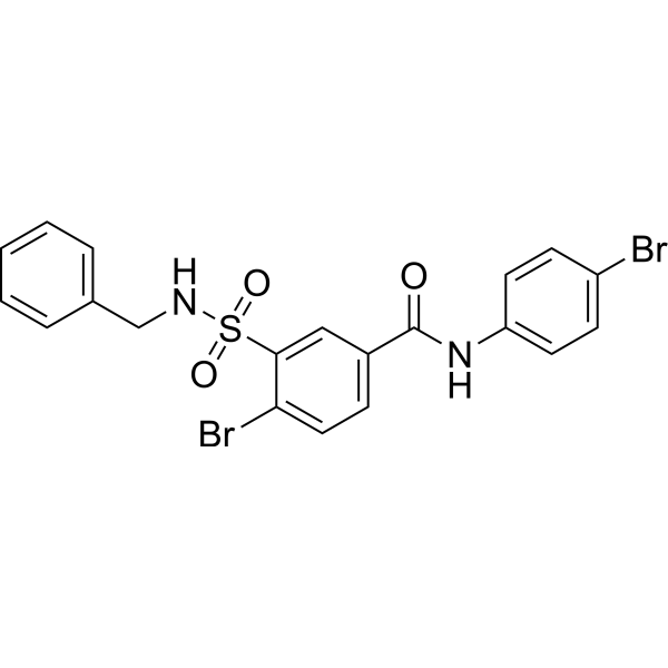
HY-19793
RS-1RAD51 Activator -
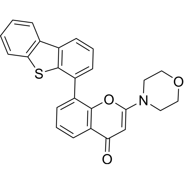
HY-11006
KU-57788DNA-PK Inhibitor -
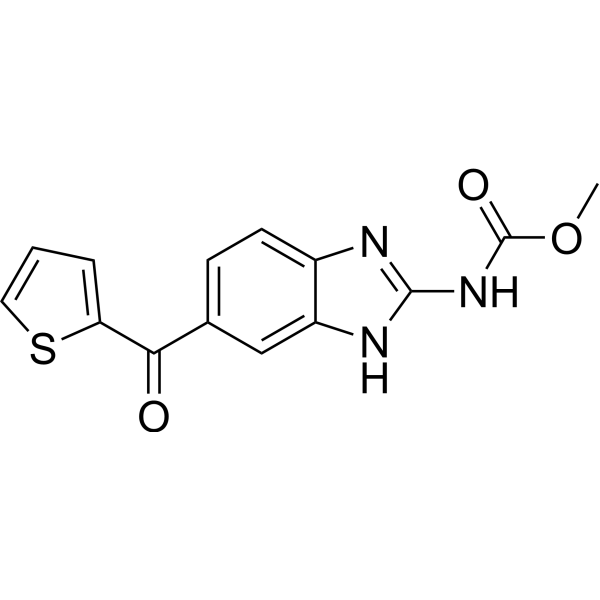
HY-13520
NocodazoleMicrotubule Inhibitor -
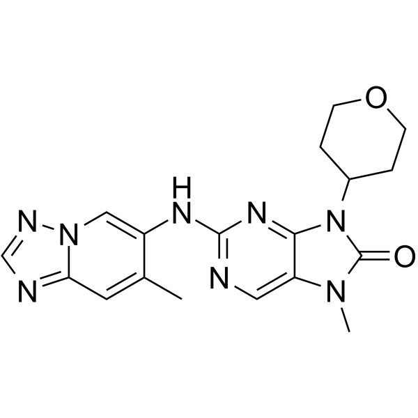
HY-111783
AZD-7648DNA-PK Inhibitor -
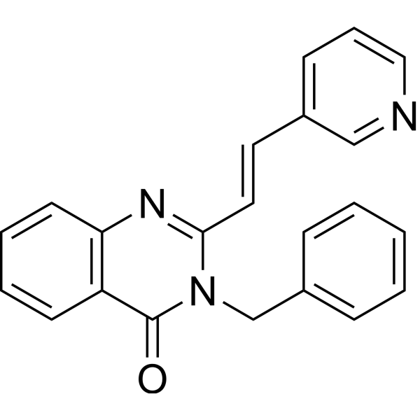
HY-101462
RAD51 Inhibitor B02RAD51 Inhibitor -
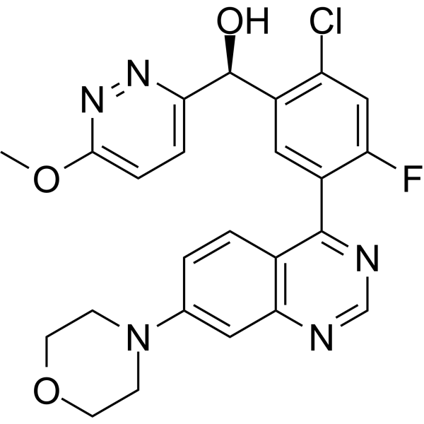
HY-101570
NedisertibDNA-PK Inhibitor -
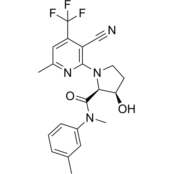
HY-141520
ART558Polθ Inhibitor -
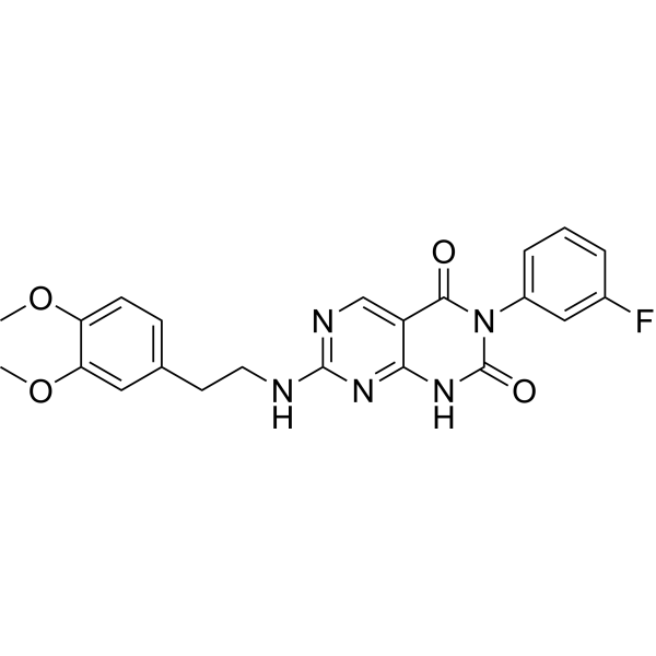
HY-122727
STL127705Ku 70/80 Heterodimer Protein Inhibitor -

HY-10071
Y-27632ROCK Inhibitor -
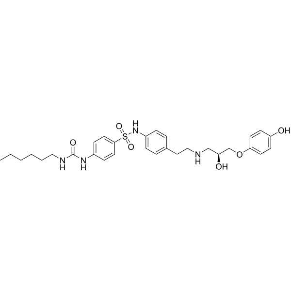
HY-19334
L755507β3-AR Agonist -

HY-B0015
PaclitaxelAntitumor Agent -
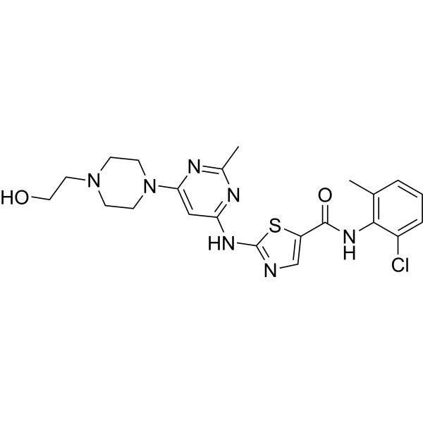
HY-10181
DasatinibSrc/Bcr-Abl Inhibitor
MedChemExpress(MCE)の使用を引用している文献 SCR7 pyrazine
More- Nat Commun. 2025 Jul 15;16(1):6502. [Abstract]
- Trends Biotechnol. 2025 Aug 30:S0167-7799(25)00314-2. [Abstract]
- Trends Biotechnol. 2025 Apr 7:S0167-7799(25)00083-6. [Abstract]
- Sci Adv. 2025 Jul 11;11(28):eadw1720. [Abstract]
- Mol Cancer Ther. 2026 Jul 15:10.1158/1535-7163.MCT-26-0182.
- Int J Mol Sci. 2025 May 3;26(9):4361. [Abstract]
- Int J Mol Sci. 2022 Jul 7;23(14):7518. [Abstract]
- Virology. 2025 May:606:110504. [Abstract]
- University of Georgia. 2025.
 Customer Validation & Images

-
Cell Proliferation/Viability Assay
DNA/RNA Synthesis アイソフォーム固有の製品をすべて表示
More

生物活性
製品説明
IC50 & Target
|
DNA Ligase |
体外実験
SCR7 pyrazine (20-100 μM; 24 hours; MCF7 cells) treatment interferes with NHEJ in cells, leading to accumulation of unrepaired double-strand breaks (DSBs)[1].
SCR7 pyrazine treatment shows a dose-dependent decrease in cell proliferation with IC50 values of 40 μM, 34 μM, 44 μM, 8.5 μM, 120 μM, 10 μM and 50 μM for MCF7, A549, HeLa, T47D, A2780, HT1080 and Nalm6 cells, respectively[1].
In MCF7 cells, SCR7 pyrazine (20, 40 μM) treatment increases phosphorylation of ATM and activates p53, decreases MDM2, BCL2, resulting in activation of proapoptotic proteins, PUMA and BAX. And the shorter fragments of MCL1, PARP1, Caspase 3, and Caspase 9 cleavage are upregulated in a dose-dependent manner[1].
MedChemExpress (MCE) has not independently confirmed the accuracy of these methods. They are for reference only. Further protocols information, click here.
-
Cell Line:MCF7 cells
-
Concentration:20 μM, 40 μM, 100 μM
-
Incubation Time:24 hours
-
Result:Showed an increase in levels of gH2AX foci and protein.
体内実験
MedChemExpress (MCE) has not independently confirmed the accuracy of these methods. They are for reference only.
-
Animal Model:BALB/c mice injected with breast adenocarcinoma cells[1]
-
Dosage:10 mg/kg
-
Administration:Intramuscular injection; on alternate days (0, 2, 4, 6, 8, and 10)
-
Result:Significantly reduced breast adenocarcinoma-induced tumor and increased lifespan.


化学情報
-
CAS 番号 14892-97-8
-
性状 Solid
-
分子量 332.38
-
分子式 C18H12N4OS
-
Color Light yellow to yellow
-
SMILES
O=C1NC(NC2=NC(C3=CC=CC=C3)=C(C4=CC=CC=C4)N=C12)=S
-
別名
SCR7ピラジン
-
輸送条件
Room temperature in continental US; may vary elsewhere.
-
保管条件
Powder -20°C 3 years 4°C 2 years In solvent -80°C 2 years -20°C 1 year


Publications (9)
-
Journal Impact Factor
-
Most Recent
-
Nat Commun
2025 Jul 15;16(1):6502. PMID: 40664653 -
Trends Biotechnol
Highly efficient prime editors for mammalian genome editing based on porcine retrovirus reverse transcriptase. [Abstract]2025 Aug 30:S0167-7799(25)00314-2. PMID: 40885667 -
Trends Biotechnol
Long-offset paired nicking-based efficient and precise strategy for in vivo targeted insertion. [Abstract]2025 Apr 7:S0167-7799(25)00083-6. PMID: 40199626 -
Sci Adv
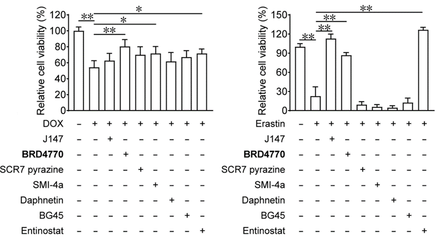
BRD4770 protects against DOX-induced cardiotoxicity by inhibiting apoptosis and ferroptosis. [Abstract]2025 Jul 11;11(28):eadw1720. PMID: 40644536
SCR7 pyrazine purchased from MedChemExpress. Usage Cited in: Sci Adv. 2025 Jul 11;11(28):eadw1720. [Abstract]
The effects of J147 (5 μM), BRD4770 (5 μM), SCR7 pyrazine (5 μM), SMI-4a (5 μM), daphnetin (5 μM), BG45 (5 μM), and entinostat (5 μM) on cell viability in H9C2 cells after 24 hours of DOX or erastin treatment.
-
-
Int J Mol Sci
CRISPR/Cas9 Ribonucleoprotein Delivery Enhanced by Lipo-Xenopeptide Carriers and Homology-Directed Repair Modulators: Insights from Reporter Cell Lines. [Abstract]2025 May 3;26(9):4361. PMID: 40362595 -
Int J Mol Sci
Chloroquine-Induced DNA Damage Synergizes with Nonhomologous End Joining Inhibition to Cause Ovarian Cancer Cell Cytotoxicity. [Abstract]2022 Jul 7;23(14):7518. PMID: 35886866 -
Virology
ISG15 depletion enhances oHSV-1 replication and antitumor efficacy in oral squamous cell carcinoma. [Abstract]2025 May:606:110504. PMID: 40121989 -


溶剤 & 溶解度
体外:
DMSO : 100 mg/mL (300.86 mM; Need ultrasonic; Hygroscopic DMSO has a significant impact on the solubility of product, please use newly opened DMSO)
Please refer to the solubility information to select the appropriate solvent. Once prepared, please aliquot and store the solution to prevent product inactivation from repeated freeze-thaw cycles.
Storage method and period of stock solution: -80°C, 2 years; -20°C, 1 year. When stored at -80°C, please use it within 2 years. When stored at -20°C, please use it within 1 year.
Please refer to the solubility information to select the appropriate solvent. Once prepared, please aliquot and store the solution to prevent product inactivation from repeated freeze-thaw cycles.
Storage method and period of stock solution: -80°C, 2 years; -20°C, 1 year. When stored at -80°C, please use it within 2 years. When stored at -20°C, please use it within 1 year.
濃度 (開始) × 体積 (開始) = 濃度 (終了) × 体積 (終了)
体内:
Select the appropriate dissolution method based on your experimental animal and administration route.
- For the following dissolution methods, please ensure to first prepare a clear stock solution using an In Vitro approach and then sequentially add co-solvents:
- To ensure reliable experimental results, the clarified stock solution can be appropriately stored based on storage conditions. As for the working solution for In Vivo experiments, it is recommended to prepare freshly and use it on the same day.
- The percentages shown for the solvents indicate their volumetric ratio in the final prepared solution. If precipitation or phase separation occurs during preparation, heat and/or sonication can be used to aid dissolution.
Add each solvent one by one: 10% DMSO 40% PEG300 5% Tween-80 45% Saline
Solubility: ≥ 2.5 mg/mL (7.52 mM); Clear solution
This protocol yields a clear solution of ≥ 2.5 mg/mL (saturation unknown).
Taking 1 mL working solution as an example, add 100 μL DMSO stock solution (25.0 mg/mL) to 400 μL PEG300, and mix evenly; then add 50 μL Tween-80 and mix evenly; then add 450 μL Saline to adjust the volume to 1 mL.
Preparation of Saline: Dissolve 0.9 g sodium chloride in ddH₂O and dilute to 100 mL to obtain a clear Saline solution.
Add each solvent one by one: 10% DMSO 90% (20% SBE-β-CD in Saline)
Solubility: ≥ 2.5 mg/mL (7.52 mM); Clear solution
This protocol yields a clear solution of ≥ 2.5 mg/mL (saturation unknown).
Taking 1 mL working solution as an example, add 100 μL DMSO stock solution (25.0 mg/mL) to 900 μL 20% SBE-β-CD in Saline, and mix evenly.
Preparation of 20% SBE-β-CD in Saline (4°C, storage for one week): 2 g SBE-β-CD powder is dissolved in 10 mL Saline, completely dissolve until clear.
In Vivo Dissolution Calculator
Please enter the basic information of animal experiments:
-
-
-
-
Recommended: Prepare an additional quantity of animals to account for potential losses during experiments.
Please enter your animal formula composition:
-
%DMSO +
Recommended: Keep the proportion of DMSO in working solution below 2% if your animal is weak.
-
%+
-
+%Tween-80 + +
-
%Saline +
The co-solvents required include: DMSO, . All of co-solvents are available by MedChemExpress (MCE). , Tween 80. All of co-solvents are available by MedChemExpress (MCE).
Working solution concentration: 0.22 mg/mL
Method for preparing stock solution: mg drug dissolved in μL DMSO. Stock solution concentration: mg/mL.
1. Take μL DMSO stock solution;
2. Add μL .
μL , mix evenly;
3. Then add μL Tween 80, mix evenly;
4. Then add μL
Please ensure that the stock solution in the first step is dissolved to a clear state, and add co-solvents in sequence. You can use ultrasonic heating (ultrasonic cleaner, recommended frequency 20-40 kHz), vortexing, etc. to assist dissolution.

プロトコル
-
Gene Editing
Gene editing modify specific sites within the genome through gene deletions, insertions or conversions to study functionally unknown genes or conduct gene therapy. It is also used to change the biological traits of organisms to establish new varieties. Gene editing techniques include zinc finger nuclease (ZFN), transcription activator-like effector nuclease (TALEN), and clustered regularly interspaced short palindromic repeats (CRISPR)/CRISPR-associated protein 9 (Cas 9) (CRISPR/Cas9).
-
CRISPR-Cas9 HDR knock-in/precise editing
CRISPR-Cas9 HDR knock-in uses a guide RNA to direct Cas9 to a genomic target adjacent to a PAM, where Cas9 creates a double-strand break; if a donor DNA template with homology to the cut region is present, cellular HDR can copy the donor sequence into the genome, producing a precise substitution, tag, reporter, or insertion rather than an indel. The readout is the fraction of alleles or cells carrying the intended donor-derived edit, measured by junction PCR, restriction-fragment analysis, Sanger sequencing, amplicon deep sequencing, flow cytometry for reporter knock-in, or clone genotyping; NHEJ indels and partial or non-HDR insertions are measured in parallel because they compete with or confound precise HDR outcomes.
-
Apoptosis
Apoptosis, also called programmed cell death, is generally characterized by distinct morphological characteristics.
-
TUNEL staining for apoptotic DNA fragmentation
TUNEL staining detects DNA strand breaks by using terminal deoxynucleotidyl transferase to add labeled nucleotides to exposed 3′-OH DNA termini, generating either microscopic staining in fixed cells or tissue sections, or fluorescence/cytometric signal in cell suspensions. TUNEL positivity reflects DNA fragmentation but should not be interpreted alone as definitive apoptosis, because TUNEL can also label necrotic, autolytic, mechanically damaged, or DNA-repair-associated DNA breaks.
-
Annexin V plus membrane-impermeant dye apoptosis staining
Annexin V-based apoptosis assays rely on the detection of phosphatidylserine (PS) externalization from the inner leaflet of the plasma membrane to the outer leaflet, an early biochemical hallmark of apoptosis. Fluorescently labeled Annexin V binds PS in a calcium-dependent manner, enabling identification of early apoptotic cells by flow cytometry or fluorescence microscopy. When combined with a membrane-impermeant DNA-binding dye (e. g. , propidium iodide), this approach allows discrimination between viable (Annexin V−/dye−), early apoptotic (Annexin V+/dye−), and late apoptotic or necrotic (Annexin V+/dye+) cell populations by assessing membrane integrity and PS exposure.
-
Apoptosis Solutions
Apoptosis is a regulated, generally non-lytic cell-death pathway that removes unwanted, damaged, infected, or abnormal cells through coordinated morphological changes, caspase activation, DNA fragmentation, and membrane remodeling. The intrinsic apoptosis pathway is controlled mainly by mitochondrial outer membrane permeabilization, BCL-2 family proteins, cytochrome c release, apoptosome formation, caspase-9 activation, and downstream executioner caspase-3/7 activation. The extrinsic apoptosis pathway is initiated by death receptors such as Fas, TNFR, and TRAIL receptors, which recruit adaptor proteins and activate caspase-8 before engaging executioner caspases or mitochondrial amplification through BID cleavage. Apoptosis is linked to many phenotypes, including cancer cell killing, tissue homeostasis, immune regulation, neurodegeneration, infection response, and treatment-induced cytotoxicity; unresolved questions include how apoptosis interacts with necroptosis, pyroptosis, ferroptos


純度とドキュメンテーション
-
データシート (280 KB)
-
SDS (393 KB)
- English - EN (393 KB)
- Français - FR (393 KB)
- Deutsch - DE (393 KB)
- Norwegian - NO (393 KB)
- Español - ES (393 KB)
- Swedish - SV (393 KB)
- Italian - IT (393 KB)
- Korean - KR (393 KB)
- Portuguese - PT (393 KB)
-
取扱説明書 (2659 KB)
参考文献
[1]. Srivastava M, et al. An inhibitor of nonhomologous end-joining abrogates double-strand break repair and impedes cancer progression. Cell. 2012 Dec 21;151(7):1474-87. [Content Brief]
[2]. Lin C, et al. Increasing the Efficiency of CRISPR/Cas9-mediated Precise Genome Editing of HSV-1 Virus in Human Cells. Sci Rep. 2016 Oct 7;6:34531. [Content Brief]


Complete Stock Solution Preparation Table
Please refer to the solubility information to select the appropriate solvent. Once prepared, please aliquot and store the solution to prevent product inactivation from repeated freeze-thaw cycles.
Storage method and period of stock solution: -80°C, 2 years; -20°C, 1 year. When stored at -80°C, please use it within 2 years. When stored at -20°C, please use it within 1 year.
| Optional Solvent | Concentration Solvent Mass | 1 mg | 5 mg | 10 mg | 25 mg |
|---|---|---|---|---|---|
| DMSO | 1 mM | 3.0086 mL | 15.0430 mL | 30.0860 mL | 75.2151 mL |
| 5 mM | 0.6017 mL | 3.0086 mL | 6.0172 mL | 15.0430 mL | |
| 10 mM | 0.3009 mL | 1.5043 mL | 3.0086 mL | 7.5215 mL | |
| 15 mM | 0.2006 mL | 1.0029 mL | 2.0057 mL | 5.0143 mL | |
| 20 mM | 0.1504 mL | 0.7522 mL | 1.5043 mL | 3.7608 mL | |
| 25 mM | 0.1203 mL | 0.6017 mL | 1.2034 mL | 3.0086 mL | |
| 30 mM | 0.1003 mL | 0.5014 mL | 1.0029 mL | 2.5072 mL | |
| 40 mM | 0.0752 mL | 0.3761 mL | 0.7522 mL | 1.8804 mL | |
| 50 mM | 0.0602 mL | 0.3009 mL | 0.6017 mL | 1.5043 mL | |
| 60 mM | 0.0501 mL | 0.2507 mL | 0.5014 mL | 1.2536 mL | |
| 80 mM | 0.0376 mL | 0.1880 mL | 0.3761 mL | 0.9402 mL | |
| 100 mM | 0.0301 mL | 0.1504 mL | 0.3009 mL | 0.7522 mL |


SCR7 pyrazine 関連分類
HY-107845 関連分類
 Powered by Bioz