



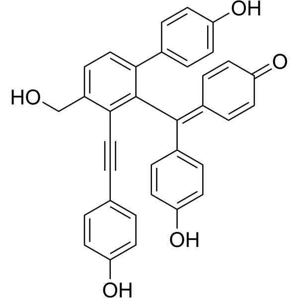
Selaginellin
Selaginellin is an inhibitor of Reactive Oxygen Species and an activator of SIRT1. Selaginellin protects endothelial cells against homocysteine-induced senescence by inhibitng reactive oxygen species and upregulating SIRT1 gene expression.
商品は「研究用試薬」です。人や動物の医療用・臨床診断用・食品用の製品ではありません。
研究用途以外に使用した場合、当社は一切の責任を負いかねます。
- CAS 番号: 941269-84-7
- 分子式: C34H24O5
- 分子量:512.55
-
保管条件:
Please store the product under the recommended conditions in the Certificate of Analysis.

To place orders, for customer services and technical support, please contact: MedChemExpress USA
Tel: 609-228-6898 E-mail: [email protected] [email protected]


-
 生物活性
生物活性
-
化学情報
- プロトコル
- 純度とドキュメンテーション
- 参考文献
-
 Help & FAQs




生物活性
製品説明


化学情報
-
CAS 番号 941269-84-7
-
分子量 512.55
-
分子式 C34H24O5
-
SMILES
O=C1C=C/C(C=C1)=C(C2=C(C(CO)=CC=C2C3=CC=C(O)C=C3)C#CC4=CC=C(C=C4)O)\C5=CC=C(C=C5)O
-
Structure Classification
-
Initial Source
-
輸送条件
Room temperature in continental US; may vary elsewhere.
-
保管条件
Please store the product under the recommended conditions in the Certificate of Analysis.

プロトコル
-
RT-PCR
Reverse transcription technology uses RNA as a template to synthesize DNA. RT-PCR is simple, specific and sensitive, and can be used to detect gene expression levels and expression differences in cells; detect RNA virus content; clone cDNA sequences of specific genes.
-
RNA extraction experimental
By lysing cells, releasing RNA, and removing impurities such as proteins and DNA, high-purity RNA products are finally obtained. The commonly used traditional method is the guanidine isothiocyanate/phenol/chloroform method (Trizol), which is suitable for a variety of animal materials including animal tissues, microorganisms, cultured cells, etc., and most plant materials.
-
ROS/oxidative-stress fluorescent staining
ROS/oxidative-stress fluorescent staining uses cell-permeant fluorogenic probes that become fluorescent after oxidation inside cells or tissues; commonly used examples include DCFH-DA/DCFDA for broad cellular oxidant detection, DHE for superoxide-related signal detection, MitoSOX for mitochondrial superoxide-related signal detection, and CellROX probes for oxidative-stress-associated fluorescence readouts. The assay detects probe oxidation rather than a single ROS species unless the probe and analysis method have been chemically validated for that species. DCFH-DA enters cells, is deacetylated by intracellular esterases to DCFH, and produces fluorescent DCF after oxidation, so the readout is used as an operational measure of total cellular oxidative stress rather than a species-specific ROS measurement. DHE and MitoSOX can report superoxide-related oxidation, but red fluorescence alone can include non-specific ethidium-like oxidation products; HPLC or optimized spectral approaches are
-
Endothelial Tube Formation Assay
Endothelial tube formation assay evaluates the ability of endothelial cells to attach, migrate, align, and organize into capillary-like networks when cultured on gelled basement membrane extract or Matrigel; the readout is the morphology and quantity of tube-like networks, which reflects an in vitro endothelial morphogenesis step related to angiogenesis. Basement membrane extract/Matrigel provides laminin-rich extracellular matrix cues that support endothelial differentiation into capillary-like structures, but it can contain biologically active growth factors, so growth-factor-reduced matrix is preferred when testing defined angiogenic stimulators or inhibitors.
-
Senescence-associated β-galactosidase staining
Senescence-associated β-galactosidase staining detects β-galactosidase activity that is histochemically visible at pH 6. 0 in senescent cells, where X-gal cleavage produces an insoluble blue precipitate observable by bright-field microscopy. This activity reflects increased lysosomal β-galactosidase/lysosomal mass rather than a senescence-essential enzyme, because GLB1 depletion or genetic lysosomal β-galactosidase deficiency can abolish SA-β-gal staining while cells still undergo senescence. SA-β-gal was originally reported in senescent but not presenescent fibroblasts and keratinocytes, absent from quiescent fibroblasts and terminally differentiated keratinocytes, and increased with donor age in human skin samples. Because SA-β-gal can also appear in some non-senescent or tissue-specific contexts, interpretation should be paired with experimental controls and, when possible, independent senescence markers.
-
Real Time qPCR (Q-PCR)
Real-time quantitative PCR (qPCR) quantifies an amplifiable nucleic-acid target by monitoring fluorescence during PCR cycling rather than measuring product only after amplification. The increase in fluorescence tracks accumulation of PCR product, and the quantification cycle (Cq; historically also Ct/CP) is related to the initial amount of target: samples containing more starting target generally reach the defined fluorescence threshold in fewer cycles.


純度とドキュメンテーション
参考文献

Calculators
濃度 (開始) × 体積 (開始) = 濃度 (終了) × 体積 (終了)


Selaginellin 関連分類
HY-N10083 関連分類
 Powered by Bioz