



Spliceostatin A

Based on 1 Customer Validation
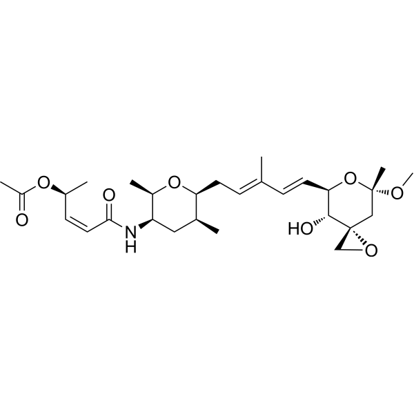
Spliceostatin A, the FR901464 (HY-16212) methylated derivative, is a potent anti-tumor agent. Spliceostatin A inhibits splicing and promotes pre-mRNA accumulation by binding SF3B1. SF3B1 is a subcomplex of U2 small nuclear ribonucleoprotein in the spliceosome. Spliceostatin A induces Apoptosis in chronic lymphocytic leukemia (CLL) cells.
商品は「研究用試薬」です。人や動物の医療用・臨床診断用・食品用の製品ではありません。
研究用途以外に使用した場合、当社は一切の責任を負いかねます。
- 純度 : 96.64%
- CAS 番号: 391611-36-2
- 分子式: C28H43NO8
- 分子量:521.64
-
保管条件:
-80°C, protect from light

To place orders, for customer services and technical support, please contact: MedChemExpress USA
Tel: 609-228-6898 E-mail: [email protected] [email protected]


-
 生物活性
生物活性
-
化学情報
-
溶剤 & 溶解度
- プロトコル
- 純度とドキュメンテーション
- 参考文献
-
 Help & FAQs


-
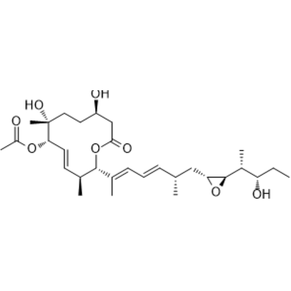
HY-16399
Pladienolide BSplicing Inhibitor -
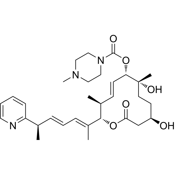
HY-111517
H3B-8800SF3B Aplicing Modulator -
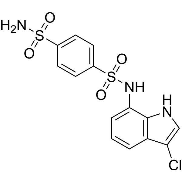
HY-13650
IndisulamCarbonic Anhydrase Inhibitor/RBM39 Molecular Glue Degrader -
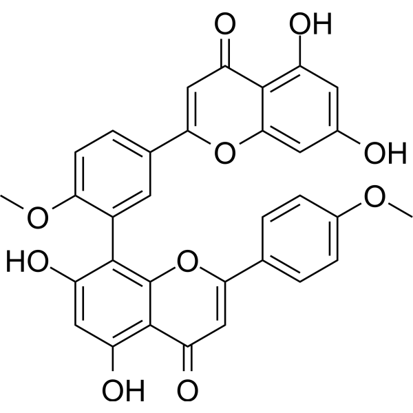
HY-N2117
Isoginkgetinpre-mRNA Splicing Inhibitor -
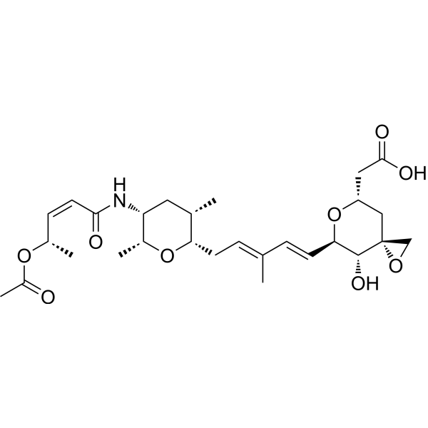
HY-129589
Thailanstatin ARNA Splicing Inhibitor -
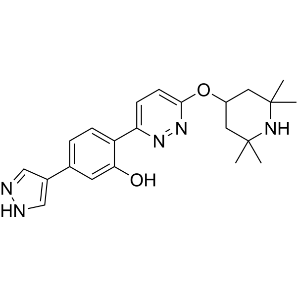
HY-19620
BranaplamSMN2 Splicing Modulator -
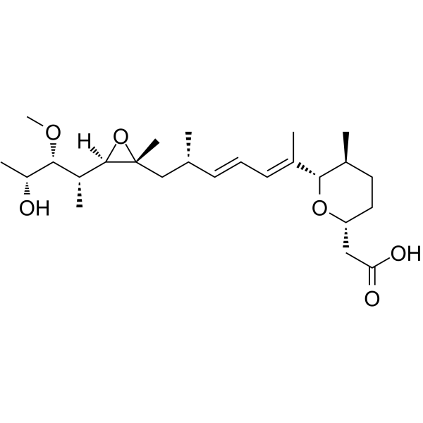
HY-19828
HerboxidienePre-mRNA Splicing Inhibitor -
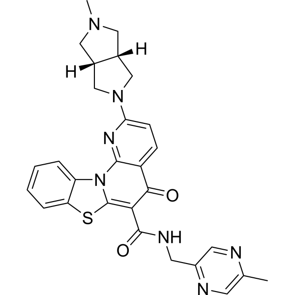
HY-148953
MTR-106G-quadruplex Stabilizer -

HY-13259
MG-132Proteasome Inhibitor -

HY-100558
Bafilomycin A1V-ATPase Inhibitor -
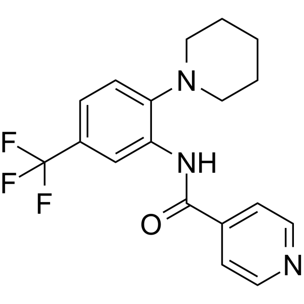
HY-13949
SRPIN340Virus Protease Inhibitor -
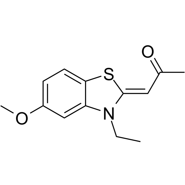
HY-15338
TG003CLK1 Inhibitor


生物活性
製品説明
IC50 & Target
IC50: 5.5 nM (wild-type SF3B1), 4.9 nM (SF3B1mutatant)[1]
Cellular Effect
|
Cell Line

|
Type | Value | Description | References |
|---|---|---|---|---|
| CWR22R | IC50 |
0.6 nM
Compound: 1
|
Suppression of AR-V7 expression in human CWR22Rv1 cells expressing GFP measured after 3 days by FACS assay
Suppression of AR-V7 expression in human CWR22Rv1 cells expressing GFP measured after 3 days by FACS assay
|
[PMID: 32551017] |
| CWR22R | IC50 |
0.6 nM
Compound: 1; SSA
|
Inhibition of AR-V7 expression in human 22Rv1 cells
Inhibition of AR-V7 expression in human 22Rv1 cells
|
[PMID: 37207846] |
体外実験
Spliceostatin A (2.5-20 nM; 0-24 h) induces caspase-dependent apoptosis of CLL cells in a dose- and time-dependent manner[1].
Spliceostatin A (10 nM, 20 nM; 24 h) in combination with ABT-199 or ABT-263 synergises, augments apoptosis in CLL cells following IL4/CD40L (10 ng/mL, 300 ng/mL; 6 h) treatment, and reduce phosphorylation levels of related proteins[1].
Spliceostatin A inhibits the viability of normal B (CD19+) and T (CD3+) lymphocytes with IC50s of 12.1 nM, and 61.7 nM, respectively[1].
MedChemExpress (MCE) has not independently confirmed the accuracy of these methods. They are for reference only. Further protocols information, click here.
-
Cell Line:CLL cells
-
Concentration:0, 2.5 nM, 5 nM, 10 nM, and 20 nM
-
Incubation Time:0, 6 h, 12 h, and 24 h
-
Result:Increased cell apoptosis.
-
Cell Line:CLL cells
-
Concentration:10 nM, and 20 nM
-
Incubation Time:24 h; treated after 10 ng/mL IL4 and/or 300 ng/mL CD40L for 6 h
-
Result:Decreased the level of PARP, phosphorylated STAT6 (pSTAT6), phosphorylated IκBα (pIκBα), Mcl-1L, Bcl-xL , Bcl-2 and Hsc70.


化学情報
-
CAS 番号 391611-36-2
-
性状 Solid
-
分子量 521.64
-
分子式 C28H43NO8
-
Color White to off-white
-
SMILES
C[C@H]1C[C@@H](NC(/C=C\[C@@H](OC(C)=O)C)=O)[C@@H](C)O[C@H]1C/C=C(C)/C=C/[C@@H]2[C@@H](O)[C@]3(CO3)C[C@@](C)(OC)O2
-
輸送条件
Shipping with dry ice.
-
保管条件
-80°C, protect from light


溶剤 & 溶解度
体外:
DMSO : 50 mg/mL (95.85 mM; Need ultrasonic; Hygroscopic DMSO has a significant impact on the solubility of product, please use newly opened DMSO)
Please refer to the solubility information to select the appropriate solvent. Once prepared, please aliquot and store the solution to prevent product inactivation from repeated freeze-thaw cycles.
Storage method and period of stock solution: -80°C, 6 months (protect from light). When stored at -80°C, please use it within 6 months.
Please refer to the solubility information to select the appropriate solvent. Once prepared, please aliquot and store the solution to prevent product inactivation from repeated freeze-thaw cycles.
Storage method and period of stock solution: -80°C, 6 months (protect from light). When stored at -80°C, please use it within 6 months.
濃度 (開始) × 体積 (開始) = 濃度 (終了) × 体積 (終了)

プロトコル
-
RNA extraction experimental
By lysing cells, releasing RNA, and removing impurities such as proteins and DNA, high-purity RNA products are finally obtained. The commonly used traditional method is the guanidine isothiocyanate/phenol/chloroform method (Trizol), which is suitable for a variety of animal materials including animal tissues, microorganisms, cultured cells, etc., and most plant materials.
-
Detection of Apoptosis
Apoptosis, also called programmed cell death, is generally characterized by distinct morphological characteristics.
-
TUNEL staining for apoptotic DNA fragmentation
TUNEL staining detects DNA strand breaks by using terminal deoxynucleotidyl transferase to add labeled nucleotides to exposed 3′-OH DNA termini, generating either microscopic staining in fixed cells or tissue sections, or fluorescence/cytometric signal in cell suspensions. TUNEL positivity reflects DNA fragmentation but should not be interpreted alone as definitive apoptosis, because TUNEL can also label necrotic, autolytic, mechanically damaged, or DNA-repair-associated DNA breaks.
-
Annexin V plus membrane-impermeant dye apoptosis staining
Annexin V-based apoptosis assays rely on the detection of phosphatidylserine (PS) externalization from the inner leaflet of the plasma membrane to the outer leaflet, an early biochemical hallmark of apoptosis. Fluorescently labeled Annexin V binds PS in a calcium-dependent manner, enabling identification of early apoptotic cells by flow cytometry or fluorescence microscopy. When combined with a membrane-impermeant DNA-binding dye (e. g. , propidium iodide), this approach allows discrimination between viable (Annexin V−/dye−), early apoptotic (Annexin V+/dye−), and late apoptotic or necrotic (Annexin V+/dye+) cell populations by assessing membrane integrity and PS exposure.
-
Apoptosis Solutions
Apoptosis is a regulated, generally non-lytic cell-death pathway that removes unwanted, damaged, infected, or abnormal cells through coordinated morphological changes, caspase activation, DNA fragmentation, and membrane remodeling. The intrinsic apoptosis pathway is controlled mainly by mitochondrial outer membrane permeabilization, BCL-2 family proteins, cytochrome c release, apoptosome formation, caspase-9 activation, and downstream executioner caspase-3/7 activation. The extrinsic apoptosis pathway is initiated by death receptors such as Fas, TNFR, and TRAIL receptors, which recruit adaptor proteins and activate caspase-8 before engaging executioner caspases or mitochondrial amplification through BID cleavage. Apoptosis is linked to many phenotypes, including cancer cell killing, tissue homeostasis, immune regulation, neurodegeneration, infection response, and treatment-induced cytotoxicity; unresolved questions include how apoptosis interacts with necroptosis, pyroptosis, ferroptos
-
CRISPR-Cas9 RNP editing of primary immune cells
CRISPR-Cas9 RNP editing uses preassembled Cas9 protein and guide RNA to direct sequence-specific DNA cleavage at a genomic target, after which cellular DNA repair generates insertions/deletions for knockout or uses an added donor template for knock-in. In primary immune cells, published protocols deliver Cas9 RNPs mainly by electroporation or nucleofection because these methods can introduce protein-RNA complexes into difficult-to-transfect T cells, B cells, NK cells, monocytes, myeloid cells, and innate lymphoid cells without viral Cas9 expression.


純度とドキュメンテーション
-
データシート (272 KB)
-
SDS (394 KB)
- English - EN (394 KB)
- Français - FR (394 KB)
- Deutsch - DE (394 KB)
- Norwegian - NO (394 KB)
- Español - ES (394 KB)
- Swedish - SV (394 KB)
- Italian - IT (394 KB)
- Korean - KR (394 KB)
- Portuguese - PT (394 KB)
-
取扱説明書 (2659 KB)
参考文献
[1]. Larrayoz M, et al. The SF3B1 inhibitor spliceostatin A (SSA) elicits apoptosis in chronic lymphocytic leukaemia cells through downregulation of Mcl-1. Leukemia. 2016 Feb;30(2):351-60. [Content Brief]
[2]. Kaida D, et al. Spliceostatin A targets SF3b and inhibits both splicing and nuclear retention of pre-mRNA. Nat Chem Biol. 2007 Sep;3(9):576-83. [Content Brief]
[3]. Roybal GA, et al. Spliceostatin A inhibits spliceosome assembly subsequent to prespliceosome formation. Nucleic Acids Res. 2010 Oct;38(19):6664-72. [Content Brief]


Complete Stock Solution Preparation Table
Please refer to the solubility information to select the appropriate solvent. Once prepared, please aliquot and store the solution to prevent product inactivation from repeated freeze-thaw cycles.
Storage method and period of stock solution: -80°C, 6 months (protect from light). When stored at -80°C, please use it within 6 months.
| Optional Solvent | Concentration Solvent Mass | 1 mg | 5 mg | 10 mg | 25 mg |
|---|---|---|---|---|---|
| DMSO | 1 mM | 1.9170 mL | 9.5852 mL | 19.1703 mL | 47.9258 mL |
| 5 mM | 0.3834 mL | 1.9170 mL | 3.8341 mL | 9.5852 mL | |
| 10 mM | 0.1917 mL | 0.9585 mL | 1.9170 mL | 4.7926 mL | |
| 15 mM | 0.1278 mL | 0.6390 mL | 1.2780 mL | 3.1951 mL | |
| 20 mM | 0.0959 mL | 0.4793 mL | 0.9585 mL | 2.3963 mL | |
| 25 mM | 0.0767 mL | 0.3834 mL | 0.7668 mL | 1.9170 mL | |
| 30 mM | 0.0639 mL | 0.3195 mL | 0.6390 mL | 1.5975 mL | |
| 40 mM | 0.0479 mL | 0.2396 mL | 0.4793 mL | 1.1981 mL | |
| 50 mM | 0.0383 mL | 0.1917 mL | 0.3834 mL | 0.9585 mL | |
| 60 mM | 0.0320 mL | 0.1598 mL | 0.3195 mL | 0.7988 mL | |
| 80 mM | 0.0240 mL | 0.1198 mL | 0.2396 mL | 0.5991 mL |


Spliceostatin A 関連分類
HY-16466 関連分類
 Powered by Bioz