



Kansuinine B

Based on 1 Customer Validation
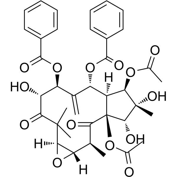
Kansuinine B is a natural diterpenoid compound with anti-inflammatory, anti-cancer and anti-viral activities, which can be isolated from the roots of Euphorbia kansui. Kansuinine B induces sustained ERK1/2 activation, inhibits IL-6-induced Stat3 activation and tyrosine phosphorylation, and enhances serine phosphorylation of Stat3. Kansuinine B negatively regulates the IL-6 signaling pathway by upregulating the expression of SOCS-3. Kansuinine B promotes the proliferation of mouse splenic lymphocytes in vitro and stimulates rat peritoneal macrophages to produce NO. Kansuinine B can be used in studies related to liver cancer and COVID-19.
Nur für Forschungszwecke. Wir verkaufen nicht an Patienten.
- Reinheit : 99.0%
- CAS. Nr.: 57685-46-8
- Formel: C38H42O14
- Molecular Weight:722.73
-
Speicherung:
4°C, sealed storage, away from moisture
* In solvent : -80°C, 6 months; -20°C, 1 month (sealed storage, away from moisture)

To place orders, for customer services and technical support, please contact: MedChemExpress USA
Tel: 609-228-6898 E-mail: [email protected] [email protected]


-
 Biologische Aktivität
-
Chemical Information
-
Lösungsmittel & Löslichkeit
- Protokoll
- Reinheit & Dokumentation
- Verweise
-
 Help & FAQs




Biologische Aktivität
Beschreibung
IC50 & Target
[1]|
IL-6 |
ERK1 |
ERK2 |
STAT3 |
Cellular Effect
|
Cell Line

|
Type | Value | Description | References |
|---|---|---|---|---|
| RAW264.7 | IC50 |
34.9 μM
Compound: 4
|
Antiinflammatory activity in mouse RAW264.7 cells assessed as inhibition of LPS-induced NO production incubated for 24 hrs by Griess method
Antiinflammatory activity in mouse RAW264.7 cells assessed as inhibition of LPS-induced NO production incubated for 24 hrs by Griess method
|
[PMID: 27246615] |
In Vitro
Kansuinine B (1-10 μM; 20 min co-treatment with IL-6) dose-dependently inhibits IL-6-induced Stat3Tyr705 phosphorylation (but not Ser727 phosphorylation) in HepG2 cells, and this effect is reversed by inhibition of the MEK/ERK pathway[1].
Kansuinine B (6 μM; 0-120 min) induces sustained phosphorylation of ERK1/2 and sustained phosphorylation of Stat3Ser727, as well as time-dependent upregulation of SOCS-3 mRNA expression in HepG2 cells, and this effect depends on the activation of PKC and MEK/ERK pathways[1].
MedChemExpress (MCE) has not independently confirmed the accuracy of these methods. They are for reference only.
-
Cell Line:human hepatoma HepG2 cells
-
Concentration:1, 5, 6, 10 μM
-
Incubation Time:20 min co-treatment with IL-6; 1 h PD98059 (HY-12028) preincubation followed by 20 min co-treatment with IL-6
-
Result:Dramatically inhibited IL-6-induced Stat3 tyrosine (Tyr705) phosphorylation in a dose-dependent manner.
Did not block IL-6-induced Stat3 serine (Ser727) phosphorylation.
Inhibitory effect on Stat3 tyrosine phosphorylation was reversed by preincubation with PD98059.
-
Cell Line:human hepatoma HepG2 cells
-
Concentration:6 μM
-
Incubation Time:5, 10, 15, 30, 60 min; 1 h Bisindolylmaleimide II (HY-108604) preincubation followed by 1 h treatment
-
Result:Induced phosphorylation of ERK1/2 starting at 5 minutes, with the effect sustained through 60 minutes.
ERK1/2 phosphorylation was blocked by preincubation with the PKC inhibitor bisindolylmaleimide II.
-
Cell Line:human hepatoma HepG2 cells
-
Concentration:6 μM
-
Incubation Time:5, 10, 15, 30, 60 min; 1 h Bisindolylmaleimide II or PD98059 preincubation followed by 1 h treatment
-
Result:Induced Stat3 serine (Ser727) phosphorylation starting at 5 minutes, with the effect sustained through 60 minutes.
Stat3 serine phosphorylation was completely inhibited by preincubation with bisindolylmaleimide II or PD98059.
-
Cell Line:human hepatoma HepG2 cells
-
Concentration:6 μM
-
Incubation Time:15, 30, 60, 120 min; 1 h Bisindolylmaleimide II or PD98059 preincubation followed by 1 h treatment
-
Result:Increased SOCS-3 mRNA expression in a time-dependent manner, inducing an ~19-fold increase at 60 minutes.
Induction of SOCS-3 mRNA was significantly blocked by preincubation with bisindolylmaleimide II or PD98059.
-
Cell Line:mice splenic lymphocytes
-
Concentration:0.78, 1.56, 3.13, 6.25, 12.5 μg/mL
-
Incubation Time:1 day
-
Result:Potently promoted proliferation of exoteric mice splenic lymphocytes, with the highest effect observed at 3.13 μg/mL.


Chemical Information
-
CAS. Nr. 57685-46-8
-
Appearance Solid
-
Molecular Weight 722.73
-
Formel C38H42O14
-
Color White to off-white
-
SMILES
O=C(C1(C)C)[C@H](O)[C@@H](OC(C2=CC=CC=C2)=O)C([C@H](OC(C3=CC=CC=C3)=O)[C@]([C@@H](OC(C)=O)[C@](C)(O)[C@@H]4O)([H])[C@@]4(OC(C)=O)C([C@@H](C)[C@@]5([H])[C@@]1([H])O5)=O)=C
-
Structure Classification
-
Versand
Room temperature in continental US; may vary elsewhere.
-
Speicherung
4°C, sealed storage, away from moisture
* In solvent : -80°C, 6 months; -20°C, 1 month (sealed storage, away from moisture)


Lösungsmittel & Löslichkeit
In Vitro:
DMSO : 25 mg/mL (34.59 mM; Need ultrasonic; Hygroscopic DMSO has a significant impact on the solubility of product, please use newly opened DMSO)
Please refer to the solubility information to select the appropriate solvent. Once prepared, please aliquot and store the solution to prevent product inactivation from repeated freeze-thaw cycles.
Storage method and period of stock solution: -80°C, 6 months; -20°C, 1 month (sealed storage, away from moisture). When stored at -80°C, please use it within 6 months. When stored at -20°C, please use it within 1 month.
Please refer to the solubility information to select the appropriate solvent. Once prepared, please aliquot and store the solution to prevent product inactivation from repeated freeze-thaw cycles.
Storage method and period of stock solution: -80°C, 6 months; -20°C, 1 month (sealed storage, away from moisture). When stored at -80°C, please use it within 6 months. When stored at -20°C, please use it within 1 month.
Konzentration (Stammlösung) × Volumen (Stammlösung) = Konzentration (Ziellösung) × Volumen (Ziellösung)
In Vivo:
Select the appropriate dissolution method based on your experimental animal and administration route.
- For the following dissolution methods, please ensure to first prepare a clear stock solution using an In Vitro approach and then sequentially add co-solvents:
- To ensure reliable experimental results, the clarified stock solution can be appropriately stored based on storage conditions. As for the working solution for In Vivo experiments, it is recommended to prepare freshly and use it on the same day.
- The percentages shown for the solvents indicate their volumetric ratio in the final prepared solution. If precipitation or phase separation occurs during preparation, heat and/or sonication can be used to aid dissolution.
Add each solvent one by one: 10% DMSO 40% PEG300 5% Tween-80 45% Saline
Solubility: ≥ 1.25 mg/mL (1.73 mM); Clear solution
This protocol yields a clear solution of ≥ 1.25 mg/mL (saturation unknown).
Taking 1 mL working solution as an example, add 100 μL DMSO stock solution (12.5 mg/mL) to 400 μL PEG300, and mix evenly; then add 50 μL Tween-80 and mix evenly; then add 450 μL Saline to adjust the volume to 1 mL.
Preparation of Saline: Dissolve 0.9 g sodium chloride in ddH₂O and dilute to 100 mL to obtain a clear Saline solution.
In Vivo Dissolution Calculator
Please enter the basic information of animal experiments:
-
-
-
-
Recommended: Prepare an additional quantity of animals to account for potential losses during experiments.
Please enter your animal formula composition:
-
%DMSO +
Recommended: Keep the proportion of DMSO in working solution below 2% if your animal is weak.
-
%+
-
+%Tween-80 + +
-
%Saline +
The co-solvents required include: DMSO, . All of co-solvents are available by MedChemExpress (MCE). , Tween 80. All of co-solvents are available by MedChemExpress (MCE).
Working solution concentration: 0.22 mg/mL
Method for preparing stock solution: mg drug dissolved in μL DMSO. Stock solution concentration: mg/mL. * In solvent : -80°C, 6 months; -20°C, 1 month (sealed storage, away from moisture)
1. Take μL DMSO stock solution;
2. Add μL .
μL , mix evenly;
3. Then add μL Tween 80, mix evenly;
4. Then add μL
Please ensure that the stock solution in the first step is dissolved to a clear state, and add co-solvents in sequence. You can use ultrasonic heating (ultrasonic cleaner, recommended frequency 20-40 kHz), vortexing, etc. to assist dissolution.


Reinheit & Dokumentation
-
Data Sheet (296 KB)
-
SDS (252 KB)
- English - EN (252 KB)
- Français - FR (252 KB)
- Deutsch - DE (252 KB)
- Norwegian - NO (252 KB)
- Español - ES (252 KB)
- Swedish - SV (252 KB)
- Italian - IT (252 KB)
- Korean - KR (252 KB)
- Portuguese - PT (252 KB)
-
Handling Instructions (2659 KB)
Verweise
[1]. Chang JS, et al. Kansuinine A and Kansuinine B from Euphorbia kansui L. inhibit IL-6-induced Stat3 activation. Planta medica. 2010 Oct;76(14):1544-9. [Content Brief]
[2]. Zhang L, et al. Bio-guided isolation of the cytotoxic terpenoids from the roots of Euphorbia kansui against human normal cell lines L-O2 and GES-1. Int J Mol Sci. 2012;13(9):11247-11259. [Content Brief]
[3]. Shu X, et al. Bioassay-guided separation of the proinflammatory constituents from the roots of Euphorbia kansui. Journal of natural medicines. 2010 Jan;64(1):98-103. [Content Brief]


Complete Stock Solution Preparation Table
Please refer to the solubility information to select the appropriate solvent. Once prepared, please aliquot and store the solution to prevent product inactivation from repeated freeze-thaw cycles.
Storage method and period of stock solution: -80°C, 6 months; -20°C, 1 month (sealed storage, away from moisture). When stored at -80°C, please use it within 6 months. When stored at -20°C, please use it within 1 month.
| Optional Solvent | Concentration Solvent Mass | 1 mg | 5 mg | 10 mg | 25 mg |
|---|---|---|---|---|---|
| DMSO | 1 mM | 1.3836 mL | 6.9182 mL | 13.8364 mL | 34.5911 mL |
| 5 mM | 0.2767 mL | 1.3836 mL | 2.7673 mL | 6.9182 mL | |
| 10 mM | 0.1384 mL | 0.6918 mL | 1.3836 mL | 3.4591 mL | |
| 15 mM | 0.0922 mL | 0.4612 mL | 0.9224 mL | 2.3061 mL | |
| 20 mM | 0.0692 mL | 0.3459 mL | 0.6918 mL | 1.7296 mL | |
| 25 mM | 0.0553 mL | 0.2767 mL | 0.5535 mL | 1.3836 mL | |
| 30 mM | 0.0461 mL | 0.2306 mL | 0.4612 mL | 1.1530 mL |


Kansuinine B Related Classifications
HY-126420 Related Classifications
 Powered by Bioz