



I-BOP
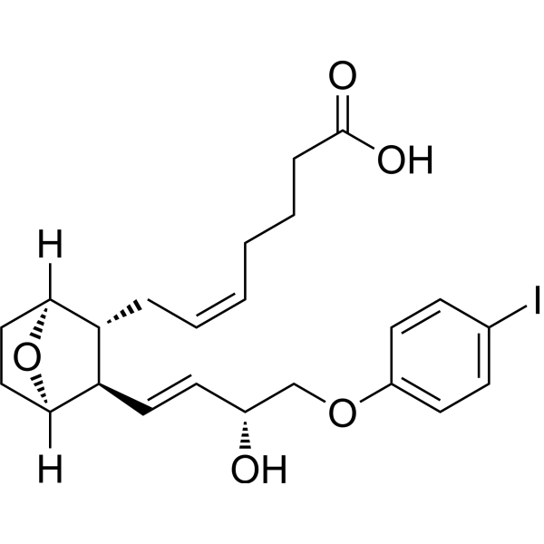
I-BOP is an agonist for thromboxane A2 receptor (TP) with a KD of 0.61 nM. I-BOP promotes proliferation through activation of PI3K pathway in vascular smooth muscle. I-BOP dose-dependently biphasicly affects the excitatory postsynaptic potential (e.p.s.p.) in hippocampal neurons.
연구목적의 판매만을 진행합니다. 환자를 대상으로 한 판매는 하지 않습니다.
- CAS No.: 128719-90-4
- 화학식: C23H29IO5
- 분자량:512.38
-
보관:
Please store the product under the recommended conditions in the Certificate of Analysis.

To place orders, for customer services and technical support, please contact: MedChemExpress USA
Tel: 609-228-6898 E-mail: [email protected] [email protected]


-
 Biological Activity
-
Chemical Information
- Protocol
- 순도&문서
- References
-
 Help & FAQs


-
HY-113096
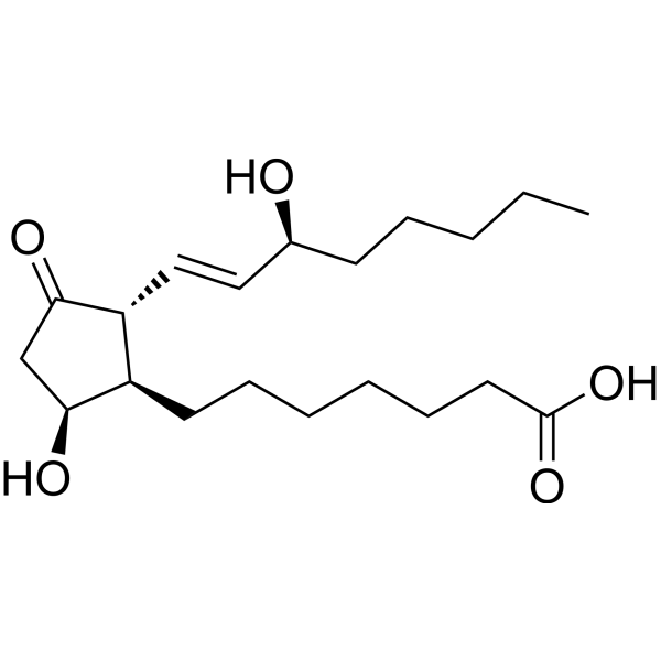
Prostaglandin D1CatSper Activator -
HY-113164
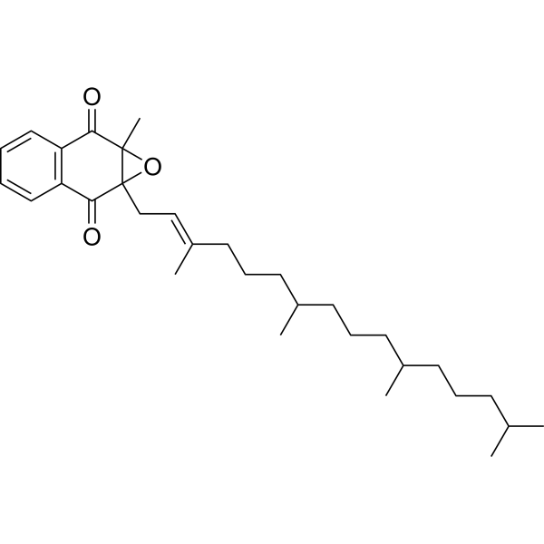
Vitamin K1 2,3-epoxideEndogenous Metabolite -
HY-113113
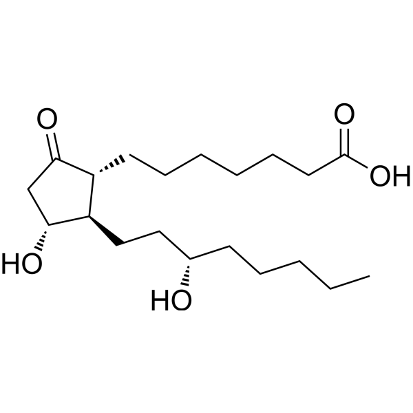
13,14-Dihydro PGE1PGE1 Metabolite -
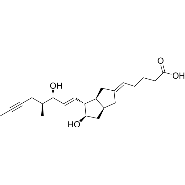
HY-118670
16(S)-IloprostStereoisomer -
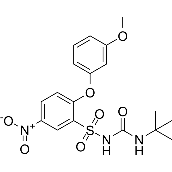
HY-118014
CAY10535FP/TP Receptor Inhibitor -
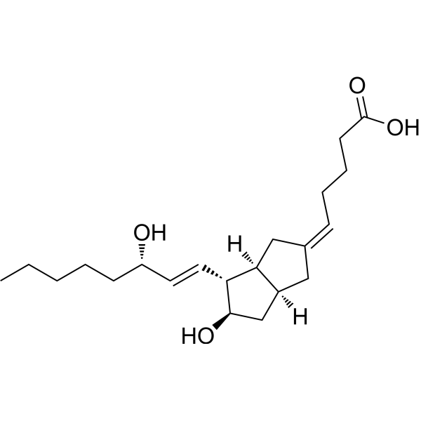
HY-124363
5-cis CarbaprostacyclinPGI2 Analog -
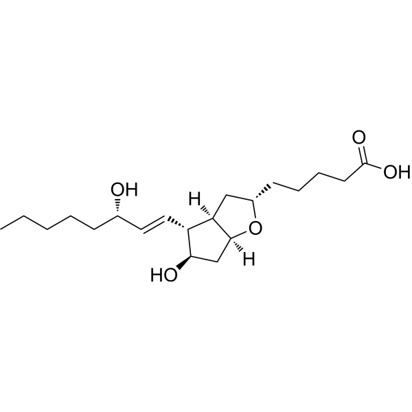
HY-130522
6β-Prostaglandin I1Prostacycline Analogue -
HY-130594
6α-Prostaglandin I1PGI2 Analog -

HY-120975
2,3-Dinor thromboxane b1Thromboxane B2 Metabolite -

HY-114660
9,11-Methane-epoxy prostaglandin F1αDerivative of Prostaglandin H1 -
HY-118670A
16(R)-IloprostStereoisomer


Biological Activity
제품 설명
In Vitro
I-BOP (1-100 nM) activates the MAP kinase, stimulates the tyrosine phosphorylation, and thus causes the activation of PI3K signaling pathway[1].
I-BOP (0.1-3 μM) enhances the synaptic transmission in hippocampal CA1 neuron at low concentration by increasing the neurotransmitter release, inhibits the excitatory synaptic transmission in hippocampal CA1 neuron through reduction of membrane input resistance at high concentration[2].
MedChemExpress (MCE) has not independently confirmed the accuracy of these methods. They are for reference only. Further protocols information, click here.
-
Cell Line:A7r5.HEL.TP cell
-
Concentration:1-100 nM
-
Incubation Time:24 h
-
Result:Increased phosphorylated tyrosine.


Chemical Information
-
CAS No. 128719-90-4
-
분자량 512.38
-
화학식 C23H29IO5
-
SMILES
O=C(O)CCC/C=C\C[C@H]1[C@](O2)([H])CC[C@]2([H])[C@@H]1/C=C/[C@@H](O)COC3=CC=C(I)C=C3
-
선적
Room temperature in continental US; may vary elsewhere.
-
보관
Please store the product under the recommended conditions in the Certificate of Analysis.

Protocol
-
Primary Embryonic Hippocampal Neuron Culture
Primary embryonic hippocampal neuron culture is an in vitro method in which hippocampi from embryonic rodents are dissected, enzymatically or mechanically dissociated, plated on adhesive substrates, and maintained in defined neuronal medium or in low-density sandwich/co-culture formats to support neuronal attachment, neurite extension, polarity formation, dendritic arborization, and synapse formation. The main readouts are cell survival, neuronal purity, neurite outgrowth, axon-dendrite polarization, synaptic marker development, and functional neuronal activity, assessed by phase-contrast microscopy, immunocytochemistry for neuronal/glial markers, live imaging, or electrophysiology depending on the downstream experiment.
-
Cell Viability Determination by MTT Colorimetric Assay
The following protocol uses the MTT colorimetric assay as a classic literature-established method for assessing cell viability/metabolic activity in cultured mammalian cells. MTT[3-(4,5-dimethylthiazol-2-yl)-2,5-diphenyltetrazolium bromide] is reduced by metabolically active cells to a colored formazan product; the amount of formazan is quantified spectrophotometrically and provides an indirect measure of metabolically active viable cells. Importantly, MTT reduction reflects cellular oxidoreductase/metabolic activity rather than an absolute direct count of living cells, so changes in cellular metabolism can alter the signal independently of cell number.


순도&문서
References
[1]. Morinelli TA, et al., Tyrosine phosphorylation of phosphatidylinositol 3-kinase and of the thromboxane A2 (TXA2) receptor by the TXA2 mimetic I-BOP in A7r5 cells. Biochem Pharmacol. 1997 Jun 15;53(12):1823-32. [Content Brief]
[2]. Hsu KS, et al., Thromboxane A2 agonist modulation of excitatory synaptic transmission in the rat hippocampal slice. Br J Pharmacol. 1996 Aug;118(8):2220-7. [Content Brief]

Calculators
Concentration (start) × Volume (start) = Concentration (final) × Volume (final)


I-BOP Related Classifications
HY-116431 Related Classifications
 Powered by Bioz