



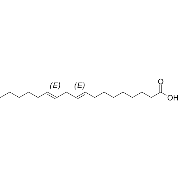
Linolelaidic acid
Based on 1 publication(s) in Google Scholar
Linolelaidic acid (Linoelaidic acid) is an omega-6 trans fatty acid (TFA) that is an essential nutrient with oral activity. Linolelaidic acid can be added to enteral nutrition (oral), parenteral nutrition (intravenous), and infant formula. Linolelaidic acid has anti-inflammatory and anti-parasitic ((Parasite)) activities, and can induce Apoptosis. Linolelaidic acid is useful for research in infections.
For research use only. We do not sell to patients.
- Purity : 99.96%
- CAS No.: 506-21-8
- Formula: C18H32O2
- Molecular Weight:280.45
-
Storage:Pure form -20°C, 3 years ; In solvent -80°C, 6 months , -20°C, 1 month

To place orders, for customer services and technical support, please contact: MedChemExpress USA
Tel: 609-228-6898 E-mail: [email protected] [email protected]


-
 Biological Activity
-
Chemical Information
-
Solvent & Solubility
- Protocols
- Purity & Documentation
- References
-
 Help & FAQs


-
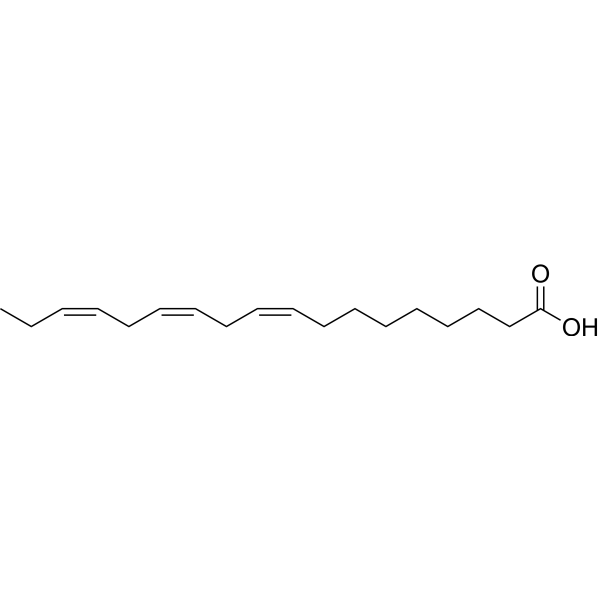
HY-N0728
α-Linolenic acidEssential Fatty Acid -
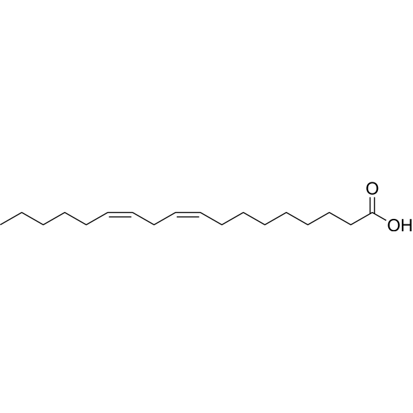
HY-N0729
Linoleic acidHuman Endogenous Metabolite -
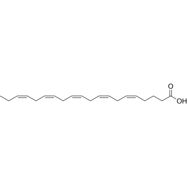
HY-B0660
Eicosapentaenoic AcidLC-PUFA -
HY-109590
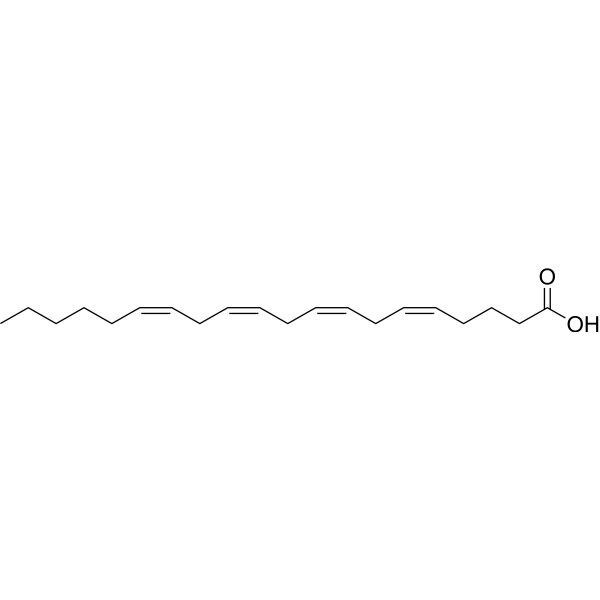
Arachidonic acidPolyunsaturated Fatty Acid -
HY-B2167
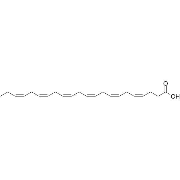
Docosahexaenoic acidomega-3 Fatty Acid -
HY-W011873
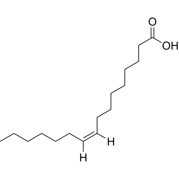
Palmitoleic acid -
HY-113016
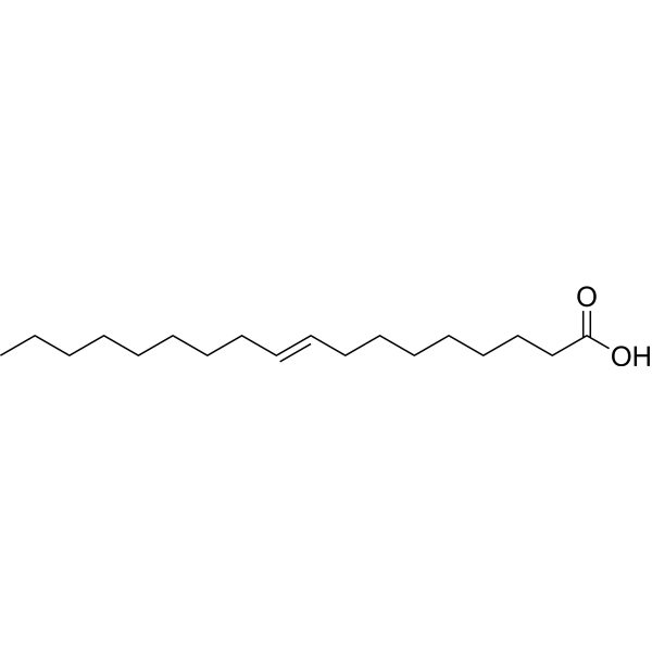
Elaidic acidTrans Fatty Acid -
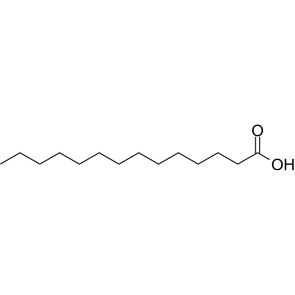
HY-N2041
Myristic acidEndogenous Metabolite -
HY-Y0366
Lauric acidMiddle Chain-free Fatty Acid -
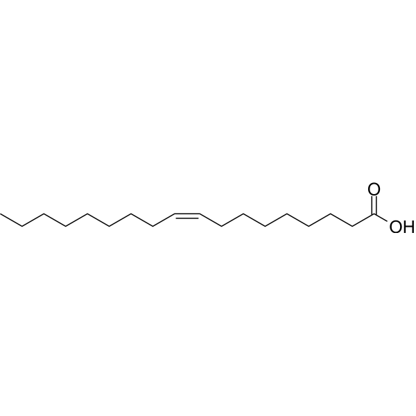
HY-N1446
Oleic acidMonounsaturated Fatty Acid -
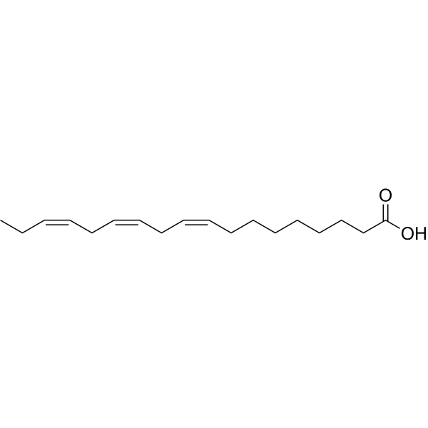
HY-N0728R
α-Linolenic acid (Standard)Essential Fatty Acid
Publications Citing Use of MedChemExpress (MCE) Linolelaidic acid
MoreAll Endogenous Metabolite Isoforms
MoreAll Parasite Isoforms
More

Biological Activity
Description
In Vitro
Linolenic Acid shows inhibitory activity against P. falciparum strains D10 (the chloroquine sensitive strain) and Dd2 (the chloroquine resistant strain), with IC50 values of 4.12 μg/mL and 5.04 μg/mL, respectively[2]. Linolelaidic acid (50 μM, 24 h) induces apoptosis, cell cycle arrest andinflammation in human umbilicalvein endothelial cells through lipid rafts[3].
MedChemExpress (MCE) has not independently confirmed the accuracy of these methods. They are for reference only. Further protocols information, click here.
-
Cell Line:Human umbilicalvein endothelial cells (HUVECs) (Methyl-β-cyclodextrin (HY-101461)-induced lipid raft disruption model)
-
Concentration:50 μM
-
Incubation Time:24 h
-
Result:Resulted in a significant decrease in cell viability.
-
Cell Line:Human umbilicalvein endothelial cells (HUVECs) (Methyl-β-cyclodextrin (HY-101461)-induced lipid raft disruption model)
-
Concentration:50 μM
-
Incubation Time:24 h
-
Result:Resulted in a significant increase in the number of apoptotic cells and the cell population in the G1 phase.
-
Cell Line:Human umbilicalvein endothelial cells (HUVECs) (Methyl-β-cyclodextrin (HY-101461)-induced lipid raft disruption model)
-
Concentration:50 μM
-
Incubation Time:24 h
-
Result:Led to a significant increase in the expression levels of pro-apoptotic proteins (caspase-3, -8, Bax, p53) and inflammatory factors (vascular cell adhesion molecule-1, intercellular adhesion molecule, E-selectin, and nitric oxide), while the expression level of the anti-apoptotic protein Bcl-2 was significantly decreased.
In Vivo
MedChemExpress (MCE) has not independently confirmed the accuracy of these methods. They are for reference only.
-
Animal Model:P. berghei (ANKA) strain-induced malaria C57BL/6 mice model (7-to-10-week old)[3]
-
Dosage:10 mg/kg
-
Administration:Oral gavage (p.o.), once daily for 4 days
-
Result:Inhibited the growth of P. berghei by 70%.
-
Animal Model:High-fat diet-fed streptozotocin (HFD-STZ) rat model[2]
-
Dosage:500 μg/kg
-
Administration:Oral gavage (p.o.), once daily for 4 weeks, 30 min myocardial ischemia followed by 4 or 6 h reperfusion after 4 weeks
-
Result:Significantly improved the instantaneous first derivation of left ventricle pressure, reduced infarct size, plasma creatine kinase and lactate dehydrogenase activities, and apotosis at the end of reperfusion in HFD-STZ diabetic rats. Not only significantly reduced tumor necrosis factor-α (TNF-α) and interleukin-6 (IL-6) concentrations but reduced the increase in superoxide production and malonaldialdehyde formation and simultaneously enhanced the antioxidant capacity in the diabetic hearts. Increased myocardial PI3K expression and Akt phosphorylation in diabetic but not normal rats.


Chemical Information
-
CAS No. 506-21-8
-
Appearance Liquid (Density: 0.89 g/cm3)
-
Molecular Weight 280.45
-
Formula C18H32O2
-
Color Colorless to light yellow
-
SMILES
CCCCC/C=C/C/C=C/CCCCCCCC(O)=O
-
Synonyms
Linoelaidic acid
-
Structure Classification
-
Initial Source
-
Shipping
Room temperature in continental US; may vary elsewhere.
-
Storage
Pure form -20°C 3 years In solvent -80°C 6 months -20°C 1 month


Publications (1)
-
Journal Impact Factor
-
Most Recent
-
Nature
2025 Jul;643(8070):192-200. PMID: 39695227


Solvent & Solubility
In Vitro:
DMSO : 125 mg/mL (445.71 mM; Need ultrasonic; Hygroscopic DMSO has a significant impact on the solubility of product, please use newly opened DMSO)
Please refer to the solubility information to select the appropriate solvent. Once prepared, please aliquot and store the solution to prevent product inactivation from repeated freeze-thaw cycles.
Storage method and period of stock solution: -80°C, 6 months; -20°C, 1 month. When stored at -80°C, please use it within 6 months. When stored at -20°C, please use it within 1 month.
Please refer to the solubility information to select the appropriate solvent. Once prepared, please aliquot and store the solution to prevent product inactivation from repeated freeze-thaw cycles.
Storage method and period of stock solution: -80°C, 6 months; -20°C, 1 month. When stored at -80°C, please use it within 6 months. When stored at -20°C, please use it within 1 month.
Concentration (start) × Volume (start) = Concentration (final) × Volume (final)

Protocols
-
How to Select the Route of Administration for Mammals
Route-of-administration selection in mammals is a pharmacokinetic, pharmacodynamic, formulation, animal-welfare, and translational decision, not a default technical choice. The selected route should match the study goal: intravenous dosing is most useful when complete systemic exposure and rapid onset are required, oral dosing is most translational for orally intended medicines but is affected by absorption and first-pass metabolism, subcutaneous or intramuscular dosing can provide slower systemic exposure, and intraperitoneal dosing can be useful in rodent proof-of-concept studies but may have limited clinical translation. Published route-comparison studies show that the same compound can produce different exposure, onset, bioavailability, tissue distribution, and tolerability depending on route; therefore, route choice should be supported by pilot pharmacokinetic or pharmacodynamic evidence when the literature is insufficient. Unresolved questions include how to standardize route sel
-
Apoptosis
Apoptosis, also called programmed cell death, is generally characterized by distinct morphological characteristics.
-
TUNEL staining for apoptotic DNA fragmentation
TUNEL staining detects DNA strand breaks by using terminal deoxynucleotidyl transferase to add labeled nucleotides to exposed 3′-OH DNA termini, generating either microscopic staining in fixed cells or tissue sections, or fluorescence/cytometric signal in cell suspensions. TUNEL positivity reflects DNA fragmentation but should not be interpreted alone as definitive apoptosis, because TUNEL can also label necrotic, autolytic, mechanically damaged, or DNA-repair-associated DNA breaks.
-
Annexin V plus membrane-impermeant dye apoptosis staining
Annexin V-based apoptosis assays rely on the detection of phosphatidylserine (PS) externalization from the inner leaflet of the plasma membrane to the outer leaflet, an early biochemical hallmark of apoptosis. Fluorescently labeled Annexin V binds PS in a calcium-dependent manner, enabling identification of early apoptotic cells by flow cytometry or fluorescence microscopy. When combined with a membrane-impermeant DNA-binding dye (e. g. , propidium iodide), this approach allows discrimination between viable (Annexin V−/dye−), early apoptotic (Annexin V+/dye−), and late apoptotic or necrotic (Annexin V+/dye+) cell populations by assessing membrane integrity and PS exposure.
-
Apoptosis Solutions
Apoptosis is a regulated, generally non-lytic cell-death pathway that removes unwanted, damaged, infected, or abnormal cells through coordinated morphological changes, caspase activation, DNA fragmentation, and membrane remodeling. The intrinsic apoptosis pathway is controlled mainly by mitochondrial outer membrane permeabilization, BCL-2 family proteins, cytochrome c release, apoptosome formation, caspase-9 activation, and downstream executioner caspase-3/7 activation. The extrinsic apoptosis pathway is initiated by death receptors such as Fas, TNFR, and TRAIL receptors, which recruit adaptor proteins and activate caspase-8 before engaging executioner caspases or mitochondrial amplification through BID cleavage. Apoptosis is linked to many phenotypes, including cancer cell killing, tissue homeostasis, immune regulation, neurodegeneration, infection response, and treatment-induced cytotoxicity; unresolved questions include how apoptosis interacts with necroptosis, pyroptosis, ferroptos
-
Research Protocol for Inflammation-related Diseases
The NLRP3 inflammasome is a cytosolic innate immune signaling platform that integrates priming signals and danger-signal activation to promote caspase-1 activation, maturation of IL-1β and IL-18, and gasdermin D-mediated pyroptotic cell death. The core experimental logic is to determine whether inflammatory disease phenotypes are driven by increased NLRP3 expression, ASC-containing inflammasome assembly, caspase-1 cleavage, GSDMD cleavage, and extracellular release of IL-1β/IL-18 rather than by nonspecific cell injury alone. The pathway is strongly linked to inflammation-related disease phenotypes because monosodium urate crystals activate NALP3/NLRP3 inflammasome signaling in gout-like crystal inflammation, cholesterol crystals activate NLRP3 inflammasomes in atherogenesis models, and DSS-induced intestinal inflammation has been reported to involve NLRP3 inflammasome activity. However, experimental colitis studies also show context-dependent protective effects of NLRP3 inflammasome co


Purity & Documentation
-
Data Sheet (274 KB)
-
SDS (394 KB)
- English - EN (394 KB)
- Français - FR (394 KB)
- Deutsch - DE (394 KB)
- Norwegian - NO (394 KB)
- Español - ES (394 KB)
- Swedish - SV (394 KB)
- Italian - IT (394 KB)
- Korean - KR (394 KB)
- Portuguese - PT (394 KB)
-
Handling Instructions (2659 KB)
References


Complete Stock Solution Preparation Table
Please refer to the solubility information to select the appropriate solvent. Once prepared, please aliquot and store the solution to prevent product inactivation from repeated freeze-thaw cycles.
Storage method and period of stock solution: -80°C, 6 months; -20°C, 1 month. When stored at -80°C, please use it within 6 months. When stored at -20°C, please use it within 1 month.
| Optional Solvent | Concentration Solvent Mass | 1 mg | 5 mg | 10 mg | 25 mg |
|---|---|---|---|---|---|
| DMSO | 1 mM | 3.5657 mL | 17.8285 mL | 35.6570 mL | 89.1424 mL |
| 5 mM | 0.7131 mL | 3.5657 mL | 7.1314 mL | 17.8285 mL | |
| 10 mM | 0.3566 mL | 1.7828 mL | 3.5657 mL | 8.9142 mL | |
| 15 mM | 0.2377 mL | 1.1886 mL | 2.3771 mL | 5.9428 mL | |
| 20 mM | 0.1783 mL | 0.8914 mL | 1.7828 mL | 4.4571 mL | |
| 25 mM | 0.1426 mL | 0.7131 mL | 1.4263 mL | 3.5657 mL | |
| 30 mM | 0.1189 mL | 0.5943 mL | 1.1886 mL | 2.9714 mL | |
| 40 mM | 0.0891 mL | 0.4457 mL | 0.8914 mL | 2.2286 mL | |
| 50 mM | 0.0713 mL | 0.3566 mL | 0.7131 mL | 1.7828 mL | |
| 60 mM | 0.0594 mL | 0.2971 mL | 0.5943 mL | 1.4857 mL | |
| 80 mM | 0.0446 mL | 0.2229 mL | 0.4457 mL | 1.1143 mL | |
| 100 mM | 0.0357 mL | 0.1783 mL | 0.3566 mL | 0.8914 mL |


Linolelaidic acid Related Classifications
HY-W071746 Related Classifications
 Powered by Bioz
Keywords