



Lucatumumab

Based on 1 Customer Validation
Lucatumumab (HCD122) is a fully human anti-CD40 antagonist monoclonal antibody, which blocks CD40/CD40L-mediated signaling. Lucatumumab efficiently mediates antibody-dependent cell-mediated cytotoxicity (ADCC) and clearance of tumor cells, can be used for refractory lymphomas, CLL and multiple myeloma research.
연구목적의 판매만을 진행합니다. 환자를 대상으로 한 판매는 하지 않습니다.
- Purity : ≥99.0%
- CAS No.: 903512-50-5
- 분자량:144.5 kDa
-
보관:
Please store the product under the recommended conditions in the Certificate of Analysis.


To place orders, for customer services and technical support, please contact: MedChemExpress USA
Tel: 609-228-6898 E-mail: [email protected] [email protected]


-
 Biological Activity
-
Chemical Information
- Protocol
- 순도&문서
- References
-
 Help & FAQs


-

HY-P99056
Utomilumab4-1BB/CD137 Agonist -

HY-P99670
Iscalimab (WT IgG1)Anti-CD40 mAb -

HY-P99742
MitazalimabAnti-CD40 Antibody -

HY-P99015
DacetuzumabAnti-CD40 Antibody -
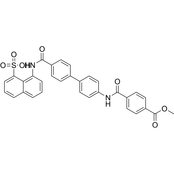
HY-120323
DRI-C21045CD40-CD40L Interaction Inhibitor -
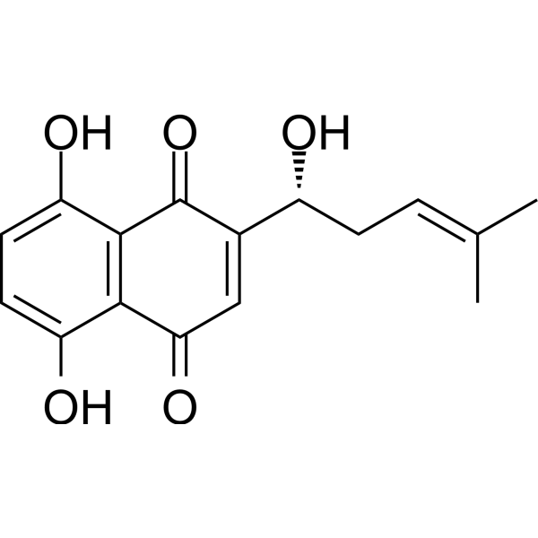
HY-N0822
ShikoninChloride Channel Inhibitor -

HY-P9908
AdalimumabTNF-α Inhibitor -

HY-P990007
TulisokibartAnti-TNFSf15/TL1A Antibody -
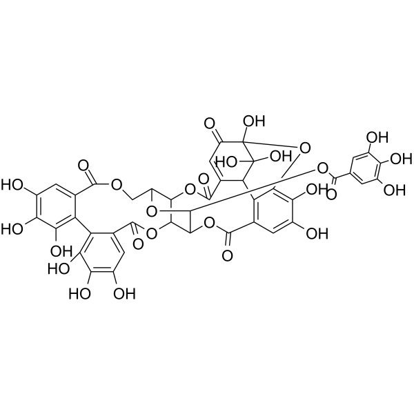
HY-N0472
GeraniinTNF Receptor Inhibitor -
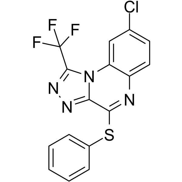
HY-110203
R-7050TNF Receptor Antagonist -

HY-P9980
BelantamabAnti-BCMA/TNFRSF17 Monoclonal Antibody -
HY-N0029
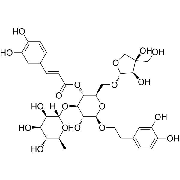
Forsythoside BTNF Inhibitor


Biological Activity
제품 설명
Isotype
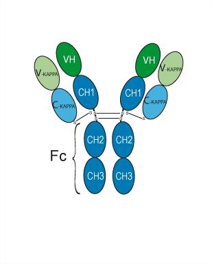
Human IgG1 kappa
Recommend Isotype Controls
Species Reactivity
Human
IC50 & Target
|
CD40 |
In Vitro
Lucatumumab (0.001-10 μg/mL; 4 h; 37 °C) inhibit B-cell chronic lymphocytic leukemia (B-CLL) growth by ADCC-mediated cell lysis, with the average maximal lysis of B-CLL cells of 49%[1].
Lucatumumab (0.1-10 μg/mL; 3 d) inhibits CD40L-induced signaling and viability of B-CLL cells[1].
Lucatumumab (10 μg/mL; 24 h) inhibits cytokine secretion by B-CLL cells[1].
MedChemExpress (MCE) has not independently confirmed the accuracy of these methods. They are for reference only.
-
Cell Line:B-cell chronic lymphocytic leukemia (B-CLL)
-
Concentration:0.001 to 10 μg/mL
-
Incubation Time:72 hours; 37 ℃
-
Result:Inhibited B-CLL growth with an ED50 of 14 pM and a average maximal lysis of B-CLL cells of 49%.
Clinical Trial
| NCT Number | Sponsor | Condition | Start Date |
Phase

|
|---|---|---|---|---|
| NCT01329991 | 2011-05 | PHASE1 |
Gene ID
Accession
Conjugated
Unconjugated
Reconsititution
The product can be reconstituted/diluted with sterile PBS or saline.
Format
-
Human IgG1 kappa
신청
ELISA, FACS, Functional assay
Verified Bioactivity
-
 Immobilized CD40 Protein, Human (HEK293, His, HY-P73499A) can bind Lucatumumab. The EC50 for this effect is 4.606 ng/mL.
 -
 Flow Cytometry analysis of Raji cells labelling TNFRSF5/CD40 (red) with Lucatumumab (anti-TNFRSF5/CD40) (HY-P99167). Goat Anti-Human IgG (Alexa Fluor 488) (HY-P83776) at a dilution of 1/1000 was used as the secondary antibody. Blue-Human IgG1 kappa (HY-P99001). Black-Unlabelled control, cells without incubation with primary antibody.


Chemical Information
-
CAS No. 903512-50-5
-
Appearance Liquid
-
분자량 144.5 kDa
-
Color Colorless to light yellow
-
SMILES
[Lucatumumab]
-
Synonyms
HCD122
-
선적
Shipping with dry ice.
-
Formulation
Please refer to the lot-specific COA for specific buffer information.
-
보관
Please store the product under the recommended conditions in the Certificate of Analysis.


순도&문서
-
Data Sheet (259 KB)
-
SDS (251 KB)
- English - EN (251 KB)
- Français - FR (251 KB)
- Deutsch - DE (251 KB)
- Norwegian - NO (251 KB)
- Español - ES (251 KB)
- Swedish - SV (251 KB)
- Italian - IT (251 KB)
- Korean - KR (251 KB)
- Portuguese - PT (251 KB)
-
Inhibitory Antibodies User Guide (603 KB)
References
[1]. Luqman M, et al. The antileukemia activity of a human anti-CD40 antagonist antibody, HCD122, on human chronic lymphocytic leukemia cells. Blood. 2008 Aug 1;112(3):711-20. [Content Brief]
[2]. Drgona L, et al. ESCMID Study Group for Infections in Compromised Hosts (ESGICH) Consensus Document on the safety of targeted and biological therapies: an infectious diseases perspective (Agents targeting lymphoid or myeloid cells surface antigens [II]: CD22, CD30, CD33, CD38, CD40, SLAMF-7 and CCR4). Clin Microbiol Infect. 2018 Jun;24 Suppl 2:S83-S94. [Content Brief]

Calculators
Concentration (start) × Volume (start) = Concentration (final) × Volume (final)


Lucatumumab Related Classifications
HY-P99167 Related Classifications
 Powered by Bioz