



Lysophosphatidylcholines
Based on 7 publication(s) in Google Scholar
Lysophosphatidylcholines is an orally active lysolipid and a component of oxidized low density lipoprotein (LDL). Lysophosphatidylcholines induces cell injury, the production of IL-1β and apoptosis. Lysophosphatidylcholines has a proactive effect on sepsis.
For research use only. We do not sell to patients.
- Purity : 99.9%
- CAS No.: 9008-30-4
-
Storage:Powder -20°C, 3 years ; In solvent -80°C, 6 months , -20°C, 1 month


To place orders, for customer services and technical support, please contact: MedChemExpress USA
Tel: 609-228-6898 E-mail: [email protected] [email protected]


-
 Biological Activity
-
Chemical Information
-
Solvent & Solubility
- Protocols
- Purity & Documentation
- References
-
 Help & FAQs


-
HY-125854
Phosphatidylcholines, eggPhosphatidylcholine -

HY-156041
Lysophosphatidylethanolamines, eggEndogenous Metabolite -
HY-125853
Phosphatidylcholines,soyaPhosphatidylcholine -
HY-B2235
LecithinEndogenous Metabolite -
HY-W251428
Phosphatidylglycerols (egg) sodium saltBiochemical Assay Reagent -
HY-W250118
Cephalin form bovine brainBiochemical Assay Reagent -

HY-42990
Hydrogenated soya phosphatidylcholinePhosphatidylcholine -
HY-B2227
Lactic acidGlycolysis Production -
HY-113498
SphingomyelinEndogenous Metabolite -

HY-B0015
PaclitaxelAntitumor Agent -
HY-90006
5-FluorouracilThymidylate Synthase Inhibitor -
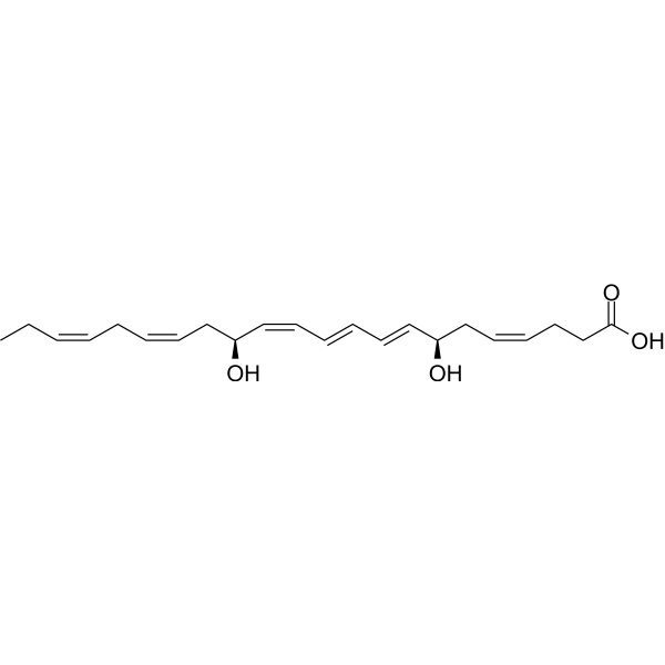
HY-116429
Maresin 1Ca2+ Stimulator
Publications Citing Use of MedChemExpress (MCE) Lysophosphatidylcholines
More- Adv Mater. 2026 Jun;38(33):e21108. [Abstract]
- J Clin Invest. 2025 Dec 15;135(24):e193082. [Abstract]
- Adv Sci (Weinh). 2025 Nov 14:e17330. [Abstract]
- Cell Death Discov. 2025 Apr 21;11(1):190. [Abstract]
- J Infect Dis. 2024 Aug 16;230(2):298-308. [Abstract]
- Anim Reprod Sci. 2026 Jan 12;287:108108.
- SSRN. 2026 Mar 5.
 Customer Validation & Images

-
Cell Proliferation/Viability Assay
-
IHC
-
IF
-
WB
-
In Vivo Efficacy Study


Biological Activity
Description
IC50 & Target
|
IL-1β |
ERK1 |
ERK2 |
In Vitro
Lysophosphatidylcholine (3-100 μM, 24 h) reduced HUVEC viability[1].
Lysophosphatidylcholines (12.5μM, 4 h) upregulates gene expression of IL-1β in human peripheral blood monocytes[2].
Lysophosphatidylcholines (75μM, 24 h) induces apoptosis in HUVEC through a p38-mitogen-activated protein kinase-dependent mechanism[3].
MedChemExpress (MCE) has not independently confirmed the accuracy of these methods. They are for reference only. Further protocols information, click here.
-
Cell Line:HUVEC
-
Concentration:3 μM, 10μM, 30 μM, 100 μM
-
Incubation Time:24 h
-
Result:Reduced HUVEC viability in a concentration-dependent manner.
-
Cell Line:HUVEC
-
Concentration:75 μM
-
Incubation Time:24 h
-
Result:Showed both ERK1/2 and p38-MAPK phosphorylation.
In Vivo
Lysophosphatidylcholines (0.1-20 mg/kg, Oral, single dose) enhances bacterial clearance, blocks cecal ligation and puncture (CLP)-induced neutrophil deactivation and increases bactericidal activity of neutrophils[4].
MedChemExpress (MCE) has not independently confirmed the accuracy of these methods. They are for reference only.
-
Animal Model:Albino ICR mice [4]
-
Dosage:0.1 mg/kg, 1 mg/kg, 10 mg/kg, 20 mg/kg
-
Administration:Oral
-
Result:Provided significant protection against cecal ligation and puncture (CLP)-induced lethality at a dose of 1 mg/kg.


Chemical Information
-
CAS No. 9008-30-4
-
Appearance Solid
-
Color White to off-white
-
SMILES
[Lysophosphatidylcholines]
-
Shipping
Room temperature in continental US; may vary elsewhere.
-
Storage
Powder -20°C 3 years In solvent -80°C 6 months -20°C 1 month


Publications (7)
-
Journal Impact Factor
-
Most Recent
-
Adv Mater
Conformationally Variable Peptides Trap and Detoxify Ox-LDL in Plaques for Attenuating Atherosclerosis in Multiple Species. [Abstract]2026 Jun;38(33):e21108. PMID: 42116761 -
J Clin Invest
Leukemia-expanded splenic CD81+ erythroblasts potentiate disease progression in mice by reshaping leukemic cell metabolism. [Abstract]2025 Dec 15;135(24):e193082. PMID: 41392984 -
Adv Sci (Weinh)
Obesity-Associated TRIM15 Promotes the Proliferation of Esophageal Adenocarcinoma Through the YY2/FOXRED1 Axis. [Abstract]2025 Nov 14:e17330. PMID: 41237333
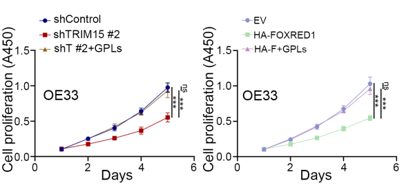
Lysophosphatidylcholines purchased from MedChemExpress. Usage Cited in: Adv Sci (Weinh). 2025 Nov 14:e17330. [Abstract]
Addition of GPLs (PCs, PEs, LysoPCs, and LysoPAs; each at 10 µg/mL) to EAC cells for 5 days attenuated the inhibitory effect of TRIM15 knockdown or FOXRED1 overexpression on cell proliferation.
-
Cell Death Discov
APOE4 impairs macrophage lipophagy and promotes demyelination of spiral ganglion neurons in mouse cochleae. [Abstract]2025 Apr 21;11(1):190. PMID: 40258814
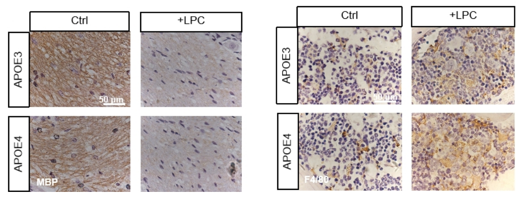
Lysophosphatidylcholines purchased from MedChemExpress. Usage Cited in: Cell Death Discov. 2025 Apr 21;11(1):190. [Abstract]
LPC (1 µL of 1% solution, single round-window administration) induced decreased MBP expression and an increase in F4/80-positive cells, indicating demyelination of spiral ganglion neurons (SGNs).
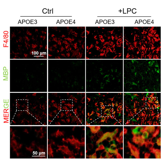
Lysophosphatidylcholines purchased from MedChemExpress. Usage Cited in: Cell Death Discov. 2025 Apr 21;11(1):190. [Abstract]
In the cochleae of mice treated with LPC (1 µL of 1% solution, single round-window administration), MBP was co-localized with F4/80-labeled RCMs in both APOE3 and APOE4 mice; however, APOE4 mice exhibited a lower proportion of MBP-positive RCMs with reduced MBP fluorescence intensity.
Lysophosphatidylcholines purchased from MedChemExpress. Usage Cited in: Cell Death Discov. 2025 Apr 21;11(1):190. [Abstract]
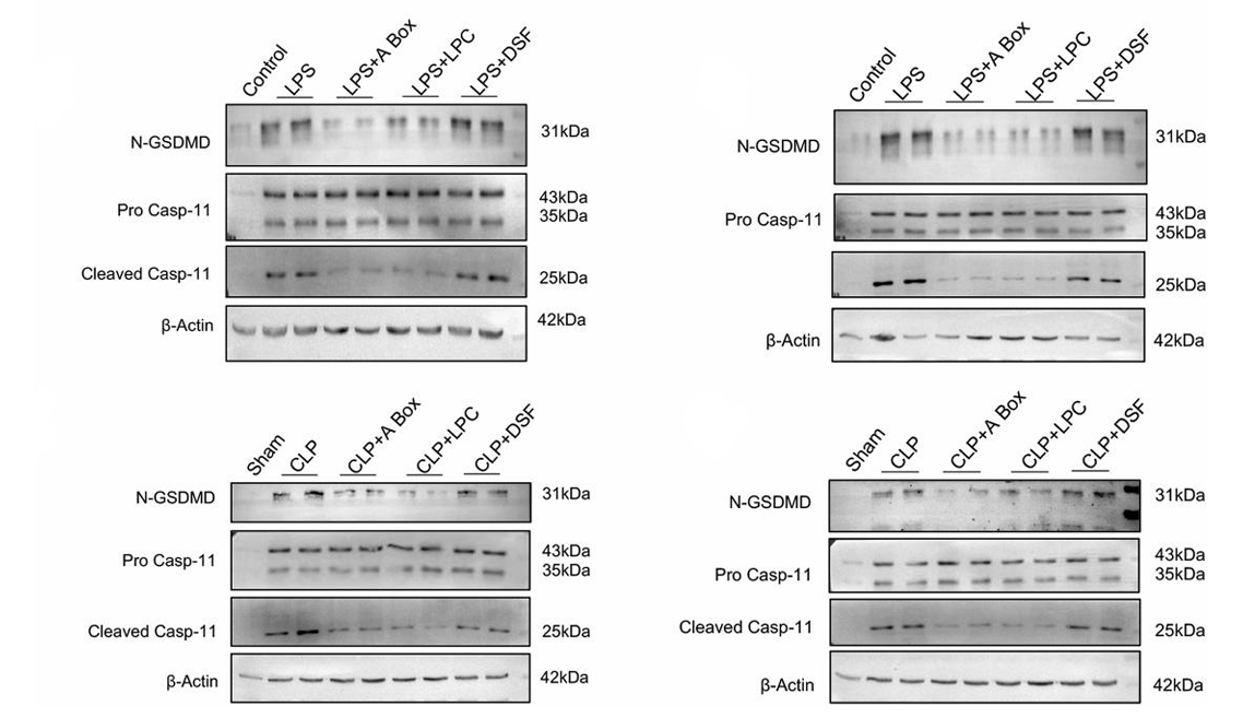
Western blot analysis of N-GSDMD, cleaved caspase-11, pro-caspase-11, and β-actin in peripheral blood monocytes (left) and peritoneal macrophages (right) from LPS and CLP model mice following treatment with 150 μg A box, 10 mg/kg LPC, or 50 mg/kg DSF. All experiments were performed at least three times. CLP, cecum ligation and puncture; DSF, disulfiram; LPC, lysophosphatidylcholine; LPS, lipopolysaccharide.
-
J Infect Dis
Role of Histiocyte-Derived frHMGB1 as a Facilitator in Non-Canonical Pyroptosis of Monocytes/Macrophages in Lethal Sepsis. [Abstract]2024 Aug 16;230(2):298-308. PMID: 38243905
Lysophosphatidylcholines purchased from MedChemExpress. Usage Cited in: J Infect Dis. 2024 Aug 16;230(2):298-308. [Abstract]
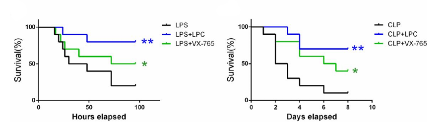
Survival curves of LPS (left) and CLP (right) model mice after treatment with 50 mg/kg VX-765 or 10 mg/kg LPC (n = 10). CLP + LPC vs. CLP, **P < 0.01; LPS + LPC vs. LPS, **P < 0.01. All data were collected from at least three independent experiments. CLP, cecum ligation and puncture; LPC, lysophosphatidylcholine; LPS, lipopolysaccharide.
-
-


Solvent & Solubility
In Vitro:
H2O : 100 mg/mL (Need ultrasonic)
Methanol : 25 mg/mL (Need ultrasonic)
DMSO : < 1 mg/mL (insoluble or slightly soluble)

Protocols
-
Apoptosis
Apoptosis, also called programmed cell death, is generally characterized by distinct morphological characteristics.
-
TUNEL staining for apoptotic DNA fragmentation
TUNEL staining detects DNA strand breaks by using terminal deoxynucleotidyl transferase to add labeled nucleotides to exposed 3′-OH DNA termini, generating either microscopic staining in fixed cells or tissue sections, or fluorescence/cytometric signal in cell suspensions. TUNEL positivity reflects DNA fragmentation but should not be interpreted alone as definitive apoptosis, because TUNEL can also label necrotic, autolytic, mechanically damaged, or DNA-repair-associated DNA breaks.
-
LPS-Induced Endotoxemia/Systemic Inflammation
Lipopolysaccharide (LPS)-induced endotoxemia is a widely used in vivo model of acute systemic inflammation in which LPS, a Gram-negative bacterial endotoxin, activates innate immune signaling primarily through TLR4, leading to rapid and transient induction of pro-inflammatory cytokines such as TNF-α, IL-6, and IL-1β in circulation and tissues. This cytokine surge is commonly used as a measurable readout of systemic inflammatory activation and immune dysregulation, and is typically assessed within hours after intraperitoneal LPS administration in mouse models of endotoxemia. The model captures key features of systemic inflammatory response syndrome, including cytokine release, immune cell activation, and downstream tissue responses, and has been used to evaluate anti-inflammatory interventions such as cytokine modulation, lipid mediators, and immune cell-targeting therapies.
-
Annexin V plus membrane-impermeant dye apoptosis staining
Annexin V-based apoptosis assays rely on the detection of phosphatidylserine (PS) externalization from the inner leaflet of the plasma membrane to the outer leaflet, an early biochemical hallmark of apoptosis. Fluorescently labeled Annexin V binds PS in a calcium-dependent manner, enabling identification of early apoptotic cells by flow cytometry or fluorescence microscopy. When combined with a membrane-impermeant DNA-binding dye (e. g. , propidium iodide), this approach allows discrimination between viable (Annexin V−/dye−), early apoptotic (Annexin V+/dye−), and late apoptotic or necrotic (Annexin V+/dye+) cell populations by assessing membrane integrity and PS exposure.
-
Apoptosis Solutions
Apoptosis is a regulated, generally non-lytic cell-death pathway that removes unwanted, damaged, infected, or abnormal cells through coordinated morphological changes, caspase activation, DNA fragmentation, and membrane remodeling. The intrinsic apoptosis pathway is controlled mainly by mitochondrial outer membrane permeabilization, BCL-2 family proteins, cytochrome c release, apoptosome formation, caspase-9 activation, and downstream executioner caspase-3/7 activation. The extrinsic apoptosis pathway is initiated by death receptors such as Fas, TNFR, and TRAIL receptors, which recruit adaptor proteins and activate caspase-8 before engaging executioner caspases or mitochondrial amplification through BID cleavage. Apoptosis is linked to many phenotypes, including cancer cell killing, tissue homeostasis, immune regulation, neurodegeneration, infection response, and treatment-induced cytotoxicity; unresolved questions include how apoptosis interacts with necroptosis, pyroptosis, ferroptos
-
Pyroptosis Solutions
Pyroptosis is a lytic inflammatory cell-death pathway executed by gasdermin pores, most classically through inflammasome-mediated activation of caspase-1, cleavage of gasdermin D, membrane pore formation, LDH release, and secretion of IL-1β and IL-18. The canonical pathway is commonly modeled by priming cells with an inflammatory signal such as LPS to induce pro-IL-1β and inflammasome components, followed by an activation signal such as ATP or nigericin to activate NLRP3, ASC speck formation, caspase-1 cleavage, GSDMD cleavage, cytokine release, and pyroptotic membrane rupture. The non-canonical pathway is triggered when cytosolic LPS activates mouse caspase-11 or human caspase-4/5, leading to GSDMD cleavage and pyroptosis, and this can secondarily activate NLRP3-dependent IL-1β release. Pyroptosis is linked to inflammatory injury, infection, cancer, liver disease, ocular disease, placental inflammation, and other disease phenotypes, but unresolved questions include which gasdermin fam


Purity & Documentation
-
Data Sheet (272 KB)
-
SDS (394 KB)
- English - EN (394 KB)
- Français - FR (394 KB)
- Deutsch - DE (394 KB)
- Norwegian - NO (394 KB)
- Español - ES (394 KB)
- Swedish - SV (394 KB)
- Italian - IT (394 KB)
- Korean - KR (394 KB)
- Portuguese - PT (394 KB)
-
Handling Instructions (2659 KB)
References
[1]. Kim E A, et al. Lysophosphatidylcholine induces endothelial cell injury by nitric oxide production through oxidative stress [J]. The Journal of Maternal-Fetal & Neonatal Medicine, 2009, 22(4): 325-331. [Content Brief]
[2]. Liu-Wu Y, et al. Lysophosphatidylcholine induces the production of IL-1β by human monocytes [J]. Atherosclerosis, 1998, 137(2): 351-357. [Content Brief]
[3]. Takahashi M, et al. Lysophosphatidylcholine induces apoptosis in human endothelial cells through a p38-mitogen-activated protein kinase-dependent mechanism [J]. Atherosclerosis, 2002, 161(2): 387-394. [Content Brief]
[4]. Yan J J, et al. Therapeutic effects of lysophosphatidylcholine in experimental sepsis [J]. Nature medicine, 2004, 10(2): 161-167. [Content Brief]

Calculators
Concentration (start) × Volume (start) = Concentration (final) × Volume (final)


Lysophosphatidylcholines Related Classifications
HY-139414 Related Classifications
 Powered by Bioz