



mTOR inhibitor-1
Based on 3 publication(s) in Google Scholar
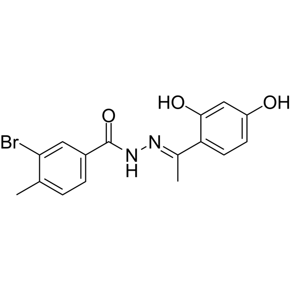
mTOR inhibitor-1 (Compound C-4) is an ATP-Competitive mTOR inhibitor which can suppress cells proliferation and inducing autophagy.
연구목적의 판매만을 진행합니다. 환자를 대상으로 한 판매는 하지 않습니다.
- Purity : 98.82%
- CAS No.: 468747-17-3
- 화학식: C16H15BrN2O3
- 분자량:363.21
-
보관:Powder -20°C, 3 years , 4°C, 2 years ; In solvent -80°C, 2 years , -20°C, 1 year

To place orders, for customer services and technical support, please contact: MedChemExpress USA
Tel: 609-228-6898 E-mail: [email protected] [email protected]


-
 Biological Activity
-
Chemical Information
-
용액&용해도
- Protocol
- 순도&문서
- References
-
 Help & FAQs


-
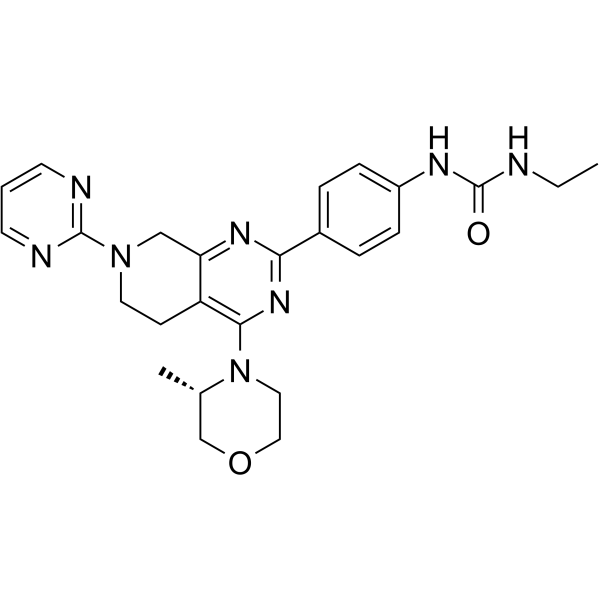
HY-18353
mTOR inhibitor-3mTOR Inhibitor -

HY-10219
RapamycinmTOR Inhibitor -
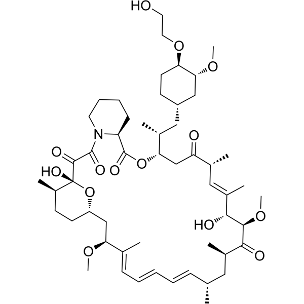
HY-10218
EverolimusmTORC Inhibitor -
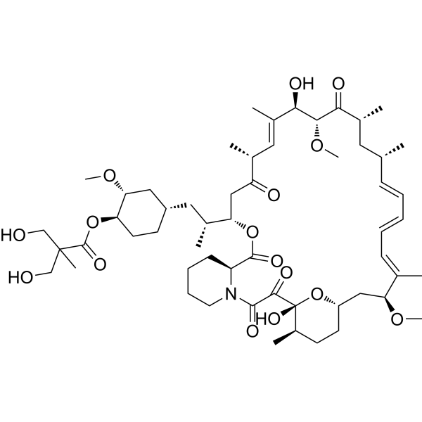
HY-50910
TemsirolimusmTOR Inhibitor -
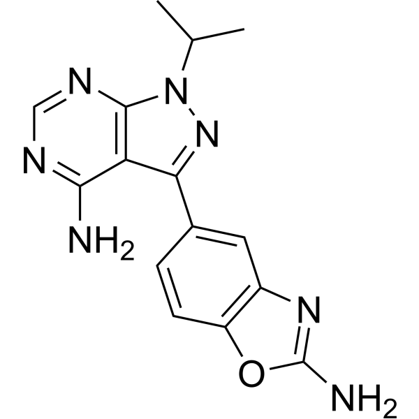
HY-13328
SapanisertibmTORC1/mTORC2 Inhibitor -
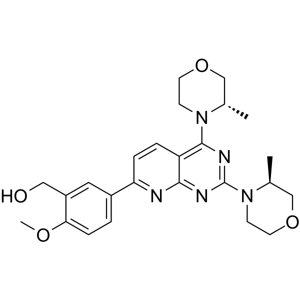
HY-10422
AZD-8055mTOR Inhibitor -
HY-15247
VistusertibmTORC Inhibitor -
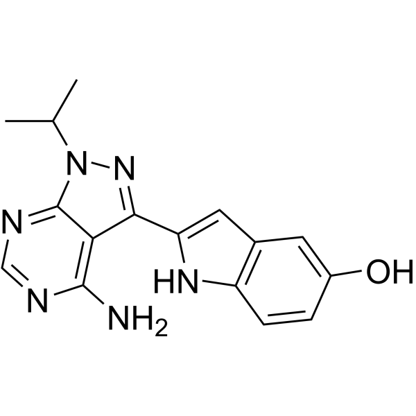
HY-10474
TorkinibmTOR Inhibitor -
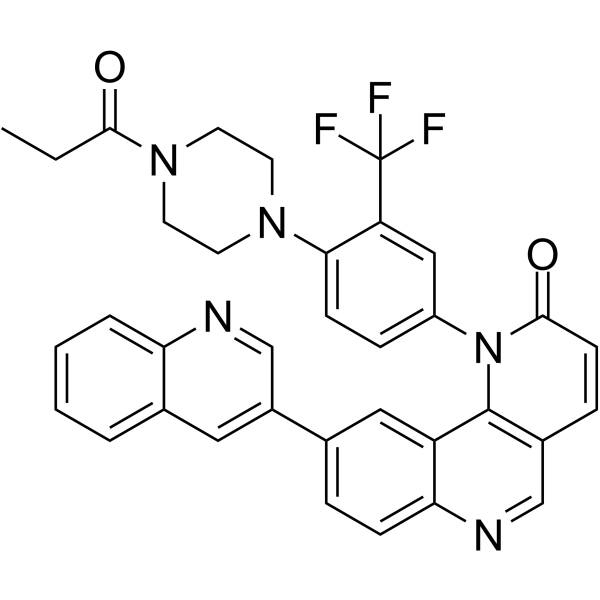
HY-13003
Torin 1mTOR Inhibitor -
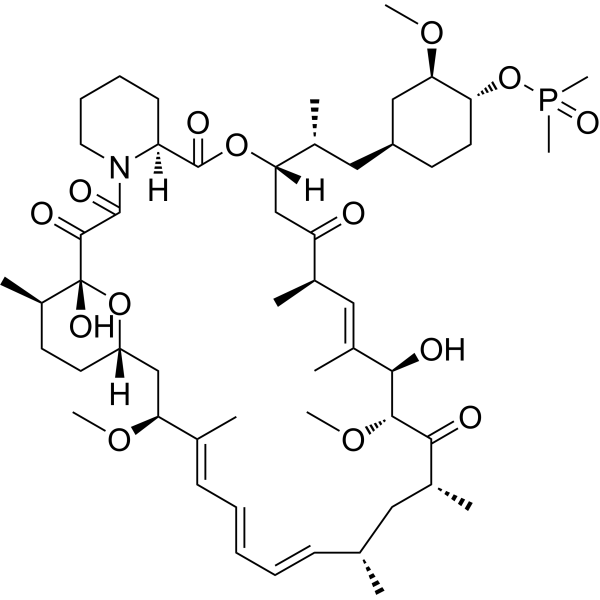
HY-50908
RidaforolimusmTOR Inhibitor -
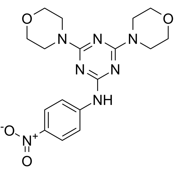
HY-B0795
MHY1485mTOR Activator -
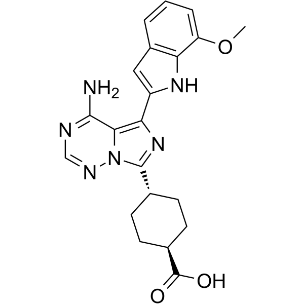
HY-10423
OSI-027mTOR Inhibitor
Publications Citing Use of MedChemExpress (MCE) mTOR inhibitor-1
More

Biological Activity
제품 설명
IC50 & Target
mTOR[1]
In Vitro
mTOR inhibitor-1 (48 h) inhibits proliferation of A549 cells (IC50: 3.176 μM), and delays S phase progression[1].
mTOR inhibitor-1 (0-12.8 μg/mL, 24 h) decreases the level of activated mTOR (p-mTORser2448) in A549 cells[1].
mTOR inhibitor-1 (3.6 μg/mL, 24 h) induces autophagosomes production in the cytoplasm of A549 cells, and accumulates LC3 puncta[1].
MedChemExpress (MCE) has not independently confirmed the accuracy of these methods. They are for reference only. Further protocols information, click here.


Chemical Information
-
CAS No. 468747-17-3
-
Appearance Solid
-
분자량 363.21
-
화학식 C16H15BrN2O3
-
Color Light yellow to yellow
-
SMILES
O=C(N/N=C(C1=CC=C(O)C=C1O)\C)C2=CC=C(C)C(Br)=C2
-
선적
Room temperature in continental US; may vary elsewhere.
-
보관
Powder -20°C 3 years 4°C 2 years In solvent -80°C 2 years -20°C 1 year


Publications (3)
-
Journal Impact Factor
-
Most Recent
-
J Am Soc Nephrol
mTOR Inhibitors Prevent CMV Infection through the Restoration of Functional αβ and γδ T cells in Kidney Transplantation. [Abstract]2022 Jan;33(1):121-137. PMID: 34725108 -
Front Pharmacol
CC-223, NSC781406, and BGT226 Exerts a Cytotoxic Effect Against Pancreatic Cancer Cells via mTOR Signaling. [Abstract]2020 Nov 11:11:580407. PMID: 33343350 -
J Ovarian Res
Baicalin improves the functions of granulosa cells and the ovary in aged mice through the mTOR signaling pathway. [Abstract]2022 Mar 17;15(1):34. PMID: 35300716


용액&용해도
In Vitro:
DMSO : 83.33 mg/mL (229.43 mM; Need ultrasonic; Hygroscopic DMSO has a significant impact on the solubility of product, please use newly opened DMSO)
Please refer to the solubility information to select the appropriate solvent. Once prepared, please aliquot and store the solution to prevent product inactivation from repeated freeze-thaw cycles.
Storage method and period of stock solution: -80°C, 2 years; -20°C, 1 year. When stored at -80°C, please use it within 2 years. When stored at -20°C, please use it within 1 year.
Please refer to the solubility information to select the appropriate solvent. Once prepared, please aliquot and store the solution to prevent product inactivation from repeated freeze-thaw cycles.
Storage method and period of stock solution: -80°C, 2 years; -20°C, 1 year. When stored at -80°C, please use it within 2 years. When stored at -20°C, please use it within 1 year.
Concentration (start) × Volume (start) = Concentration (final) × Volume (final)
In Vivo:
Select the appropriate dissolution method based on your experimental animal and administration route.
- For the following dissolution methods, please ensure to first prepare a clear stock solution using an In Vitro approach and then sequentially add co-solvents:
- To ensure reliable experimental results, the clarified stock solution can be appropriately stored based on storage conditions. As for the working solution for In Vivo experiments, it is recommended to prepare freshly and use it on the same day.
- The percentages shown for the solvents indicate their volumetric ratio in the final prepared solution. If precipitation or phase separation occurs during preparation, heat and/or sonication can be used to aid dissolution.
Add each solvent one by one: 10% DMSO 40% PEG300 5% Tween-80 45% Saline
Solubility: ≥ 2.08 mg/mL (5.73 mM); Clear solution
This protocol yields a clear solution of ≥ 2.08 mg/mL (saturation unknown).
Taking 1 mL working solution as an example, add 100 μL DMSO stock solution (20.8 mg/mL) to 400 μL PEG300, and mix evenly; then add 50 μL Tween-80 and mix evenly; then add 450 μL Saline to adjust the volume to 1 mL.
Preparation of Saline: Dissolve 0.9 g sodium chloride in ddH₂O and dilute to 100 mL to obtain a clear Saline solution.
Add each solvent one by one: 10% DMSO 90% (20% SBE-β-CD in Saline)
Solubility: ≥ 2.08 mg/mL (5.73 mM); Clear solution
This protocol yields a clear solution of ≥ 2.08 mg/mL (saturation unknown).
Taking 1 mL working solution as an example, add 100 μL DMSO stock solution (20.8 mg/mL) to 900 μL 20% SBE-β-CD in Saline, and mix evenly.
Preparation of 20% SBE-β-CD in Saline (4°C, storage for one week): 2 g SBE-β-CD powder is dissolved in 10 mL Saline, completely dissolve until clear.
In Vivo Dissolution Calculator
Please enter the basic information of animal experiments:
-
-
-
-
Recommended: Prepare an additional quantity of animals to account for potential losses during experiments.
Please enter your animal formula composition:
-
%DMSO +
Recommended: Keep the proportion of DMSO in working solution below 2% if your animal is weak.
-
%+
-
+%Tween-80 + +
-
%Saline +
The co-solvents required include: DMSO, . All of co-solvents are available by MedChemExpress (MCE). , Tween 80. All of co-solvents are available by MedChemExpress (MCE).
Working solution concentration: 0.22 mg/mL
Method for preparing stock solution: mg drug dissolved in μL DMSO. Stock solution concentration: mg/mL.
1. Take μL DMSO stock solution;
2. Add μL .
μL , mix evenly;
3. Then add μL Tween 80, mix evenly;
4. Then add μL
Please ensure that the stock solution in the first step is dissolved to a clear state, and add co-solvents in sequence. You can use ultrasonic heating (ultrasonic cleaner, recommended frequency 20-40 kHz), vortexing, etc. to assist dissolution.

Protocol
-
Autophagy
Autophagy is a process in which eukaryotic cells use lysosomes to degrade their own cytoplasmic proteins and damaged organelles under the regulation of autophagy related gene (Atg). Microtubule-associated proteins light chain 3 (LC3) is recognized as autophagy marker, which transfers from cytoplasmic LC3 (LC3-I) to membrane type (LC3-II). LC3-II/I ratio could be detected by Western Blot and fluorescence microscopy.
-
Lysosome and acidic-vesicle live-cell staining
Lysosome and acidic-vesicle live-cell staining detects acidic intracellular compartments by using membrane-permeant acidotropic probes that accumulate in low-pH vesicles, including lysosomes, late endosomes, autolysosomes, and acidic phagosomes. LysoTracker staining is commonly used as an intensity-based readout of acidic lysosomal compartment abundance or enlargement, while acridine orange produces green fluorescence in less concentrated compartments and red fluorescence after concentration-dependent accumulation in acidic vesicular organelles. Loss or reduction of acridine-orange red signal can be used as a readout of lysosomal membrane permeabilization or reduced acidic-vesicle integrity. This protocol is designed for live cultured cells and can be adapted for fluorescence microscopy, high-content imaging, plate-reader readout, or flow cytometry when the selected literature supports the readout. Because these dyes report acidotropic accumulation rather than lysosome identity alone,
-
Macroautophagy Solutions
Macroautophagy is a conserved lysosome-dependent degradation pathway in which cytoplasmic material is sequestered into double-membrane autophagosomes and delivered to lysosomes for degradation and recycling. The pathway supports cellular homeostasis during nutrient limitation, organelle stress, protein-aggregate accumulation, infection, differentiation, and tissue remodeling by coupling cargo sequestration, autophagosome maturation, lysosomal fusion, and degradation of cargo-derived macromolecules. The core molecular sequence includes initiation by nutrient- and stress-regulated autophagy machinery, autophagosome nucleation, LC3/ATG8-family conjugation to autophagosomal membranes, cargo selection through receptors such as SQSTM1/p62, autophagosome-lysosome fusion, and lysosomal degradation. LC3 was identified as a mammalian homolog of yeast Atg8 that localizes to autophagosomal membranes after processing, and p62/SQSTM1 was shown to connect ubiquitinated cargo with autophagic degradati
-
Cell Viability Determination by MTT Colorimetric Assay
The following protocol uses the MTT colorimetric assay as a classic literature-established method for assessing cell viability/metabolic activity in cultured mammalian cells. MTT[3-(4,5-dimethylthiazol-2-yl)-2,5-diphenyltetrazolium bromide] is reduced by metabolically active cells to a colored formazan product; the amount of formazan is quantified spectrophotometrically and provides an indirect measure of metabolically active viable cells. Importantly, MTT reduction reflects cellular oxidoreductase/metabolic activity rather than an absolute direct count of living cells, so changes in cellular metabolism can alter the signal independently of cell number.


순도&문서
-
Data Sheet (272 KB)
-
SDS (393 KB)
- English - EN (393 KB)
- Français - FR (393 KB)
- Deutsch - DE (393 KB)
- Norwegian - NO (393 KB)
- Español - ES (393 KB)
- Swedish - SV (393 KB)
- Italian - IT (393 KB)
- Korean - KR (393 KB)
- Portuguese - PT (393 KB)
-
Handling Instructions (2659 KB)
References


Complete Stock Solution Preparation Table
Please refer to the solubility information to select the appropriate solvent. Once prepared, please aliquot and store the solution to prevent product inactivation from repeated freeze-thaw cycles.
Storage method and period of stock solution: -80°C, 2 years; -20°C, 1 year. When stored at -80°C, please use it within 2 years. When stored at -20°C, please use it within 1 year.
| Optional Solvent | Concentration Solvent Mass | 1 mg | 5 mg | 10 mg | 25 mg |
|---|---|---|---|---|---|
| DMSO | 1 mM | 2.7532 mL | 13.7661 mL | 27.5323 mL | 68.8307 mL |
| 5 mM | 0.5506 mL | 2.7532 mL | 5.5065 mL | 13.7661 mL | |
| 10 mM | 0.2753 mL | 1.3766 mL | 2.7532 mL | 6.8831 mL | |
| 15 mM | 0.1835 mL | 0.9177 mL | 1.8355 mL | 4.5887 mL | |
| 20 mM | 0.1377 mL | 0.6883 mL | 1.3766 mL | 3.4415 mL | |
| 25 mM | 0.1101 mL | 0.5506 mL | 1.1013 mL | 2.7532 mL | |
| 30 mM | 0.0918 mL | 0.4589 mL | 0.9177 mL | 2.2944 mL | |
| 40 mM | 0.0688 mL | 0.3442 mL | 0.6883 mL | 1.7208 mL | |
| 50 mM | 0.0551 mL | 0.2753 mL | 0.5506 mL | 1.3766 mL | |
| 60 mM | 0.0459 mL | 0.2294 mL | 0.4589 mL | 1.1472 mL | |
| 80 mM | 0.0344 mL | 0.1721 mL | 0.3442 mL | 0.8604 mL | |
| 100 mM | 0.0275 mL | 0.1377 mL | 0.2753 mL | 0.6883 mL |


mTOR inhibitor-1 Related Classifications
HY-112914 Related Classifications
 Powered by Bioz