



Monotropein
Based on 3 publication(s) in Google Scholar
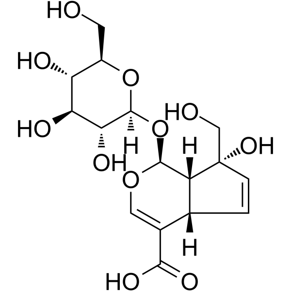
Monotropein is an iridoid glycoside that can be isolated from the roots of Morinda officinalis. Monotropein inhibits the expression of inflammatory mediators in dextran sulfate sodium (DSS)-induced colitis mouse model. Monotropein exerts protective effects against IL-1β-induced apoptosis and catabolic responses on osteoarthritis chondrocytes. Monotropein has cartilage protective activity. Monotropein can alleviate Cisplatin (HY-17394)-induced acute kidney injury by inhibiting oxidative damage, inflammation and apoptosis through activation of Nrf2/HO-1 pathway and inhibition of NF-κB signaling. Monotropein can be studied in research for osteoarthritis, acute kidney injury and acute lung injury.
For research use only. We do not sell to patients.
- Purity : 99.80%
- CAS No.: 5945-50-6
- Formula: C16H22O11
- Molecular Weight:390.34
-
Storage:Powder -20°C, 3 years , 4°C, 2 years ; In solvent -80°C, 6 months , -20°C, 1 month

To place orders, for customer services and technical support, please contact: MedChemExpress USA
Tel: 609-228-6898 E-mail: [email protected] [email protected]


-
 Biological Activity
-
Chemical Information
-
Solvent & Solubility
- Protocols
- Purity & Documentation
- References
-
 Help & FAQs



Publications Citing Use of MedChemExpress (MCE) Monotropein
More Customer Validation & Images

-
ELISA

-
IF

-
Histological Imaging/Staining

-
WB

-
RT-PCR


Biological Activity
Description
In Vitro
Monotropein (100 μg/mL, 24 h) decreases the apoptotic rate of chondrocytes[1].
Monotropein (25-100 μg/mL, 24 h) significantly decreases the mRNA levels of MMP-3 and MMP-13 in chondrocytes[1].
Monotropein (25-100 μg/mL, 48 h) exhibits anti-apoptosis and anti-catabolic activity and counteracts the effect of IL-1β[1].
Monotropein (Compound Mon) (0-200 μg/mL, 24 h) enhances cell viability and reduces apoptosis in LPS-treated MLE-12 cells[3].
Monotropein (12.5-100 μg/mL, 24 h) reduces inflammatory response and fibrosis induced by LPS in MLE-12 cells[3].
Monotropein (100 μg/mL, 24 h) inhibits NF-κB pathway in LPS-elicited MLE-12 cells[3].
MedChemExpress (MCE) has not independently confirmed the accuracy of these methods. They are for reference only. Further protocols information, click here.
-
Cell Line:LPS-elicited MLE-12 cells
-
Concentration:100 μg/mL
-
Incubation Time:24 h
-
Result:Decreased the concentrations of TNF-α, IL-1β, and IL-6.
Decreased relative protein expression of -SMA, FN, and Collagen I.
In Vivo
Monotropein (100 mg/kg, i.g., single dose) exhibits chondroprotective effects on knee joint tissue of OA rat model[1].
Monotropein (20 mg/kg, i.p., 1 h pre-treatment and 24-48 h after) attenuates cisplatin-induced renal dysfunction and histopathological damage in AKI mouse model[2].
MedChemExpress (MCE) has not independently confirmed the accuracy of these methods. They are for reference only.
-
Animal Model:AKI BALB/c mouse model (20-25 g) injected (i.p.) with 20 mg/kg Cisplatin[2]
-
Dosage:20 mg/kg
-
Administration:Intraperitoneal injection (i.p.), 1 h pre-treatment and 24-48 h after
-
Result:Reduced the level on BUN compared to control group.
Attenuated cisplatin-induced oxidative stress in kidney.
Attenuated inflammation in kidney.
Reversed the anti-apoptotic protein Bcl-2 and pro-apoptotic proteins including Bax.


Chemical Information
-
CAS No. 5945-50-6
-
Appearance Solid
-
Molecular Weight 390.34
-
Formula C16H22O11
-
Color Off-white to yellow
-
SMILES
O[C@H]([C@@H](O)[C@@H]1O)[C@](O[C@@H]1CO)([H])O[C@H]2[C@@]([C@@]3(O)CO)([H])[C@@](C=C3)([H])C(C(O)=O)=CO2
-
Structure Classification
-
Shipping
Room temperature in continental US; may vary elsewhere.
-
Storage
Powder -20°C 3 years 4°C 2 years In solvent -80°C 6 months -20°C 1 month


Publications (3)
-
Journal Impact Factor
-
Most Recent
-
Int Immunopharmacol
The active ingredient Monotropein in Morinda officinalis alleviates neuroinflammation via inhibiting cGAS/STING signaling pathway. [Abstract]2026 Aug 15:183:116862. PMID: 42150287
Monotropein purchased from MedChemExpress. Usage Cited in: Int Immunopharmacol. 2026 Aug 15:183:116862. [Abstract]
IL-1β, TNF-α, IL-6, IFN-β, and IL-10 levels in mouse hippocampal tissue treated with Monotropein (Mon) (20, 80 mg/kg, i.p.).
Monotropein purchased from MedChemExpress. Usage Cited in: Int Immunopharmacol. 2026 Aug 15:183:116862. [Abstract]
Representative fluorescence staining of Iba-1 in each group of mice treated with Monotropein (Mon) (20, 80 mg/kg, i.p.).
Monotropein purchased from MedChemExpress. Usage Cited in: Int Immunopharmacol. 2026 Aug 15:183:116862. [Abstract]
H&E staining of mouse hippocampal tissue treated with Monotropein (Mon) (20, 80 mg/kg, i.p.).
Monotropein purchased from MedChemExpress. Usage Cited in: Int Immunopharmacol. 2026 Aug 15:183:116862. [Abstract]
Bands of key proteins in the cGAS/STING signaling pathway treated with Monotropein (Mon) (20, 80 mg/kg, i.p.).
Monotropein purchased from MedChemExpress. Usage Cited in: Int Immunopharmacol. 2026 Aug 15:183:116862. [Abstract]
The expression levels of the Ifnb1, Il-6, Tnf-α, Ifit1, Cxcl10, and Isg15 genes in hippocampal tissue treated with Monotropein (Mon) (20, 80 mg/kg, i.p.).
-
Viruses
Anthraquinone-2-Carboxylic Acid Is a Potential Antiviral Candidate Against Influenza Viruses In Vitro and In Vivo. [Abstract]2025 Apr 27;17(5):628. PMID: 40431640 -


Solvent & Solubility
In Vitro:
H2O : 31.25 mg/mL (80.06 mM; Need ultrasonic)
Please refer to the solubility information to select the appropriate solvent. Once prepared, please aliquot and store the solution to prevent product inactivation from repeated freeze-thaw cycles.
Storage method and period of stock solution: -80°C, 6 months; -20°C, 1 month. When stored at -80°C, please use it within 6 months. When stored at -20°C, please use it within 1 month.
* Note: If you choose water as the stock solution, please dilute it to the working solution, then filter and sterilize it with a 0.22 μm filter before use.
Please refer to the solubility information to select the appropriate solvent. Once prepared, please aliquot and store the solution to prevent product inactivation from repeated freeze-thaw cycles.
Storage method and period of stock solution: -80°C, 6 months; -20°C, 1 month. When stored at -80°C, please use it within 6 months. When stored at -20°C, please use it within 1 month.
* Note: If you choose water as the stock solution, please dilute it to the working solution, then filter and sterilize it with a 0.22 μm filter before use.
Concentration (start) × Volume (start) = Concentration (final) × Volume (final)

Protocols
-
RNA extraction experimental
By lysing cells, releasing RNA, and removing impurities such as proteins and DNA, high-purity RNA products are finally obtained. The commonly used traditional method is the guanidine isothiocyanate/phenol/chloroform method (Trizol), which is suitable for a variety of animal materials including animal tissues, microorganisms, cultured cells, etc., and most plant materials.
-
Nephrotoxicity Study
This protocol assesses nephrotoxicity by combining functional kidney injury readouts, urinary/tissue injury biomarkers, and renal histopathology. Serum creatinine and BUN reflect impaired kidney function, while KIM-1, NGAL, clusterin, osteopontin, IL-18, cystatin C, nephrin, Oat5, urinary protein, glucose, and alkaline phosphatase have been used to detect tubular injury in cisplatin-, gentamicin-, and acetaminophen-induced nephrotoxicity models.
-
Annexin V plus membrane-impermeant dye apoptosis staining
Annexin V-based apoptosis assays rely on the detection of phosphatidylserine (PS) externalization from the inner leaflet of the plasma membrane to the outer leaflet, an early biochemical hallmark of apoptosis. Fluorescently labeled Annexin V binds PS in a calcium-dependent manner, enabling identification of early apoptotic cells by flow cytometry or fluorescence microscopy. When combined with a membrane-impermeant DNA-binding dye (e. g. , propidium iodide), this approach allows discrimination between viable (Annexin V−/dye−), early apoptotic (Annexin V+/dye−), and late apoptotic or necrotic (Annexin V+/dye+) cell populations by assessing membrane integrity and PS exposure.
-
ROS/oxidative-stress fluorescent staining
ROS/oxidative-stress fluorescent staining uses cell-permeant fluorogenic probes that become fluorescent after oxidation inside cells or tissues; commonly used examples include DCFH-DA/DCFDA for broad cellular oxidant detection, DHE for superoxide-related signal detection, MitoSOX for mitochondrial superoxide-related signal detection, and CellROX probes for oxidative-stress-associated fluorescence readouts. The assay detects probe oxidation rather than a single ROS species unless the probe and analysis method have been chemically validated for that species. DCFH-DA enters cells, is deacetylated by intracellular esterases to DCFH, and produces fluorescent DCF after oxidation, so the readout is used as an operational measure of total cellular oxidative stress rather than a species-specific ROS measurement. DHE and MitoSOX can report superoxide-related oxidation, but red fluorescence alone can include non-specific ethidium-like oxidation products; HPLC or optimized spectral approaches are
-
Research Protocol for Inflammation-related Diseases
The NLRP3 inflammasome is a cytosolic innate immune signaling platform that integrates priming signals and danger-signal activation to promote caspase-1 activation, maturation of IL-1β and IL-18, and gasdermin D-mediated pyroptotic cell death. The core experimental logic is to determine whether inflammatory disease phenotypes are driven by increased NLRP3 expression, ASC-containing inflammasome assembly, caspase-1 cleavage, GSDMD cleavage, and extracellular release of IL-1β/IL-18 rather than by nonspecific cell injury alone. The pathway is strongly linked to inflammation-related disease phenotypes because monosodium urate crystals activate NALP3/NLRP3 inflammasome signaling in gout-like crystal inflammation, cholesterol crystals activate NLRP3 inflammasomes in atherogenesis models, and DSS-induced intestinal inflammation has been reported to involve NLRP3 inflammasome activity. However, experimental colitis studies also show context-dependent protective effects of NLRP3 inflammasome co
-
How to Choose the Right Model Animal
Choosing the right model animal is a validity-driven decision in which the species, strain, sex, age, genetic background, disease-induction method, outcome measures, and welfare burden must match the scientific question rather than laboratory tradition or convenience. A model should be selected by judging face validity, construct validity, and predictive validity: whether it resembles the human phenotype, whether it reproduces relevant mechanisms, and whether results are likely to predict human biology or treatment response. Animal studies often fail to translate because of species differences, weak disease resemblance, poor experimental design, inadequate reporting, publication bias, and underuse of randomization, blinding, and sample-size justification. Unresolved questions include how to rank competing models objectively, how much human-disease complexity must be reproduced for a given objective, and when non-animal systems such as organoids, ex vivo tissue, or computational models
-
Apoptosis
Apoptosis, also called programmed cell death, is generally characterized by distinct morphological characteristics.
-
TUNEL staining for apoptotic DNA fragmentation
TUNEL staining detects DNA strand breaks by using terminal deoxynucleotidyl transferase to add labeled nucleotides to exposed 3′-OH DNA termini, generating either microscopic staining in fixed cells or tissue sections, or fluorescence/cytometric signal in cell suspensions. TUNEL positivity reflects DNA fragmentation but should not be interpreted alone as definitive apoptosis, because TUNEL can also label necrotic, autolytic, mechanically damaged, or DNA-repair-associated DNA breaks.
-
DSS-Induced Colitis
Dextran sulfate sodium (DSS)-induced colitis is generated by administering DSS in mouse drinking water, producing epithelial injury, barrier disruption, weight loss, diarrhea, fecal blood, colon shortening, histologic mucosal damage, and inflammatory mediator changes; the model is mainly used to study acute or chronic intestinal inflammation resembling selected features of ulcerative colitis. DSS injury is interpreted through clinical and tissue readouts rather than a single molecular endpoint: daily body weight, stool consistency, and bleeding are combined into a disease activity index, while colon length, histology, cytokines, myeloperoxidase activity, intestinal permeability, and tight-junction markers provide complementary measures of inflammation and barrier damage.
-
TNBS-Induced Colitis
TNBS-induced colitis is produced by intrarectal delivery of 2,4,6-trinitrobenzene sulfonic acid in ethanol, where ethanol disrupts the mucosal barrier and TNBS haptenates colonic proteins, generating immune-mediated colonic inflammation with weight loss, diarrhea, ulceration, transmural injury, inflammatory-cell infiltration, and cytokine responses. The model is used as an experimental intestinal inflammation model with Crohn’s disease–like features, especially when Th1-type responses, IL-12–dependent inflammation, chronic relapsing inflammation, or fibrosis-related endpoints are studied.
-
Apoptosis Solutions
Apoptosis is a regulated, generally non-lytic cell-death pathway that removes unwanted, damaged, infected, or abnormal cells through coordinated morphological changes, caspase activation, DNA fragmentation, and membrane remodeling. The intrinsic apoptosis pathway is controlled mainly by mitochondrial outer membrane permeabilization, BCL-2 family proteins, cytochrome c release, apoptosome formation, caspase-9 activation, and downstream executioner caspase-3/7 activation. The extrinsic apoptosis pathway is initiated by death receptors such as Fas, TNFR, and TRAIL receptors, which recruit adaptor proteins and activate caspase-8 before engaging executioner caspases or mitochondrial amplification through BID cleavage. Apoptosis is linked to many phenotypes, including cancer cell killing, tissue homeostasis, immune regulation, neurodegeneration, infection response, and treatment-induced cytotoxicity; unresolved questions include how apoptosis interacts with necroptosis, pyroptosis, ferroptos
-
Inhalation Toxicity Study
Inhalation toxicity studies expose rodents to a controlled aerosol, vapor, gas, or smoke atmosphere and assess respiratory and systemic toxicity using exposure-atmosphere characterization, clinical observations, body and organ weights, bronchoalveolar lavage fluid, histopathology, blood chemistry, hematology, and, when included, molecular endpoints such as transcriptomics, proteomics, lipidomics, or tissue burden analysis. The primary biological readouts are airway irritation, pulmonary inflammation, cytotoxicity, altered surfactant or lipid homeostasis, impaired particle clearance, and tissue remodeling, reflected by BALF cell differentials, BALF protein, LDH, phosphatase activities, cytokines, lung weight, microscopic respiratory-tract lesions, and retained lung burden.
-
Pyroptosis Solutions
Pyroptosis is a lytic inflammatory cell-death pathway executed by gasdermin pores, most classically through inflammasome-mediated activation of caspase-1, cleavage of gasdermin D, membrane pore formation, LDH release, and secretion of IL-1β and IL-18. The canonical pathway is commonly modeled by priming cells with an inflammatory signal such as LPS to induce pro-IL-1β and inflammasome components, followed by an activation signal such as ATP or nigericin to activate NLRP3, ASC speck formation, caspase-1 cleavage, GSDMD cleavage, cytokine release, and pyroptotic membrane rupture. The non-canonical pathway is triggered when cytosolic LPS activates mouse caspase-11 or human caspase-4/5, leading to GSDMD cleavage and pyroptosis, and this can secondarily activate NLRP3-dependent IL-1β release. Pyroptosis is linked to inflammatory injury, infection, cancer, liver disease, ocular disease, placental inflammation, and other disease phenotypes, but unresolved questions include which gasdermin fam


Purity & Documentation
-
Data Sheet (286 KB)
-
SDS (393 KB)
- English - EN (393 KB)
- Français - FR (393 KB)
- Deutsch - DE (393 KB)
- Norwegian - NO (393 KB)
- Español - ES (393 KB)
- Swedish - SV (393 KB)
- Italian - IT (393 KB)
- Korean - KR (393 KB)
- Portuguese - PT (393 KB)
-
Handling Instructions (2659 KB)
References
[1]. Wang F, et al.Monotropein exerts protective effects against IL-1β-induced apoptosis and catabolic responses on osteoarthritis chondrocytes.Int Immunopharmacol. 2014 Dec;23(2):575-80. [Content Brief]
[2]. Zhang, Y., et al., (2020). The effect of monotropein on alleviating cisplatin-induced acute kidney injury by inhibiting oxidative damage, inflammation and apoptosis. Biomedicine & pharmacotherapy = Biomedecine & pharmacotherapie, 129, 110408. [Content Brief]
[3]. Gong, Y., & Wang, J. (2023). Monotropein alleviates sepsis-elicited acute lung injury via the NF-κB pathway. The Journal of pharmacy and pharmacology, 75(9), 1249–1258. [Content Brief]


Complete Stock Solution Preparation Table
Please refer to the solubility information to select the appropriate solvent. Once prepared, please aliquot and store the solution to prevent product inactivation from repeated freeze-thaw cycles.
Storage method and period of stock solution: -80°C, 6 months; -20°C, 1 month. When stored at -80°C, please use it within 6 months. When stored at -20°C, please use it within 1 month.
| Optional Solvent | Concentration Solvent Mass | 1 mg | 5 mg | 10 mg | 25 mg |
|---|---|---|---|---|---|
| H2O | 1 mM | 2.5619 mL | 12.8093 mL | 25.6187 mL | 64.0467 mL |
| 5 mM | 0.5124 mL | 2.5619 mL | 5.1237 mL | 12.8093 mL | |
| 10 mM | 0.2562 mL | 1.2809 mL | 2.5619 mL | 6.4047 mL | |
| 15 mM | 0.1708 mL | 0.8540 mL | 1.7079 mL | 4.2698 mL | |
| 20 mM | 0.1281 mL | 0.6405 mL | 1.2809 mL | 3.2023 mL | |
| 25 mM | 0.1025 mL | 0.5124 mL | 1.0247 mL | 2.5619 mL | |
| 30 mM | 0.0854 mL | 0.4270 mL | 0.8540 mL | 2.1349 mL | |
| 40 mM | 0.0640 mL | 0.3202 mL | 0.6405 mL | 1.6012 mL | |
| 50 mM | 0.0512 mL | 0.2562 mL | 0.5124 mL | 1.2809 mL | |
| 60 mM | 0.0427 mL | 0.2135 mL | 0.4270 mL | 1.0674 mL | |
| 80 mM | 0.0320 mL | 0.1601 mL | 0.3202 mL | 0.8006 mL |
* Note: If you choose water as the stock solution, please dilute it to the working solution, then filter and sterilize it with a 0.22 μm filter before use.


Monotropein Related Classifications
HY-N0648 Related Classifications
 Powered by Bioz