



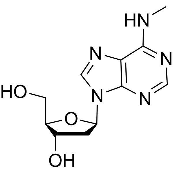
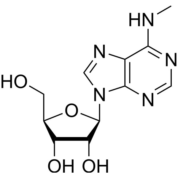
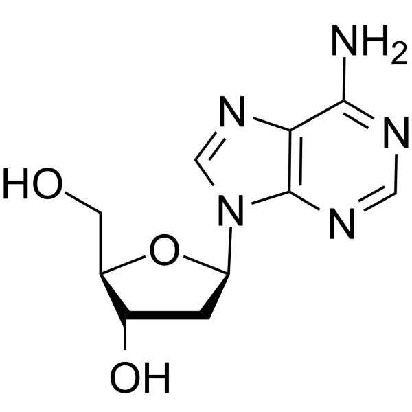
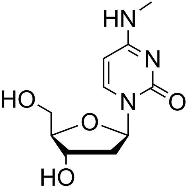
N-6-Methyl-2-deoxyadenosine

Based on 1 Customer Validation
N-6-Methyl-2-deoxyadenosine (m6dA) is an adenine nucleoside analogue. N-6-Methyl-2-deoxyadenosine targets nuclear processes and DNA replication machineries including WER, SATB1, TFAM, Jumu, SSBP1, DNA polymerase η and phage polymerase Gp90 exo−. N-6-Methyl-2-deoxyadenosine acts as a multifunctional epigenetic regulator that modulates transcription, DNA damage response, cell cycle, transposon silencing, stress adaptation, epigenetic crosstalk, and nucleosome organization in both prokaryotes and eukaryotes. N-6-Methyl-2-deoxyadenosine regulates mitochondrial epigenetic inheritance and is required for fear extinction memory in mice. N-6-Methyl-2-deoxyadenosine exhibits dysregulated levels in cancers. N-6-Methyl-2-deoxyadenosine can be used for the research of glioblastoma, triple negative breast cancer, and conditioned fear (fear extinction impairment).
For research use only. We do not sell to patients.
- Purity : 99.83%
- CAS No.: 2002-35-9
- Formula: C11H15N5O3
- Molecular Weight:265.27
-
Storage:Powder -20°C, 3 years ; In solvent -80°C, 6 months , -20°C, 1 month

To place orders, for customer services and technical support, please contact: MedChemExpress USA
Tel: 609-228-6898 E-mail: [email protected] [email protected]


-
 Biological Activity
-
Chemical Information
-
Solvent & Solubility
- Protocols
- Purity & Documentation
- References
-
 Help & FAQs



All DNA/RNA Synthesis Isoforms
More

Biological Activity
Description
IC50 & Target
[1]|
DNA Polymerase |
In Vitro
N-6-Methyl-2-deoxyadenosine causes site-specific transcriptional pausing by Saccharomyces cerevisiae RNA polymerase II[1].
N-6-Methyl-2-deoxyadenosine inhibits DNA replication by human DNA polymerase-η in vitro[1].
N-6-Methyl-2-deoxyadenosine enhances the DNA-binding affinity of human mitochondrial SSBP1, reduces TFAM-mediated mtDNA transcription, and can be catalyzed and deposited onto mammalian mitochondrial DNA by human METTL4 in vitro[1].
N-6-Methyl-2-deoxyadenosine reduces the binding affinity of Arabidopsis thaliana transcription factor WER to its target DNA motifs in vitro[1].
N-6-Methyl-2-deoxyadenosine slightly increases the binding affinity of Drosophila melanogaster transcription factor Jumu to DNA in vitro[1].
N-6-Methyl-2-deoxyadenosine inhibits DNA replication by Pseudomonas aeruginosa phage PaP1 DNA polymerase Gp90 exo− [1].
N-6-Methyl-2-deoxyadenosine (20 mM; 7 h) was measured in mouse primary cortical neurons (7 days in vitro) treated with KCl, which exhibited a significant increase in global m6dA levels[2].
MedChemExpress (MCE) has not independently confirmed the accuracy of these methods. They are for reference only. Further protocols information, click here.
In Vivo
MedChemExpress (MCE) has not independently confirmed the accuracy of these methods. They are for reference only.


Chemical Information
-
CAS No. 2002-35-9
-
Appearance Solid
-
Molecular Weight 265.27
-
Formula C11H15N5O3
-
Color White to off-white
-
SMILES
OC[C@@H]1[C@@H](O)C[C@H](N2C(N=CN=C3NC)=C3N=C2)O1
-
Synonyms
m6dA
-
Shipping
Room temperature in continental US; may vary elsewhere.
-
Storage
Powder -20°C 3 years In solvent -80°C 6 months -20°C 1 month


Solvent & Solubility
In Vitro:
DMSO : 100 mg/mL (376.97 mM; ultrasonic and warming and heat to 60°C; Hygroscopic DMSO has a significant impact on the solubility of product, please use newly opened DMSO)
Please refer to the solubility information to select the appropriate solvent. Once prepared, please aliquot and store the solution to prevent product inactivation from repeated freeze-thaw cycles.
Storage method and period of stock solution: -80°C, 6 months; -20°C, 1 month. When stored at -80°C, please use it within 6 months. When stored at -20°C, please use it within 1 month.
Please refer to the solubility information to select the appropriate solvent. Once prepared, please aliquot and store the solution to prevent product inactivation from repeated freeze-thaw cycles.
Storage method and period of stock solution: -80°C, 6 months; -20°C, 1 month. When stored at -80°C, please use it within 6 months. When stored at -20°C, please use it within 1 month.
Concentration (start) × Volume (start) = Concentration (final) × Volume (final)
In Vivo:
Select the appropriate dissolution method based on your experimental animal and administration route.
- For the following dissolution methods, please ensure to first prepare a clear stock solution using an In Vitro approach and then sequentially add co-solvents:
- To ensure reliable experimental results, the clarified stock solution can be appropriately stored based on storage conditions. As for the working solution for In Vivo experiments, it is recommended to prepare freshly and use it on the same day.
- The percentages shown for the solvents indicate their volumetric ratio in the final prepared solution. If precipitation or phase separation occurs during preparation, heat and/or sonication can be used to aid dissolution.
Add each solvent one by one: 10% DMSO 40% PEG300 5% Tween-80 45% Saline
Solubility: ≥ 2.5 mg/mL (9.42 mM); Clear solution
This protocol yields a clear solution of ≥ 2.5 mg/mL (saturation unknown).
Taking 1 mL working solution as an example, add 100 μL DMSO stock solution (25.0 mg/mL) to 400 μL PEG300, and mix evenly; then add 50 μL Tween-80 and mix evenly; then add 450 μL Saline to adjust the volume to 1 mL.
Preparation of Saline: Dissolve 0.9 g sodium chloride in ddH₂O and dilute to 100 mL to obtain a clear Saline solution.
Add each solvent one by one: 10% DMSO 90% (20% SBE-β-CD in Saline)
Solubility: ≥ 2.5 mg/mL (9.42 mM); Clear solution
This protocol yields a clear solution of ≥ 2.5 mg/mL (saturation unknown).
Taking 1 mL working solution as an example, add 100 μL DMSO stock solution (25.0 mg/mL) to 900 μL 20% SBE-β-CD in Saline, and mix evenly.
Preparation of 20% SBE-β-CD in Saline (4°C, storage for one week): 2 g SBE-β-CD powder is dissolved in 10 mL Saline, completely dissolve until clear.
In Vivo Dissolution Calculator
Please enter the basic information of animal experiments:
-
-
-
-
Recommended: Prepare an additional quantity of animals to account for potential losses during experiments.
Please enter your animal formula composition:
-
%DMSO +
Recommended: Keep the proportion of DMSO in working solution below 2% if your animal is weak.
-
%+
-
+%Tween-80 + +
-
%Saline +
The co-solvents required include: DMSO, . All of co-solvents are available by MedChemExpress (MCE). , Tween 80. All of co-solvents are available by MedChemExpress (MCE).
Working solution concentration: 0.22 mg/mL
Method for preparing stock solution: mg drug dissolved in μL DMSO. Stock solution concentration: mg/mL.
1. Take μL DMSO stock solution;
2. Add μL .
μL , mix evenly;
3. Then add μL Tween 80, mix evenly;
4. Then add μL
Please ensure that the stock solution in the first step is dissolved to a clear state, and add co-solvents in sequence. You can use ultrasonic heating (ultrasonic cleaner, recommended frequency 20-40 kHz), vortexing, etc. to assist dissolution.

Protocols
-
PCNA Immunodetection Proliferation Assay
PCNA immunodetection measures proliferative activity by detecting proliferating cell nuclear antigen, a nuclear protein associated with DNA polymerase δ function and DNA replication. The assay readout is the proportion of PCNA-positive nuclei among total counted cells, but PCNA labeling is not identical to BrdU labeling because PCNA can mark late G1/early S-associated replication competence and may persist beyond active DNA synthesis depending on fixation and extraction conditions.
-
Flow cytometric DNA-content cell-cycle staining
Flow cytometric DNA-content cell-cycle staining measures the fluorescence intensity of DNA-bound fluorochromes in single cells or nuclei to estimate DNA content distributions, allowing assignment of populations to G0/G1, S, and G2/M phases by DNA histogram deconvolution. Propidium iodide (PI) intercalates into DNA, and PI fluorescence is proportional to cellular DNA content when staining is performed under conditions that make DNA accessible and minimize non-DNA signal. Cells with G2/M DNA content are expected to show approximately twice the fluorescence intensity of G0/G1 cells, while S-phase cells occupy intermediate fluorescence values. PI-based DNA-content analysis can also detect cells with fractional DNA content, often reported as sub-G1, when DNA fragmentation and extraction during staining reduce retained DNA signal in apoptotic cells. DAPI is an alternative DNA fluorochrome for univariate DNA-content analysis, while bivariate approaches combining DNA content with proliferation
-
BrdU Incorporation Assay
Bromodeoxyuridine (BrdU) incorporation assay is based on the principle that BrdU, a thymidine analog, is incorporated into newly synthesized DNA during the S phase of the cell cycle, thereby serving as a marker of DNA replication and cellular proliferation. Incorporated BrdU can be detected using anti-BrdU antibodies following DNA denaturation, enabling visualization or quantification of proliferating cells through immunochemical detection methods such as immunofluorescence or immunohistochemistry.
-
Protocol for Cell Cycle
Cell-cycle analysis by flow cytometry measures DNA content in single cells to estimate the fraction of cells in G0/G1, S, and G2/M phases. Propidium iodide intercalates into DNA, and after RNA removal with RNase, fluorescence intensity reflects cellular DNA content: 2N cells are assigned to G0/G1, cells between 2N and 4N to S phase, and 4N cells to G2/M. DNA-content analysis alone cannot reliably separate G0 from G1 or G2 from M. Ki-67 can distinguish quiescent G0 cells from cycling cells, EdU or BrdU incorporation marks active DNA synthesis in S phase, and phospho-histone H3 staining identifies mitotic cells within the 4N population.
-
Breast Cancer Modeling
Breast cancer is a heterogeneous cancer, and it has been distinguished into four subtypes: luminal A, luminal B, HER2-positive and basal-like. Molecular mutations, epigenetic alterations, hormone exposure and immune microenvironment are related to the progression of breast cancer.
-
Genotoxicity/Mutagenicity Study
The bacterial reverse mutation assay detects point mutations that restore amino-acid prototrophy in auxotrophic Salmonella typhimurium or Escherichia coli tester strains; after exposure to a test article, mutagenic activity is read out as an increased number of revertant colonies on minimal agar compared with the vehicle control. The assay uses tester strains with different mutation targets so that base-substitution and frameshift mutagens can be detected, and testing is performed with and without exogenous mammalian metabolic activation because some chemicals require biotransformation to become mutagenic.


Purity & Documentation
-
Data Sheet (279 KB)
-
SDS (394 KB)
- English - EN (394 KB)
- Français - FR (394 KB)
- Deutsch - DE (394 KB)
- Norwegian - NO (394 KB)
- Español - ES (394 KB)
- Swedish - SV (394 KB)
- Italian - IT (394 KB)
- Korean - KR (394 KB)
- Portuguese - PT (394 KB)
-
Handling Instructions (2659 KB)
References
[1]. Boulias K, et al. Means, mechanisms and consequences of adenine methylation in DNA. Nat Rev Genet. 2022;23(7):411-428. [Content Brief]
[2]. Li X, et al. The DNA modification N6-methyl-2'-deoxyadenosine (m6dA) drives activity-induced gene expression and is required for fear extinction. Nat Neurosci. 2019;22(4):534-544. [Content Brief]


Complete Stock Solution Preparation Table
Please refer to the solubility information to select the appropriate solvent. Once prepared, please aliquot and store the solution to prevent product inactivation from repeated freeze-thaw cycles.
Storage method and period of stock solution: -80°C, 6 months; -20°C, 1 month. When stored at -80°C, please use it within 6 months. When stored at -20°C, please use it within 1 month.
| Optional Solvent | Concentration Solvent Mass | 1 mg | 5 mg | 10 mg | 25 mg |
|---|---|---|---|---|---|
| DMSO | 1 mM | 3.7697 mL | 18.8487 mL | 37.6974 mL | 94.2436 mL |
| 5 mM | 0.7539 mL | 3.7697 mL | 7.5395 mL | 18.8487 mL | |
| 10 mM | 0.3770 mL | 1.8849 mL | 3.7697 mL | 9.4244 mL | |
| 15 mM | 0.2513 mL | 1.2566 mL | 2.5132 mL | 6.2829 mL | |
| 20 mM | 0.1885 mL | 0.9424 mL | 1.8849 mL | 4.7122 mL | |
| 25 mM | 0.1508 mL | 0.7539 mL | 1.5079 mL | 3.7697 mL | |
| 30 mM | 0.1257 mL | 0.6283 mL | 1.2566 mL | 3.1415 mL | |
| 40 mM | 0.0942 mL | 0.4712 mL | 0.9424 mL | 2.3561 mL | |
| 50 mM | 0.0754 mL | 0.3770 mL | 0.7539 mL | 1.8849 mL | |
| 60 mM | 0.0628 mL | 0.3141 mL | 0.6283 mL | 1.5707 mL | |
| 80 mM | 0.0471 mL | 0.2356 mL | 0.4712 mL | 1.1780 mL | |
| 100 mM | 0.0377 mL | 0.1885 mL | 0.3770 mL | 0.9424 mL |


N-6-Methyl-2-deoxyadenosine Related Classifications
HY-W011725 Related Classifications
 Powered by Bioz