



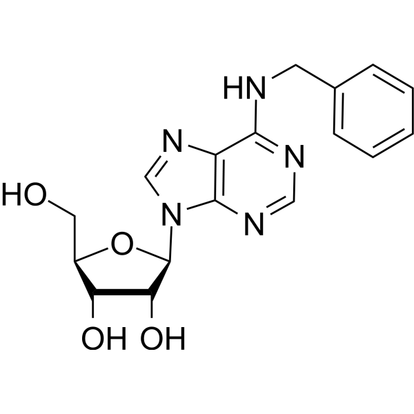
N6-Benzyladenosine

Based on 1 Customer Validation
N6-Benzyladenosine is an adenosine receptor agonist, has a cytoactive activity. N6-Benzyladenosine arrests cell cycle at G0/G1 phase and induces cell apoptosis. N6-Benzyladenosine also exerts inhibitory effect on T. gondii adenosine kinase and glioma-.
For research use only. We do not sell to patients.
- Purity : 99.72%
- CAS No.: 4294-16-0
- Formula: C17H19N5O4
- Molecular Weight:357.36
-
Storage:
4°C, protect from light
* In solvent : -80°C, 6 months; -20°C, 1 month (protect from light)

To place orders, for customer services and technical support, please contact: MedChemExpress USA
Tel: 609-228-6898 E-mail: [email protected] [email protected]


-
 Biological Activity
-
Chemical Information
-
Solvent & Solubility
- Protocols
- Purity & Documentation
- References
-
 Help & FAQs


-

HY-B2132
Tryptamine5-HT4 Receptor Agonist -
HY-101055
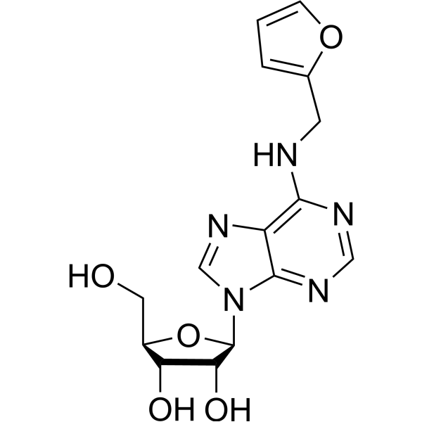
Kinetin ribosideApoptosis Activator -
HY-18775
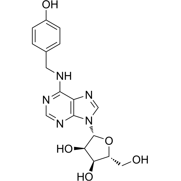
N6-(4-Hydroxybenzyl)adenosineENT1 Inhibitor/A2aR Agonist -
HY-N0086
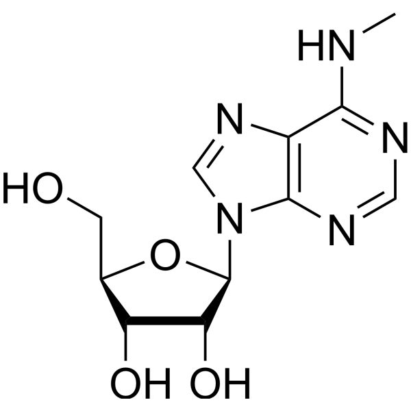
N6-MethyladenosineViral RNA Modified Agent -
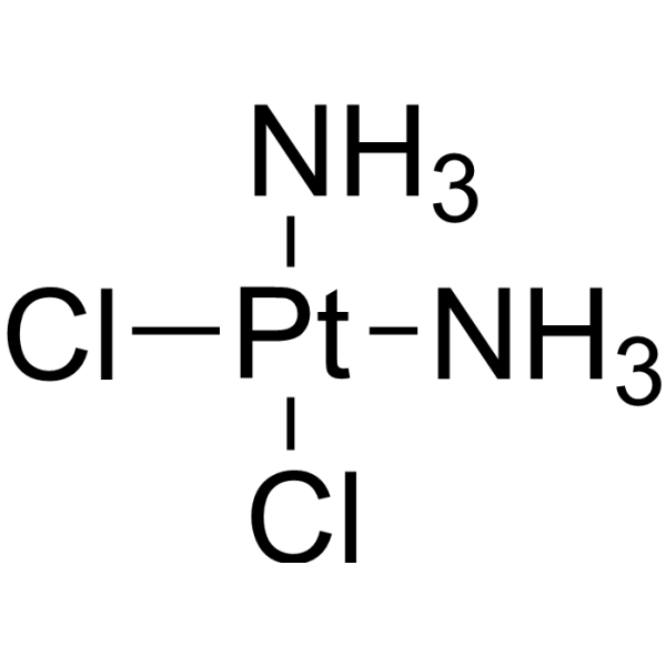
HY-17394
CisplatinDNA Alkylator -
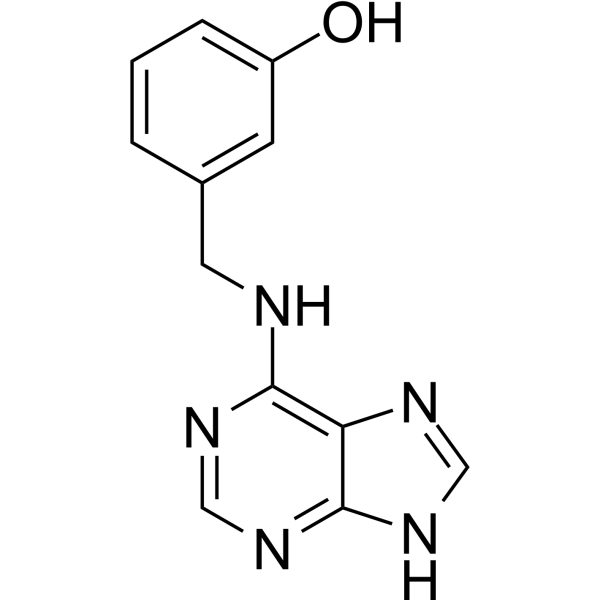
HY-112104
Meta-topolinHydroxylated Aromatic Cytokinin -
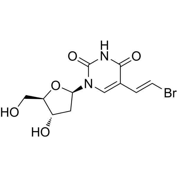
HY-13578
BrivudineCMV Inhibitor -

HY-N7844R
N6-Benzyladenosine (Standard)Adenosine Receptor Agonist -
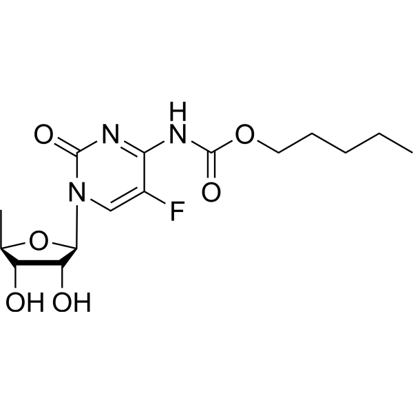
HY-B0016
CapecitabineDNA/RNA Synthesis Inhibitor -
HY-B0307
IdoxuridineAntiviral Agent -
HY-118122
FialuridineAntiviral Agent -
HY-B0017
TelbivudineHBV Replication Inhibitor
All Adenosine Receptor Isoforms
More

Biological Activity
Description
Cellular Effect
|
Cell Line

|
Type | Value | Description | References |
|---|---|---|---|---|
| B16 | IC50 |
>166.7 μM
Compound: BAPR
|
Antitumor activity against mouse B16 cells after 72 hrs by Calcein AM assay
Antitumor activity against mouse B16 cells after 72 hrs by Calcein AM assay
|
[PMID: 17418578] |
| Caco-2 | GI50 |
7.4 μM
Compound: 19, BnA
|
Antitumor activity against human CaCo2 cells after 48 hrs by MTS assay
Antitumor activity against human CaCo2 cells after 48 hrs by MTS assay
|
[PMID: 18588281] |
| CCRF-CEM | IC50 |
1.4 μM
Compound: BAPR
|
Antitumor activity against human CEM cells after 72 hrs by Calcein AM assay
Antitumor activity against human CEM cells after 72 hrs by Calcein AM assay
|
[PMID: 17418578] |
| G-361 | IC50 |
>166.7 μM
Compound: BAPR
|
Antitumor activity against human G361 cells after 72 hrs by Calcein AM assay
Antitumor activity against human G361 cells after 72 hrs by Calcein AM assay
|
[PMID: 17418578] |
| HL-60 | IC50 |
0.94 μM
Compound: BAPR
|
Antitumor activity against human HL60 cells after 72 hrs by Calcein AM assay
Antitumor activity against human HL60 cells after 72 hrs by Calcein AM assay
|
[PMID: 17418578] |
| HOS | IC50 |
>166.7 μM
Compound: BAPR
|
Antitumor activity against human HOS cells after 72 hrs by Calcein AM assay
Antitumor activity against human HOS cells after 72 hrs by Calcein AM assay
|
[PMID: 17418578] |
| HT-29 | GI50 |
22.3 μM
Compound: 19, BnA
|
Antitumor activity against human HT29 cells after 48 hrs by MTS assay
Antitumor activity against human HT29 cells after 48 hrs by MTS assay
|
[PMID: 18588281] |
| K562 | GI50 |
2.92 μM
Compound: 19, BnA
|
Growth inhibition of 0.1 uM 1,4-dihydro-2-methyl-6-phenyl-4-(phenylethyl)-3,5-pyridine-dicarboxylic acid 3-ethyl-5-[(3-nitrophenyl) methyl] ester pretreated human K562 cells after 72 hrs by MTS assay
Growth inhibition of 0.1 uM 1,4-dihydro-2-methyl-6-phenyl-4-(phenylethyl)-3,5-pyridine-dicarboxylic acid 3-ethyl-5-[(3-nitrophenyl) methyl] ester pretreated human K562 cells after 72 hrs by MTS assay
|
[PMID: 18588281] |
| K562 | GI50 |
2.92 μM
Compound: 19, BnA
|
Growth inhibition of 1 uM 1,4-dihydro-2-methyl-6-phenyl-4-(phenylethyl)-3,5-pyridine-dicarboxylic acid 3-ethyl-5-[(3-nitrophenyl) methyl] ester pretreated human K562 cells after 72 hrs by MTS assay
Growth inhibition of 1 uM 1,4-dihydro-2-methyl-6-phenyl-4-(phenylethyl)-3,5-pyridine-dicarboxylic acid 3-ethyl-5-[(3-nitrophenyl) methyl] ester pretreated human K562 cells after 72 hrs by MTS assay
|
[PMID: 18588281] |
| K562 | GI50 |
3.91 μM
Compound: 19, BnA
|
Growth inhibition of 0.1 uM 1,4-dihydro-2-methyl-6-phenyl-4-(phenylethyl)-3,5-pyridine-dicarboxylic acid 3-ethyl-5-[(3-nitrophenyl) methyl] ester pretreated human K562 cells after 48 hrs by MTS assay
Growth inhibition of 0.1 uM 1,4-dihydro-2-methyl-6-phenyl-4-(phenylethyl)-3,5-pyridine-dicarboxylic acid 3-ethyl-5-[(3-nitrophenyl) methyl] ester pretreated human K562 cells after 48 hrs by MTS assay
|
[PMID: 18588281] |
| K562 | GI50 |
4.17 μM
Compound: 19, BnA
|
Growth inhibition of 1 uM 1,4-dihydro-2-methyl-6-phenyl-4-(phenylethyl)-3,5-pyridine-dicarboxylic acid 3-ethyl-5-[(3-nitrophenyl) methyl] ester pretreated human K562 cells after 48 hrs by MTS assay
Growth inhibition of 1 uM 1,4-dihydro-2-methyl-6-phenyl-4-(phenylethyl)-3,5-pyridine-dicarboxylic acid 3-ethyl-5-[(3-nitrophenyl) methyl] ester pretreated human K562 cells after 48 hrs by MTS assay
|
[PMID: 18588281] |
| K562 | GI50 |
4.6 μM
Compound: 19, BnA
|
Antitumor activity against human K562 cells after 48 hrs by MTS assay
Antitumor activity against human K562 cells after 48 hrs by MTS assay
|
[PMID: 18588281] |
| K562 | IC50 |
5.5 μM
Compound: BAPR
|
Antitumor activity against human K562 cells after 72 hrs by Calcein AM assay
Antitumor activity against human K562 cells after 72 hrs by Calcein AM assay
|
[PMID: 17418578] |
| MCF7 | GI50 |
61 μM
Compound: 19, BnA
|
Antitumor activity against human MCF7 cells after 48 hrs by MTS assay
Antitumor activity against human MCF7 cells after 48 hrs by MTS assay
|
[PMID: 18588281] |
| NIH3T3 | IC50 |
39 μM
Compound: BAPR
|
Cytotoxicity against mouse NIH 3T3 cells after 72 hrs by Calcein AM assay
Cytotoxicity against mouse NIH 3T3 cells after 72 hrs by Calcein AM assay
|
[PMID: 17418578] |
| Platelet | IC50 |
37.12 μM
Compound: 2d
|
Inhibition of collagen-induced human platelet aggregation after 3 mins by light transmission aggregometry
Inhibition of collagen-induced human platelet aggregation after 3 mins by light transmission aggregometry
|
[PMID: 25467153] |
| RD | CC50 |
4 μM
Compound: 23
|
Cytotoxicity against human RD cells
Cytotoxicity against human RD cells
|
[PMID: 26854380] |
| RD | EC50 |
0.28 μM
Compound: 23
|
Antiviral activity against Enterovirus 71 infected in human RD cells assessed as cell viability after 3 days by MTS assay
Antiviral activity against Enterovirus 71 infected in human RD cells assessed as cell viability after 3 days by MTS assay
|
[PMID: 26854380] |
In Vitro
N6-benzyladenosine suppresses the clonogenic activity and the growth of different neoplastic cells[2].
N6-benzyladenosine results cell morphology alteration and actin cytoskeleton disorganization in T24 cell[2].
N6-benzyladenosine (10 μM; 24 h) is a potent inductor of apoptosis, and belongs to apoptotic systems with distinct caspase-3 and caspase-9 activation[3].
N6-benzyladenosine (0-100 μM; 24 h) induces chromatin condensation, formation of apoptotic bodies, and cleavage of DNA to nucleosomal fragments in a dose-dependent manner[3].
N6-benzyladenosine acts as a selective anti-toxoplasma agent with binding affinity to T. gondii adenosine kinase (apparent Km =179.8 μM), over human adenosine kinase[4].
N6-benzyladenosine (0-50 μM) shows weak inhibition against adenosine kinase deficient (TgAKS3) strains of Toxoplasma gondii[4].
N6-benzyladenosine (compound 2) (0.3-20 μM) exerts anti-glioma activity by interfering with the mevalonate pathway and inhibiting FPPS (Farnesyl pyrophosphate synthase) [5].
MedChemExpress (MCE) has not independently confirmed the accuracy of these methods. They are for reference only. Further protocols information, click here.
-
Cell Line:HL-60
-
Concentration:10 μM
-
Incubation Time:24 hours
-
Result:Induced cell apoptosis by increasing caspase-3 (DEVDase) as well as caspase-9 (LEHDase) activity, indicating an apoptotic systems with distinct caspase-3/9 activation.
-
Cell Line:U87MG human glioma cell line.
-
Concentration:0.3, 0.6, 1.2, 2.5, 5, 10, 20 μM
-
Incubation Time:48 hours
-
Result:Inhibited glioma growth by interfering with the mevalonate pathway and inhibiting FPPS.


Chemical Information
-
CAS No. 4294-16-0
-
Appearance Solid
-
Molecular Weight 357.36
-
Formula C17H19N5O4
-
Color White to off-white
-
SMILES
O[C@H]1[C@@H](O)[C@H](N2C(N=CN=C3NCC4=CC=CC=C4)=C3N=C2)O[C@@H]1CO
-
Synonyms
Benzyladenosine
-
Structure Classification
-
Shipping
Room temperature in continental US; may vary elsewhere.
-
Storage
4°C, protect from light
* In solvent : -80°C, 6 months; -20°C, 1 month (protect from light)


Solvent & Solubility
In Vitro:
DMSO : 125 mg/mL (349.79 mM; Need ultrasonic; Hygroscopic DMSO has a significant impact on the solubility of product, please use newly opened DMSO)
Please refer to the solubility information to select the appropriate solvent. Once prepared, please aliquot and store the solution to prevent product inactivation from repeated freeze-thaw cycles.
Storage method and period of stock solution: -80°C, 6 months; -20°C, 1 month (protect from light). When stored at -80°C, please use it within 6 months. When stored at -20°C, please use it within 1 month.
Please refer to the solubility information to select the appropriate solvent. Once prepared, please aliquot and store the solution to prevent product inactivation from repeated freeze-thaw cycles.
Storage method and period of stock solution: -80°C, 6 months; -20°C, 1 month (protect from light). When stored at -80°C, please use it within 6 months. When stored at -20°C, please use it within 1 month.
Concentration (start) × Volume (start) = Concentration (final) × Volume (final)

Protocols
-
Apoptosis
Apoptosis, also called programmed cell death, is generally characterized by distinct morphological characteristics.
-
TUNEL staining for apoptotic DNA fragmentation
TUNEL staining detects DNA strand breaks by using terminal deoxynucleotidyl transferase to add labeled nucleotides to exposed 3′-OH DNA termini, generating either microscopic staining in fixed cells or tissue sections, or fluorescence/cytometric signal in cell suspensions. TUNEL positivity reflects DNA fragmentation but should not be interpreted alone as definitive apoptosis, because TUNEL can also label necrotic, autolytic, mechanically damaged, or DNA-repair-associated DNA breaks.
-
Flow cytometric DNA-content cell-cycle staining
Flow cytometric DNA-content cell-cycle staining measures the fluorescence intensity of DNA-bound fluorochromes in single cells or nuclei to estimate DNA content distributions, allowing assignment of populations to G0/G1, S, and G2/M phases by DNA histogram deconvolution. Propidium iodide (PI) intercalates into DNA, and PI fluorescence is proportional to cellular DNA content when staining is performed under conditions that make DNA accessible and minimize non-DNA signal. Cells with G2/M DNA content are expected to show approximately twice the fluorescence intensity of G0/G1 cells, while S-phase cells occupy intermediate fluorescence values. PI-based DNA-content analysis can also detect cells with fractional DNA content, often reported as sub-G1, when DNA fragmentation and extraction during staining reduce retained DNA signal in apoptotic cells. DAPI is an alternative DNA fluorochrome for univariate DNA-content analysis, while bivariate approaches combining DNA content with proliferation
-
Annexin V plus membrane-impermeant dye apoptosis staining
Annexin V-based apoptosis assays rely on the detection of phosphatidylserine (PS) externalization from the inner leaflet of the plasma membrane to the outer leaflet, an early biochemical hallmark of apoptosis. Fluorescently labeled Annexin V binds PS in a calcium-dependent manner, enabling identification of early apoptotic cells by flow cytometry or fluorescence microscopy. When combined with a membrane-impermeant DNA-binding dye (e. g. , propidium iodide), this approach allows discrimination between viable (Annexin V−/dye−), early apoptotic (Annexin V+/dye−), and late apoptotic or necrotic (Annexin V+/dye+) cell populations by assessing membrane integrity and PS exposure.
-
BrdU Incorporation Assay
Bromodeoxyuridine (BrdU) incorporation assay is based on the principle that BrdU, a thymidine analog, is incorporated into newly synthesized DNA during the S phase of the cell cycle, thereby serving as a marker of DNA replication and cellular proliferation. Incorporated BrdU can be detected using anti-BrdU antibodies following DNA denaturation, enabling visualization or quantification of proliferating cells through immunochemical detection methods such as immunofluorescence or immunohistochemistry.
-
Apoptosis Solutions
Apoptosis is a regulated, generally non-lytic cell-death pathway that removes unwanted, damaged, infected, or abnormal cells through coordinated morphological changes, caspase activation, DNA fragmentation, and membrane remodeling. The intrinsic apoptosis pathway is controlled mainly by mitochondrial outer membrane permeabilization, BCL-2 family proteins, cytochrome c release, apoptosome formation, caspase-9 activation, and downstream executioner caspase-3/7 activation. The extrinsic apoptosis pathway is initiated by death receptors such as Fas, TNFR, and TRAIL receptors, which recruit adaptor proteins and activate caspase-8 before engaging executioner caspases or mitochondrial amplification through BID cleavage. Apoptosis is linked to many phenotypes, including cancer cell killing, tissue homeostasis, immune regulation, neurodegeneration, infection response, and treatment-induced cytotoxicity; unresolved questions include how apoptosis interacts with necroptosis, pyroptosis, ferroptos
-
Protocol for Cell Cycle
Cell-cycle analysis by flow cytometry measures DNA content in single cells to estimate the fraction of cells in G0/G1, S, and G2/M phases. Propidium iodide intercalates into DNA, and after RNA removal with RNase, fluorescence intensity reflects cellular DNA content: 2N cells are assigned to G0/G1, cells between 2N and 4N to S phase, and 4N cells to G2/M. DNA-content analysis alone cannot reliably separate G0 from G1 or G2 from M. Ki-67 can distinguish quiescent G0 cells from cycling cells, EdU or BrdU incorporation marks active DNA synthesis in S phase, and phospho-histone H3 staining identifies mitotic cells within the 4N population.


Purity & Documentation
-
Data Sheet (278 KB)
-
SDS (394 KB)
- English - EN (394 KB)
- Français - FR (394 KB)
- Deutsch - DE (394 KB)
- Norwegian - NO (394 KB)
- Español - ES (394 KB)
- Swedish - SV (394 KB)
- Italian - IT (394 KB)
- Korean - KR (394 KB)
- Portuguese - PT (394 KB)
-
Handling Instructions (2659 KB)
References
[2]. Castiglioni S, et al. N6-isopentenyladenosine and its analogue N6-benzyladenosine induce cell cycle arrest and apoptosis in bladder carcinoma T24 cells. Anticancer Agents Med Chem. 2013 May;13(4):672-8. [Content Brief]
[3]. Mlejnek P. Caspase inhibition and N6-benzyladenosine-induced apoptosis in HL-60 cells. J Cell Biochem. 2001;83(4):678-89. [Content Brief]
[4]. Kim YA, et al. Synthesis, biological evaluation and molecular modeling studies of N6-benzyladenosine analogues as potential anti-toxoplasma agents. Biochem Pharmacol. 2007 May 15;73(10):1558-72. [Content Brief]
[5]. Grimaldi M, et al. NMR for screening and a biochemical assay: Identification of new FPPS inhibitors exerting anticancer activity. Bioorg Chem. 2020 May;98:103449. [Content Brief]


Complete Stock Solution Preparation Table
Please refer to the solubility information to select the appropriate solvent. Once prepared, please aliquot and store the solution to prevent product inactivation from repeated freeze-thaw cycles.
Storage method and period of stock solution: -80°C, 6 months; -20°C, 1 month (protect from light). When stored at -80°C, please use it within 6 months. When stored at -20°C, please use it within 1 month.
| Optional Solvent | Concentration Solvent Mass | 1 mg | 5 mg | 10 mg | 25 mg |
|---|---|---|---|---|---|
| DMSO | 1 mM | 2.7983 mL | 13.9915 mL | 27.9830 mL | 69.9575 mL |
| 5 mM | 0.5597 mL | 2.7983 mL | 5.5966 mL | 13.9915 mL | |
| 10 mM | 0.2798 mL | 1.3991 mL | 2.7983 mL | 6.9957 mL | |
| 15 mM | 0.1866 mL | 0.9328 mL | 1.8655 mL | 4.6638 mL | |
| 20 mM | 0.1399 mL | 0.6996 mL | 1.3991 mL | 3.4979 mL | |
| 25 mM | 0.1119 mL | 0.5597 mL | 1.1193 mL | 2.7983 mL | |
| 30 mM | 0.0933 mL | 0.4664 mL | 0.9328 mL | 2.3319 mL | |
| 40 mM | 0.0700 mL | 0.3498 mL | 0.6996 mL | 1.7489 mL | |
| 50 mM | 0.0560 mL | 0.2798 mL | 0.5597 mL | 1.3991 mL | |
| 60 mM | 0.0466 mL | 0.2332 mL | 0.4664 mL | 1.1660 mL | |
| 80 mM | 0.0350 mL | 0.1749 mL | 0.3498 mL | 0.8745 mL | |
| 100 mM | 0.0280 mL | 0.1399 mL | 0.2798 mL | 0.6996 mL |


N6-Benzyladenosine Related Classifications
HY-N7844 Related Classifications
 Powered by Bioz