



Pedalitin
Based on 2 publication(s) in Google Scholar
Pedalitin is a tyrosinase and α-glucosidase inhibitor with IC50 values of 0.28 mM and 0.29 mM, respectively, and can be isolated and extracted from Rabdosia serra. Pedalitin is a xanthine oxidase (Xanthine Oxidase) (IC50 = 3.7 µM) and myeloperoxidase (MPO) (IC50 = 3.8 nM) inhibitor. Pedalitin ameliorates NAFLD by downregulating the EGFR/IRS1/AKT1/FOXO1 signaling pathway and related inflammatory and lipid metabolism factors. Pedalitin reduces the risk of urinary tract infection and stones by inhibiting the expression of the urease UreC gene in Proteus mirabilis. Pedalitin also exhibits antifungal activity, with a MIC of 3.9 mg/L against Cryptococcus neoformans. Pedalitin can be used for research on non-alcoholic fatty liver disease, chronic kidney disease, kidney stones, cryptococcosis, and abdominal pain.
For research use only. We do not sell to patients.
- Purity : 99.67%
- CAS No.: 22384-63-0
- Formula: C16H12O7
- Molecular Weight:316.26
-
Storage:
4°C, sealed storage, away from moisture
* In solvent : -80°C, 6 months; -20°C, 1 month (sealed storage, away from moisture)

To place orders, for customer services and technical support, please contact: MedChemExpress USA
Tel: 609-228-6898 E-mail: [email protected] [email protected]


-
 Biological Activity
-
Chemical Information
- Protocols
- Purity & Documentation
- References
-
 Help & FAQs


-
HY-N0466
Rebaudioside Aα-Glucosidase Inhibitor -
HY-N4195
Resveratrolosideα-Glucosidase Inhibitior -
HY-N2278
Kushenol ATyrosinase Inhibitor -
HY-14860A
1-Deoxynojirimycin hydrochlorideα-glucosidase Inhibitor -
HY-B0025
VogliboseGlucosidase Inhibitor -
HY-N7377
Butyl isobutyl phthalateα-Glucosidase Inhibitor -
HY-W009417
Cedryl acetateα-glucosidase Inhibitor -
HY-16134A
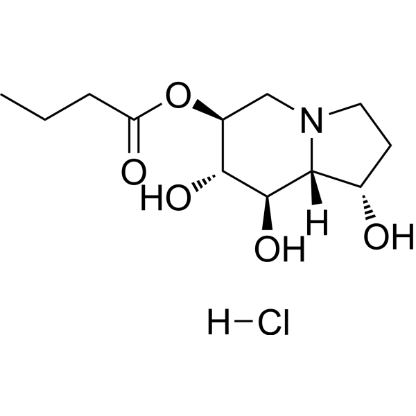
Celgosivir hydrochlorideα-Glucosidase I Inhibitor -
HY-N2574
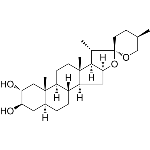
GitogeninGlucosidase Inhibitor -
HY-N1510
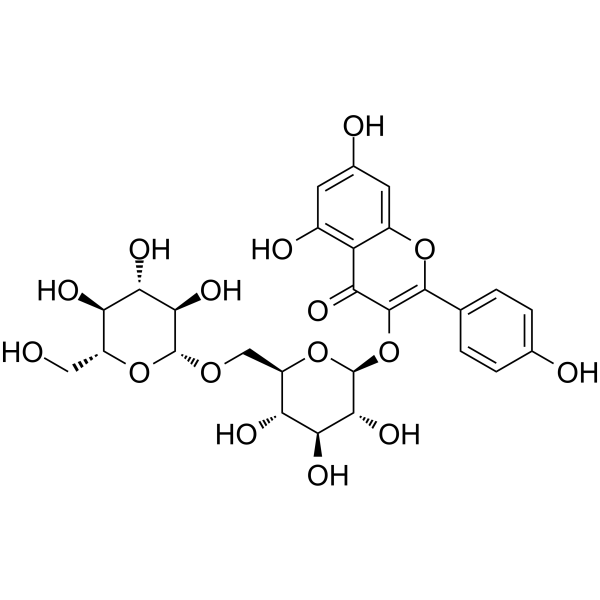
Kaempferol 3-O-gentiobiosideFlavonoid -

HY-W005598
2-Hydroxyquinolineα-Glucosidase/α-Amylase Inhibitor -
HY-N9317
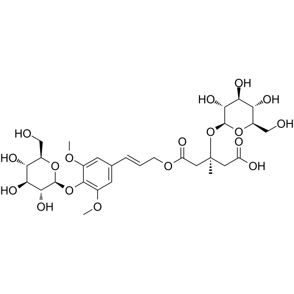
Tangshenoside Iα-glucosidase Inhibitor
Publications Citing Use of MedChemExpress (MCE) Pedalitin
MoreAll Endogenous Metabolite Isoforms
More

Biological Activity
Description
|
α-Glucosidase 0.29 mM (IC50) |
MPO 3.8 nM (IC50) |
Cellular Effect
|
Cell Line

|
Type | Value | Description | References |
|---|---|---|---|---|
| HepG2 | IC50 |
131.7 mg/L
|
Cytotoxicity against human HepG2 cells assessed as concentration at which 50% of cells were killed after 24 hrs exposure by MTT assay.
Cytotoxicity against human HepG2 cells assessed as concentration at which 50% of cells were killed after 24 hrs exposure by MTT assay.
|
27742203 |
| MRC5 | IC50 |
191.7 mg/L
|
Cytotoxicity against human MRC-5 cells assessed as concentration at which 50% of cells were killed after 24 hrs exposure by MTT assay.
Cytotoxicity against human MRC-5 cells assessed as concentration at which 50% of cells were killed after 24 hrs exposure by MTT assay.
|
27742203 |
| U-87MG ATCC | IC50 |
175 mg/L
|
Cytotoxicity against human U87-MG cells assessed as concentration at which 50% of cells were killed after 24 hrs exposure by MTT assay.
Cytotoxicity against human U87-MG cells assessed as concentration at which 50% of cells were killed after 24 hrs exposure by MTT assay.
|
27742203 |
In Vitro
Pedalitin (20-80 μM; 24 h) significantly reduced intracellular triglyceride (TG) levels and decreased cytoplasmic lipid droplet accumulation in FFA-induced LO2 cells[1].
Pedalitin (20 μM; 24 h) significantly downregulated the FFA-induced upregulation of inflammatory factors (IL-17, TNF-α), fatty acid metabolism factors (CPT2, HADH), and FOXO signaling pathway genes (EGFR, IRS-1, AKT-1, FOXO1) in LO2 cells[1].
Pedalitin (4 mg/L; 0.5-48 h) exhibits significant antifungal and time-dependent fungicidal activity in Cryptococcus neoformans strain cultures, with an MIC of 3.9 mg/L[10].
Pedalitin (1 mg/L; 30 min-2 h) combined with Amphotericin B (HY-B0221) significantly reduces Cryptococcus neoformans infection in U87-MG cells and MRC-5 cells[10].
Pedalitin (20-80 μM; 24 h) did not substantially reduce FFA-induced LO2 cell viability, maintaining cell viability at approximately 80%[1].
Pedalitin exhibited potent MPO inhibitory activity, ABTS radical scavenging activity, and DPPH radical scavenging activity, with IC50 values of 3.8 nM, 1.4 μM, and 5.2 μM, respectively[3].
Pedalitin significantly inhibits urease activity in the enzymatic reaction solution extracted from Proteus mirabilis[5].
Pedalitin (0.12-62.5 mg/L; 24 h) exhibited low cytotoxicity in HepG2, MRC-5, NOK, and U87-MG cell lines, with IC50 values of 131.7, 191.7, 119, and 175 mg/L, respectively[10].
Pedalitin (0.01-0.4 mM; pre-incubation for 2-5 min) significantly inhibited tyrosinase and α-glucosidase activities, with IC50 values of 0.28 mM and 0.29 mM, respectively[11].
MedChemExpress (MCE) has not independently confirmed the accuracy of these methods. They are for reference only. Further protocols information, click here.
-
Cell Line:LO2
-
Concentration:20, 40, 80 μM
-
Incubation Time:24 h (FFA induction); 1 h (CCK-8)
-
Result:Reduced cell viability to around 80% at 40 and 80 μM.
-
Cell Line:LO2
-
Concentration:20 μM
-
Incubation Time:24 h
-
Result:Significantly inhibited FFA-induced upregulation of TNF-α and IL-17 mRNA expression.
Restored CPT2 and HADH mRNA expression levels to near-normal ranges.
Restored expression levels of EGFR, IRS-1, AKT-1, and FOXO1 genes within the FOXO signaling pathway to a balanced state.
-
Cell Line:U87-MG, MRC-5
-
Concentration:4 mg/L
-
Incubation Time:30 min, 1 h, 2 h
-
Result:Reduced infection by approximately 50% after 30 min in both cell types.
Reduced infection to approximately 60% in lung fibroblasts after 2 h.
Reduced infection to approximately 40% in glioblastoma cells after 2 h.
In Vivo
Pedalitin (10 mg/kg; intraperitoneal injection; once daily; 40 days) combined with Amphotericin B has a synergistic effect, improving the survival rate, fungal burden, and histopathology of BALB/c mice infected with C. neoformans[8].
Pedalitin (1-10 mg/kg; intraperitoneal injection; single administration; 30 min) exhibited significant dose-dependent analgesic activity in the acetic acid-induced writhing test in mice[9].
MedChemExpress (MCE) has not independently confirmed the accuracy of these methods. They are for reference only.
-
Animal Model:CD1 (male, 25-30 g)[9]
-
Dosage:1, 5, 10 mg/kg
-
Administration:i.p.; single dose; 30 min
-
Result:Decreased the number of writhes significantly from a minimum dose of 1 mg/kg, producing at least 50% inhibition of nociception.
-
Animal Model:BALB/c mice[10]
-
Dosage:40 mg/kg
-
Administration:i.p.; once daily; 40 days
-
Result:Significantly prolonged the survival of mice.
Achieved 60% and 40% survival after 20 and 40 days, respectively, with combination therapy.
Significantly decreased the fungal burden in both lungs and brain.
Reduced fungal burden by 48% in lungs and brain after 7 days.
Reduced the number of colonies by >80% in both organs after 14 days.
Significantly reduced the number of yeasts in histopathological analysis after 14 days.


Chemical Information
-
CAS No. 22384-63-0
-
Appearance Solid
-
Molecular Weight 316.26
-
Formula C16H12O7
-
Color Light yellow to yellow
-
SMILES
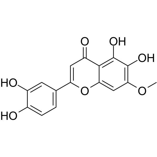
O=C1C=C(C2=CC=C(O)C(O)=C2)OC3=CC(OC)=C(O)C(O)=C13
-
Structure Classification
-
Initial Source
-
Shipping
Room temperature in continental US; may vary elsewhere.
-
Storage
4°C, sealed storage, away from moisture
* In solvent : -80°C, 6 months; -20°C, 1 month (sealed storage, away from moisture)


Publications (2)
-
Journal Impact Factor
-
Most Recent
-
Food Chem
Flavonoid-mediated metabolic underpinning quality variation in red bud-sport pear mutants. [Abstract]2025 Oct 15:489:144992. PMID: 40466530 -

Protocols
-
Research Protocol for Infectious Diseases
Infectious-disease experiments test how pathogens interact with host barriers, innate immune receptors, inflammatory signaling, pathogen replication, and tissue injury; pattern-recognition receptors such as TLRs, RIG-I-like receptors, NOD-like receptors, and inflammasomes detect microbial molecules and activate NF-κB, interferon, and cytokine responses. The central hypothesis is that infection severity reflects the balance between pathogen burden and host response: protective inflammation restricts pathogen growth, whereas excessive or mislocalized inflammation contributes to tissue damage and disease phenotype. Unresolved questions include which host pathways are protective versus pathogenic, why some infection models fail to translate to human disease, and which combined readouts best predict clinically relevant infection outcomes.
-
RNA extraction experimental
By lysing cells, releasing RNA, and removing impurities such as proteins and DNA, high-purity RNA products are finally obtained. The commonly used traditional method is the guanidine isothiocyanate/phenol/chloroform method (Trizol), which is suitable for a variety of animal materials including animal tissues, microorganisms, cultured cells, etc., and most plant materials.
-
Research Protocol for Metabolic Diseases
AMP-activated protein kinase, AMPK, is a conserved cellular energy sensor that responds to reduced cellular energy status and coordinates metabolism by increasing ATP-generating catabolic pathways while suppressing ATP-consuming anabolic processes. In metabolic disease research, the AMPK pathway is experimentally relevant because it regulates hepatic lipid synthesis, fatty acid oxidation, glucose production, skeletal-muscle glucose disposal, mTORC1-linked biosynthesis, autophagy, mitochondrial homeostasis, and whole-body energy balance. The central pathway logic is that energy stress, metformin, exercise-like stimulation, or direct AMPK activators increase AMPKα Thr172 phosphorylation and downstream substrate phosphorylation, including ACC and RAPTOR. Phosphorylation of ACC suppresses lipogenesis and supports fatty acid oxidation, whereas phosphorylation of RAPTOR suppresses mTORC1 signaling and links cellular energy status to growth and protein synthesis control. The pathway is linked


Purity & Documentation
-
Data Sheet (292 KB)
-
SDS (393 KB)
- English - EN (393 KB)
- Français - FR (393 KB)
- Deutsch - DE (393 KB)
- Norwegian - NO (393 KB)
- Español - ES (393 KB)
- Swedish - SV (393 KB)
- Italian - IT (393 KB)
- Korean - KR (393 KB)
- Portuguese - PT (393 KB)
-
Handling Instructions (2659 KB)
References
[3]. Fernandes DC, et al. Myeloperoxidase inhibitory and radical scavenging activities of flavones from Pterogyne nitens. Chemical & pharmaceutical bulletin. 2008 May;56(5):723-6. [Content Brief]
[11]. Lin L, et al. Comparative evaluation of rosmarinic acid, methyl rosmarinate and pedalitin isolated from Rabdosia serra (MAXIM.) HARA as inhibitors of tyrosinase and α-glucosidase. Food Chem. 2011 Dec 1;129(3):884-9. [Content Brief]

Calculators
Concentration (start) × Volume (start) = Concentration (final) × Volume (final)


Pedalitin Related Classifications
HY-N3101 Related Classifications
 Powered by Bioz