



PR-104 sodium
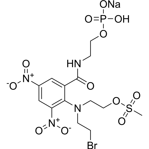
PR-104 (sodium) is a selective hypoxia-activated DNA cross-linking agent and can be used for the research of multiple tumor xenograft models. PR-104 (sodium), as a nitrogen mustard pre-proagent, is converted efficiently to the more lipophilic dinitrobenzamide mustards alcohol PR-104A.
For research use only. We do not sell to patients.
- CAS No.: 851627-80-0
- Formula: C14H19BrN4NaO12PS
- Molecular Weight:601.25
-
Storage:
Please store the product under the recommended conditions in the Certificate of Analysis.

To place orders, for customer services and technical support, please contact: MedChemExpress USA
Tel: 609-228-6898 E-mail: [email protected] [email protected]


-
 Biological Activity
-
Chemical Information
- Protocols
- Purity & Documentation
- References
-
 Help & FAQs


-
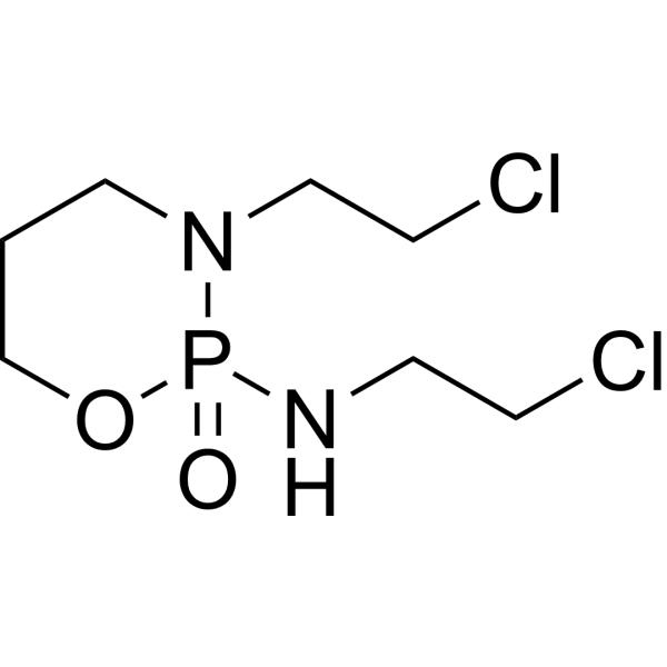
HY-17419
IfosfamideDNA Alkylator/Crosslinker -
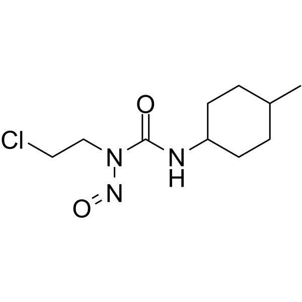
HY-13747
SemustineAntitumor Alkylating Agent -
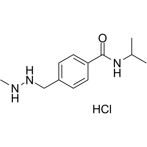
HY-13733
Procarbazine HydrochlorideAlkylating Agent -
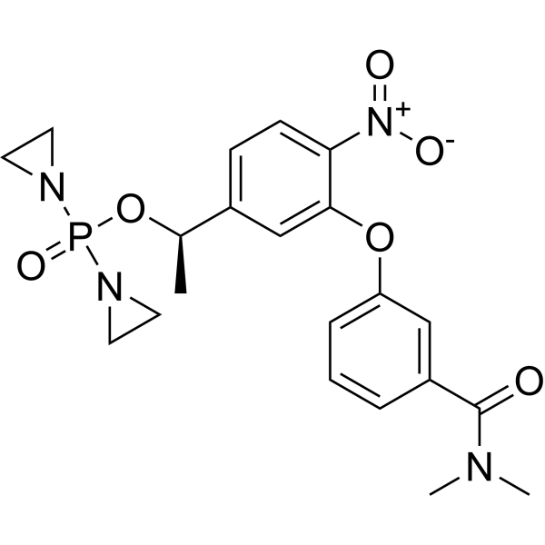
HY-124573
(R)-OdafosfamideDNA-alkylating Agent -

HY-17419R
Ifosfamide (Standard)DNA Alkylator/Crosslinker -
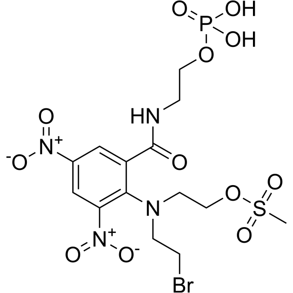
HY-16405
PR-104DNA Cross-Linking Agent -

HY-13733R
Procarbazine Hydrochloride (Standard)Alkylating Agent -
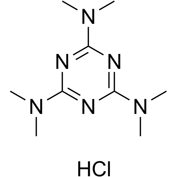
HY-B0181A
Altretamine hydrochlorideDNA Alkylator/Crosslinker


Biological Activity
Description
In Vitro
PR-104 (sodium) (80 μM; 1 hour; SiHa cells) shows greater suppression of radiation-induced DNA single-strand breaks under hypoxic than aerobic conditions. PR-104 (sodium) (100 μM; 1 hour; SiHa cells) results in phosphorylation of Ser139 of histone H2AX (gH2AX). PR-104 (sodium) (0.266 mmol/kg; 18 h; SiHa cells) shows activity against hypoxic cells after irradiation. PR-104 (sodium) varies in potency between cell lines, with the lowest IC50 (0.51 μmol/L) in H460 cells and highest (7.3 μmol/L) in PC3 prostate cells[1].
MedChemExpress (MCE) has not independently confirmed the accuracy of these methods. They are for reference only. Further protocols information, click here.
In Vivo
MedChemExpress (MCE) has not independently confirmed the accuracy of these methods. They are for reference only.
-
Animal Model:CD-1nu/nu mice
-
Dosage:0.56 mmol/kg (Pharmacokinetics Analysis)
-
Administration:I.v. or i.p.
-
Result:The plasma area under the curve.
-
Animal Model:CD1-Foxn1nu mice
-
Dosage:0.23 mmol/kg
-
Administration:I.p.
-
Result:Showed antitumor activity.


Chemical Information
-
CAS No. 851627-80-0
-
Molecular Weight 601.25
-
Formula C14H19BrN4NaO12PS
-
SMILES
O=C(NCCOP(O)(O[Na])=O)C1=CC([N+]([O-])=O)=CC([N+]([O-])=O)=C1N(CCBr)CCOS(=O)(C)=O
-
Shipping
Room temperature in continental US; may vary elsewhere.
-
Storage
Please store the product under the recommended conditions in the Certificate of Analysis.

Protocols
-
Patient-Derived Xenograft (PDX)
Patient-derived xenograft (PDX) models are generated by engrafting primary human tumor tissue directly into immunodeficient mice, allowing in vivo propagation of patient tumor biology without initial in vitro adaptation. These models are used to preserve key histopathological and molecular characteristics of the original tumor and enable assessment of tumor growth dynamics and therapeutic response in a living organism. The biological readout is tumor engraftment and subsequent growth in the murine host, which reflects the ability of human tumor cells to survive, vascularize, and expand in an immunocompromised microenvironment.
-
Splenic/Portal-Vein Liver Metastasis Xenograft
Splenic and portal-vein liver metastasis xenograft models deliver tumor cells into the portal circulation so that cells reach the liver first and form hepatic metastatic lesions; splenic injection uses the spleen as an access route to the portal system, while direct portal-vein injection introduces cells into the portal vein without requiring splenectomy. The assay detects liver colonization, intrahepatic tumor growth, tumor distribution, treatment response, survival, and liver-metastasis microenvironment changes; readouts include bioluminescence or fluorescence imaging, gross liver nodule counts, liver weight or tumor burden, histology, and survival.
-
Subcutaneous Cell-Line-Derived Xenograft
Subcutaneous cell-line-derived xenograft (CDX) models are established by implanting cultured human cancer cell lines into immunodeficient mice, where the injected cells form localized tumors that can be monitored in vivo as a measure of tumorigenic potential, growth kinetics, and treatment response. These models are widely used in oncology research because they allow reproducible tumor formation and enable comparative assessment of tumor growth between different cell lines or genetic manipulations in a controlled in vivo microenvironment. Subcutaneous implantation of cancer cells in immunodeficient mice is a standard approach for evaluating tumor growth behavior and therapeutic response across multiple cancer types, including prostate, esophageal, pancreatic, and colon cancer models.
-
Orthotopic Cell-Line Xenograft
Orthotopic cell-line xenograft models involve implantation of human cancer cell lines into the anatomically corresponding organ of immunodeficient mice to reproduce tumor growth within a native microenvironment, enabling more clinically relevant tumor behavior compared with subcutaneous models. These models are widely used because orthotopic placement better recapitulates tumor progression, including invasion and metastatic spread, which are often underrepresented in heterotopic implantation systems. Compared with conventional xenografts, orthotopic implantation is described as more technically complex but provides improved simulation of tumor-microenvironment interactions and metastatic behavior, making it particularly valuable for translational oncology research. Surgical orthotopic implantation approaches have been emphasized as enabling faithful reproduction of clinical cancer features, including metastasis and disease progression patterns that align with the tumor’s organ of origi
-
Intraperitoneal/Peritoneal Dissemination Xenograft
Intraperitoneal (IP) or peritoneal dissemination xenograft models are based on the introduction of human cancer cells into the peritoneal cavity of immunodeficient mice, where they attach to peritoneal surfaces, form multicellular aggregates or spheroids, and progressively generate disseminated tumor nodules that mimic advanced peritoneal metastatic disease. These models are widely used to study ovarian cancer progression, tumor-microenvironment interactions, and intraperitoneal therapeutic responses, often incorporating bioluminescence or fluorescence imaging to longitudinally monitor tumor burden in vivo. The biological principle relies on the capacity of tumor cells such as SKOV3 or related ovarian carcinoma lines to survive in suspension, aggregate within ascites-like fluid, adhere to mesothelial surfaces, and invade peritoneal organs, thereby recapitulating human peritoneal carcinomatosis patterns observed in advanced disease.


Purity & Documentation
References

Calculators
Concentration (start) × Volume (start) = Concentration (final) × Volume (final)


PR-104 sodium Related Classifications
HY-16406 Related Classifications
 Powered by Bioz