



PROTAC MLKL Degrader-1
Based on 1 publication(s) in Google Scholar
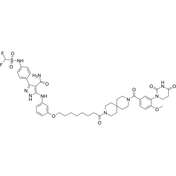
PROTAC MLKL Degrader-1 is a selective MLKL PROTAC degrader with a DC50 of 2.4 μM. PROTAC MLKL Degrader-1 blocks necroptosis without modulating the phosphorylation of RIPK1 or RIPK3, and exhibits a linear correlation between MLKL levels and necroptotic cell death.
(Pink: Mixed Lineage Kinase ligand (HY-175705); Blue: Cereblon ligand (HY-W249500); Black: linker).
연구목적의 판매만을 진행합니다. 환자를 대상으로 한 판매는 하지 않습니다.
- Purity : 99.76%
- CAS No.: 3032600-90-8
- 화학식: C46H55F2N9O9S
- 분자량:948.05
-
보관:
4°C, protect from light, stored under nitrogen
* In solvent : -80°C, 6 months; -20°C, 1 month (protect from light, stored under nitrogen)

To place orders, for customer services and technical support, please contact: MedChemExpress USA
Tel: 609-228-6898 E-mail: [email protected] [email protected]


-
 Biological Activity
-
Chemical Information
-
용액&용해도
- Protocol
- 순도&문서
- References
-
 Help & FAQs


Publications Citing Use of MedChemExpress (MCE) PROTAC MLKL Degrader-1
MoreAll PROTACs Isoforms
More

Biological Activity
제품 설명
IC50 & Target
[1]|
Cereblon |
Cellular Effect
|
Cell Line

|
Type | Value | Description | References |
|---|---|---|---|---|
| HT-29 | EC50 |
1.2 μM
Compound: 36
|
Protection against TSZ-induced necroptosis in human HT-29 cells assessed as cell viability incubated for 24 hrs by CellTiter Glo assay
Protection against TSZ-induced necroptosis in human HT-29 cells assessed as cell viability incubated for 24 hrs by CellTiter Glo assay
|
[PMID: 37535857] |
In Vitro
PROTAC MLKL Degrader-1 (Compound 36) (0.1-10 μM; 2-24 h) dose-dependently degrades MLKL in HT29 cells, with a DC50 of 2.4 μM. It achieves a degradation rate of > 90% within 5 h at a concentration of 10 μM, and this effect lasts for up to 24 h[1].
PROTAC MLKL Degrader-1 (0.1-10 μM; 24 h) dose-dependently protects HT29 cells against TSZ-induced necroptosis, with an EC50 of 1.2 μM[1].
MedChemExpress (MCE) has not independently confirmed the accuracy of these methods. They are for reference only. Further protocols information, click here.
-
Cell Line:HT29 colon cancer cells
-
Concentration:0.1, 0.3, 1, 3 and 10 μM
-
Incubation Time:24 h
-
Result:Protected cells from necroptosis in a dose-dependent manner, reaching 100% protection at 10 μM, with an EC50 of 1.2 μM.
Demonstrated anti-necroptotic efficacy that correlated linearly with the extent of MLKL degradation.


Chemical Information
-
CAS No. 3032600-90-8
-
Appearance Solid
-
분자량 948.05
-
화학식 C46H55F2N9O9S
-
Color Off-white to light yellow
-
SMILES
O=C(C1=C(NN=C1C2=CC=C(C=C2)NS(C(F)F)(=O)=O)NC3=CC=CC(OCCCCCCCC(N4CCC5(CCN(CC5)C(C6=CC=C(C(N7CCC(NC7=O)=O)=C6)OC)=O)CC4)=O)=C3)N
-
선적
Room temperature in continental US; may vary elsewhere.
-
보관
4°C, protect from light, stored under nitrogen
* In solvent : -80°C, 6 months; -20°C, 1 month (protect from light, stored under nitrogen)


Publications (1)
-
Journal Impact Factor
-
Most Recent
-
Nat Cell Biol
2026 Jun;28(6):1204-1218. PMID: 42174134


용액&용해도
In Vitro:
DMSO : 100 mg/mL (105.48 mM; Need ultrasonic; Hygroscopic DMSO has a significant impact on the solubility of product, please use newly opened DMSO)
Please refer to the solubility information to select the appropriate solvent. Once prepared, please aliquot and store the solution to prevent product inactivation from repeated freeze-thaw cycles.
Storage method and period of stock solution: -80°C, 6 months; -20°C, 1 month (protect from light, stored under nitrogen). When stored at -80°C, please use it within 6 months. When stored at -20°C, please use it within 1 month.
Please refer to the solubility information to select the appropriate solvent. Once prepared, please aliquot and store the solution to prevent product inactivation from repeated freeze-thaw cycles.
Storage method and period of stock solution: -80°C, 6 months; -20°C, 1 month (protect from light, stored under nitrogen). When stored at -80°C, please use it within 6 months. When stored at -20°C, please use it within 1 month.
Concentration (start) × Volume (start) = Concentration (final) × Volume (final)

Protocol
-
Kinase activity and phosphorylation assays
Kinase activity assays measure the ability of kinases to transfer phosphate groups from ATP to specific substrates, while phosphorylation assays detect the presence and levels of phosphorylated proteins. Common methods include radiolabeled ATP incorporation (e. g. ,), ADP release detection via bioluminescence (e. g. ,[3]), enzyme-linked immunosorbent assays (ELISA) for phospho-specific epitopes (e. g. ,[6]), and microtiter-based formats for high-throughput screening (e. g. ,[8]). The ADP-Glo assay quantifies kinase activity by measuring ADP produced during phosphorylation using a luciferase-based system. Radiometric assays involve autoradiography or scintillation counting after incorporation of 32P-labeled ATP into substrate proteins. ELISA-based approaches rely on phospho-specific antibodies to detect activated kinases in cell lysates or purified samples.
-
Western Blot
Western blotting (WB) is a commonly used experimental method in molecular biology, biochemistry, and immunogenetics for identifying and quantifying target proteins. It combines gel electrophoresis with immunoassay, enabling researchers to analyze protein expression, post-translational modifications, and molecular weight.
-
Cell Cytotoxicity Assay
Cytotoxicity assays are usually based on the assessment of cell membrane damage, which can also be indirectly detected by measuring cell viability. Detection methods include MTT assay, CKK-8 assay, LDH assay and ATP assay, etc.
-
Mammalian live/dead viability and cytotoxicity staining
Live/dead viability and cytotoxicity staining assays are based on the simultaneous detection of intracellular esterase activity in metabolically active (viable) cells and membrane integrity loss in non-viable cells. In commonly used dual-staining approaches, membrane-permeant fluorogenic substrates are converted by intracellular esterases into fluorescent products in live cells, while impermeant DNA-binding dyes selectively enter cells with compromised plasma membranes and label nucleic acids in dead or dying cells, enabling discrimination between viable and non-viable populations by fluorescence microscopy or flow cytometry.
-
Apoptosis Solutions
Apoptosis is a regulated, generally non-lytic cell-death pathway that removes unwanted, damaged, infected, or abnormal cells through coordinated morphological changes, caspase activation, DNA fragmentation, and membrane remodeling. The intrinsic apoptosis pathway is controlled mainly by mitochondrial outer membrane permeabilization, BCL-2 family proteins, cytochrome c release, apoptosome formation, caspase-9 activation, and downstream executioner caspase-3/7 activation. The extrinsic apoptosis pathway is initiated by death receptors such as Fas, TNFR, and TRAIL receptors, which recruit adaptor proteins and activate caspase-8 before engaging executioner caspases or mitochondrial amplification through BID cleavage. Apoptosis is linked to many phenotypes, including cancer cell killing, tissue homeostasis, immune regulation, neurodegeneration, infection response, and treatment-induced cytotoxicity; unresolved questions include how apoptosis interacts with necroptosis, pyroptosis, ferroptos
-
Protocol for Kinase activity and phosphorylation assays
Kinase activity assays measure transfer of phosphate from ATP to a protein or peptide substrate, generating phosphorylated substrate, ADP, or incorporated radiolabeled phosphate as the readout; phosphorylation assays measure site-specific phosphorylation in cells or tissues as a proxy for kinase-pathway activation, inhibition, or substrate regulation. Phosphorylation can be detected by phospho-specific Western blot, immunoprecipitation kinase assay, phospho-immunofluorescence, phospho-flow cytometry, luminescent ADP detection, radiolabeled ATP incorporation, or reporter-based pathway assays, and these readouts can be applied to cancer cells, primary neurons, mouse tumors, organoids, inflammatory macrophages, ferroptosis studies, and mitophagy studies when the kinase target is biologically relevant.


순도&문서
-
Data Sheet (270 KB)
-
SDS (252 KB)
- English - EN (252 KB)
- Français - FR (252 KB)
- Deutsch - DE (252 KB)
- Norwegian - NO (252 KB)
- Español - ES (252 KB)
- Swedish - SV (252 KB)
- Italian - IT (252 KB)
- Korean - KR (252 KB)
- Portuguese - PT (252 KB)
-
Handling Instructions (2659 KB)
References


Complete Stock Solution Preparation Table
Please refer to the solubility information to select the appropriate solvent. Once prepared, please aliquot and store the solution to prevent product inactivation from repeated freeze-thaw cycles.
Storage method and period of stock solution: -80°C, 6 months; -20°C, 1 month (protect from light, stored under nitrogen). When stored at -80°C, please use it within 6 months. When stored at -20°C, please use it within 1 month.
| Optional Solvent | Concentration Solvent Mass | 1 mg | 5 mg | 10 mg | 25 mg |
|---|---|---|---|---|---|
| DMSO | 1 mM | 1.0548 mL | 5.2740 mL | 10.5480 mL | 26.3699 mL |
| 5 mM | 0.2110 mL | 1.0548 mL | 2.1096 mL | 5.2740 mL | |
| 10 mM | 0.1055 mL | 0.5274 mL | 1.0548 mL | 2.6370 mL | |
| 15 mM | 0.0703 mL | 0.3516 mL | 0.7032 mL | 1.7580 mL | |
| 20 mM | 0.0527 mL | 0.2637 mL | 0.5274 mL | 1.3185 mL | |
| 25 mM | 0.0422 mL | 0.2110 mL | 0.4219 mL | 1.0548 mL | |
| 30 mM | 0.0352 mL | 0.1758 mL | 0.3516 mL | 0.8790 mL | |
| 40 mM | 0.0264 mL | 0.1318 mL | 0.2637 mL | 0.6592 mL | |
| 50 mM | 0.0211 mL | 0.1055 mL | 0.2110 mL | 0.5274 mL | |
| 60 mM | 0.0176 mL | 0.0879 mL | 0.1758 mL | 0.4395 mL | |
| 80 mM | 0.0132 mL | 0.0659 mL | 0.1318 mL | 0.3296 mL | |
| 100 mM | 0.0105 mL | 0.0527 mL | 0.1055 mL | 0.2637 mL |


PROTAC MLKL Degrader-1 Related Classifications
HY-156591 Related Classifications
 Powered by Bioz