



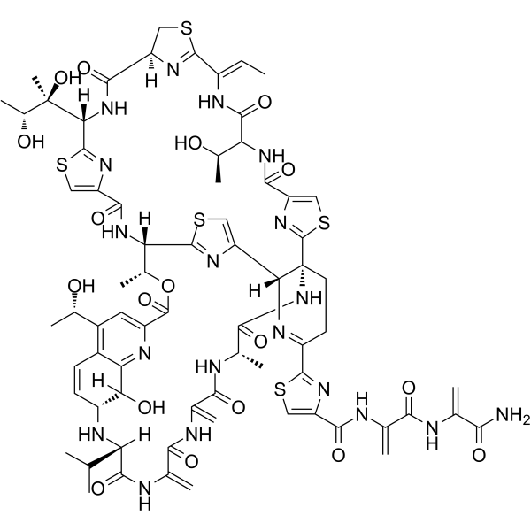
Siomycin A
Siomycin A is a thiopeptide antibiotic and is a Forkhead box M1(FOXM1) selective inhibitor without affecting other members of the Forkhead box family. Siomycin A has anti-tumor and promotes apoptosis.
Nos produits utilisent uniquement pour la recherche. Nous ne vendons pas aux patients.
- CAS No.: 12656-09-6
- Formule: C71H81N19O18S5
- Masse moléculaire:1648.84
-
Stockage:
Please store the product under the recommended conditions in the Certificate of Analysis.

To place orders, for customer services and technical support, please contact: MedChemExpress USA
Tel: 609-228-6898 E-mail: [email protected] [email protected]


-
 Activité biologique
-
Chemical Information
- Protocole
- Pureté et documentation
- Références
-
 Help & FAQs


Voir tous les produits spécifiques à Isoform Antibiotic
More

Activité biologique
Description
IC50 & Target
Forkhead box M1(FOXM1)[1]
In Vitro
Siomycin A (0-10 μM; 24-72 hours; K562, MCF7 and MiaPaCa-2 cells) treatment markedly reduces cell viability in a dose-dependent and a time-dependent association in K562, MCF7 and MiaPaCa-2 cells. Among the three cell lines, the IC50 of the human leukemia K562 cells is the lowest at 6.25 μM at 24 h, while that for the human pancreatic cancer MiaPaCa-2 cells is 6.38 μM. However, the IC50 of the human pancreatic cancer MiaPaCa-2 cells at 48 and 72 h are the lowest of the three cell lines, which are 0.76 and 0.54 μM, respectively[1].
Siomycin A (0-10 μM; MiaPaCa-2 cells) treatment has potent proapoptotic effect in MiaPaCa-2 cells[1].
Siomycin A (0-10 μM; 24 hours; MiaPaCa-2 cells) treatment significantly reduces the expression levels of MMP-2, MMP-9 and α-tubulin protein in the MiaPaCa-2 cells[1].
MedChemExpress (MCE) has not independently confirmed the accuracy of these methods. They are for reference only.
-
Cell Line:K562, MCF7 and MiaPaCa-2 cells
-
Concentration:0 µM, 0.625 µM, 1.25 µM, 2.5 µM, 5 µM or 10 µM
-
Incubation Time:24, 48 and 72 hours
-
Result:Cell viability was markedly reduced in a dose-dependent and a time-dependent association.
-
Cell Line:MiaPaCa-2 cells
-
Concentration:0 µM, 0.625 µM, 1.25 µM, 2.5 µM, 5 µM or 10 µM
-
Incubation Time:
-
Result:Promoted apoptosis of MiaPaCa-2 cells.
-
Cell Line:MiaPaCa-2 cells
-
Concentration:0 µM, 0.625 µM, 1.25 µM, 2.5 µM, 5 µM or 10 µM
-
Incubation Time:24 hours
-
Result:The expression levels of MMP-2 and MMP-9 protein in the MiaPaCa-2 cells were significantly reduced in the 2.5, 5 and 10 µM groups.
In Vivo
MedChemExpress (MCE) has not independently confirmed the accuracy of these methods. They are for reference only.
-
Animal Model:Four- to five-week old male Balb/c nude mice with IOMM-LEE cells[2]
-
Dosage:1 µM-pretreated IOMM-Lee cells
-
Administration:Subcutaneous injection; for 4 weeks
-
Result:Decreased tumour growth in an in vivo mouse model.


Chemical Information
-
CAS No. 12656-09-6
-
Masse moléculaire 1648.84
-
Formule C71H81N19O18S5
-
SMILES
O=C1N[C@]23[C@@](N=C(C4=NC(C(NC(C(NC(C(N)=O)=C)=O)=C)=O)=CS4)CC3)([H])C5=CSC([C@@]([C@H](OC(C6=CC([C@@H](O)C)=C7C([C@](O)([H])[C@@H](C=C7)N[C@](C(C)C)([H])C(NC(C(NC(C(N[C@H]1C)=O)=C)=O)=C)=O)=N6)=O)C)([H])NC(C8=CSC([C@](NC([C@]9([H])N=C(SC9)/C(NC(C([C@H](O)C)NC(C%10=CSC2=N%10)=O)=O)=C/C)=O)([H])[C@](C)(O)[C@H](O)C)=N8)=O)=N5
-
Livraison
Room temperature in continental US; may vary elsewhere.
-
Stockage
Please store the product under the recommended conditions in the Certificate of Analysis.


Pureté et documentation
Références
[1]. Wang B, et al. Effects and mechanism of siomycin A on the growth and apoptosis of MiaPaCa-2 cancer cells. Oncol Lett. 2019 Sep;18(3):2869-2876. [Content Brief]
[2]. Kim H, et al. Forkhead box M1 (FOXM1) transcription factor is a key oncogenic driver of aggressive human meningioma progression. Neuropathol Appl Neurobiol. 2019 Jun 9. [Content Brief]

Calculators
Concentration (start) × Volume (start) = Concentration (final) × Volume (final)


Siomycin A Related Classifications
HY-P1687 Related Classifications
 Powered by Bioz