



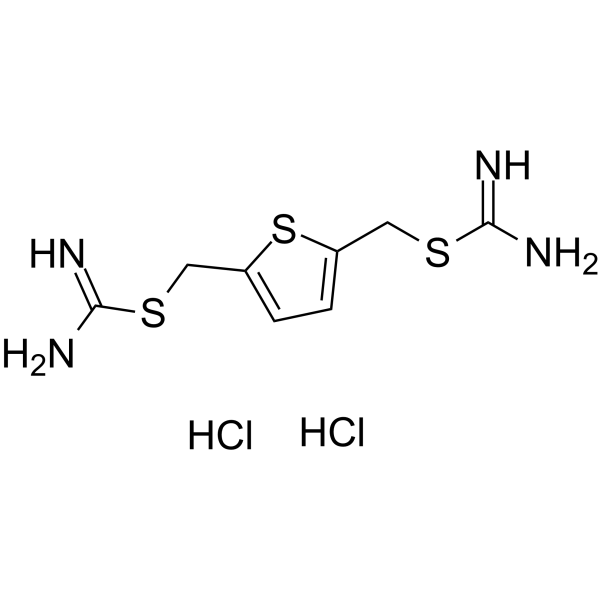
TPT-260 Dihydrochloride
Based on 2 publication(s) in Google Scholar
TPT-260 Dihydrochloride (NSC55712), a thiophene thiourea derivative, is a retromer complex stabilizer against thermal denaturation (Kd = ~5 µM). TPT-260 Dihydrochloride increases the levels of retromer proteins, shifts amyloid-precursor protein (APP) away from the endosome, and decreases the pathogenic processing of APP. TPT-260 Dihydrochloride inhibits TLR4 upregulation, IKKβ phosphorylation, NF-κB p65 nuclear translocation, and NLRP3 inflammasome formation. TPT-260 Dihydrochloride improves retromer-mediated cargo trafficking, reduces brain infarct area, and decreases amyloid plaque deposition. TPT-260 Dihydrochloride exhibits minimal cytotoxicity to primary microglia at tested concentrations. TPT-260 Dihydrochloride can be used for the research of inflammatory bowel disease, ischemic stroke and Alzheimer's disease.
For research use only. We do not sell to patients.
- Purity : 98.0%
- CAS No.: 2076-91-7
- Formula: C8H14Cl2N4S3
- Molecular Weight:333.32
-
Storage:
4°C, sealed storage, away from moisture
* In solvent : -80°C, 6 months; -20°C, 1 month (sealed storage, away from moisture)

To place orders, for customer services and technical support, please contact: MedChemExpress USA
Tel: 609-228-6898 E-mail: [email protected] [email protected]


-
 Biological Activity
-
Chemical Information
-
Solvent & Solubility
- Protocols
- Purity & Documentation
- References
-
 Help & FAQs


Publications Citing Use of MedChemExpress (MCE) TPT-260 Dihydrochloride
More

Biological Activity
Description
In Vitro
TPT-260 (10 μM) Dihydrochloride improves Occludin recycling and epithelial barrier function in Caco-2 cells but has no effect on VMP1-knockdown Caco-2 cells, indicating VMP1 is required for the Target Reagent's activity[1].
TPT-260 (5-20 μM) Dihydrochloride has no cytotoxic effect on primary mouse microglia, as measured by cell viability and LDH release assays[2].
TPT-260 (5-20 μM) Dihydrochloride protects primary mouse microglia from LPS (HY-D1056)/Nigericin (HY-127019)-induced cytotoxicity, as shown by restored cell viability and reduced LDH release[2].
TPT-260 (5-20 μM) Dihydrochloride inhibits LPS/Nigericin-induced inflammasome formation in primary mouse microglia, as measured by reduced ASC speck formation[2].
TPT-260 (5-20 μM) Dihydrochloride inhibits LPS/Nigericin-induced nuclear translocation of NF-κB p65 in primary mouse microglia, as measured by reduced nuclear p65 fluorescence intensity[2].
TPT-260 (5-20 μM) Dihydrochloride inhibits LPS (HY-D1056)/Nigericin (HY-127019)-induced upregulation of pro-inflammatory gene expression (Nlrp3, Tnfa, Il1b) in primary mouse microglia[2].
TPT-260 (5-20 μM) Dihydrochloride attenuates LPS/Nigericin-induced activation of the TLR4-IKKβ-NF-κB pathway in primary mouse microglia, as shown by reduced levels of pathway-related proteins and pro-inflammatory IL-1β[2].
TPT-260 (5 μM; 48 h) Dihydrochloride increases plasma membrane expression of NHE3 in Caco-2/bbe cells, raising surface NHE3 to 142.4% of control[3].
TPT-260 (5 μM; 48 h) Dihydrochloride increases levels of core retromer proteins VPS35 and VPS26 in Caco-2/bbe cells, and prevents the Cholera toxin (CT)-induced reduction in these retromer proteins when given either concurrently with CT or prior to CT exposure[3].
TPT-260 (5 μM; 48 h) Dihydrochloride increases plasma membrane expression of NHE3 in polarized Caco-2/bbe-HA-NHE3 cells, and reverses the CT-induced reduction in NHE3 surface expression when given either concurrently with CT or prior to CT exposure[3].
TPT-260 (5 μM; 48 h) Dihydrochloride partially reduces forskolin-induced fluid secretion in 3D human duodenal enteroids by stimulating NHE3-mediated fluid absorption[3].
TPT-260 Dihydrochloride (R55) binds to retromer complex with a Kd of ~5 μM[4].
TPT-260 Dihydrochloride (48 h) increases Vps35 levels with an EC50 of ~3.3 μM in primary hippocampal neurons[4].
TPT-260 Dihydrochloride (48 h) significantly reduces the levels of both endogenous Aβ40 and Aβ42 in hippocampal neurons[4].
TPT-260 Dihydrochloride inhibits Aβ levels with an IC50 of ~12 μM in hippocampal neurons[4].
TPT-260 Dihydrochloride significantly reduces the levels of both endogenous β-CTF and sAPPβ, and increases sAPPα levels in hippocampal neurons[4].
MedChemExpress (MCE) has not independently confirmed the accuracy of these methods. They are for reference only. Further protocols information, click here.
-
Cell Line:Caco-2/bbe cells
-
Concentration:5 μM
-
Incubation Time:48 h
-
Result:Increased amounts of VPS35 to 125% of control and VPS26 to 129% of control when used alone.
Prevented the CT-induced reduction in VPS35 and VPS26 levels, maintaining them at levels similar to untreated controls when given concurrently with CT or before CT.
In Vivo
MedChemExpress (MCE) has not independently confirmed the accuracy of these methods. They are for reference only.
-
Animal Model:C57BL/6 (2-month-old) bearing middle cerebral artery occlusion (MCAO) surgery[2]
-
Dosage:5 mg/kg
-
Administration:i.p.; single dose (24 hours prior to MCAO surgery)
-
Result:Significantly improved Bederson score (neurological function) in MCAO mice.
Significantly reduced brain infarct volume on the affected side.
Significantly suppressed brain tissue levels of pro-inflammatory factors IL-1β and TNF-α compared to untreated MCAO mice.


Chemical Information
-
CAS No. 2076-91-7
-
Appearance Solid
-
Molecular Weight 333.32
-
Formula C8H14Cl2N4S3
-
Color Light yellow to yellow
-
SMILES
NC(SCC1=CC=C(CSC(N)=N)S1)=N.Cl.Cl
-
Synonyms
NSC55712; TPU-260 Dihydrochloride
-
Shipping
Room temperature in continental US; may vary elsewhere.
-
Storage
4°C, sealed storage, away from moisture
* In solvent : -80°C, 6 months; -20°C, 1 month (sealed storage, away from moisture)


Publications (2)
-
Journal Impact Factor
-
Most Recent
-
-
J Inflamm Res
Therapeutic Potential of TPT-260 in Ischemic Stroke: An Investigation Into Its Anti-Inflammatory Effects and Impact on Microglial Activation. [Abstract]2025 Mar 1:18:3055-3066. PMID: 40046681


Solvent & Solubility
In Vitro:
DMSO : 100 mg/mL (300.01 mM; Need ultrasonic; Hygroscopic DMSO has a significant impact on the solubility of product, please use newly opened DMSO)
H2O : 33.33 mg/mL (99.99 mM; Need ultrasonic)
Please refer to the solubility information to select the appropriate solvent. Once prepared, please aliquot and store the solution to prevent product inactivation from repeated freeze-thaw cycles.
Storage method and period of stock solution: -80°C, 6 months; -20°C, 1 month (sealed storage, away from moisture). When stored at -80°C, please use it within 6 months. When stored at -20°C, please use it within 1 month.
* Note: If you choose water as the stock solution, please dilute it to the working solution, then filter and sterilize it with a 0.22 μm filter before use.
Please refer to the solubility information to select the appropriate solvent. Once prepared, please aliquot and store the solution to prevent product inactivation from repeated freeze-thaw cycles.
Storage method and period of stock solution: -80°C, 6 months; -20°C, 1 month (sealed storage, away from moisture). When stored at -80°C, please use it within 6 months. When stored at -20°C, please use it within 1 month.
* Note: If you choose water as the stock solution, please dilute it to the working solution, then filter and sterilize it with a 0.22 μm filter before use.
Concentration (start) × Volume (start) = Concentration (final) × Volume (final)
In Vivo:
Select the appropriate dissolution method based on your experimental animal and administration route.
- For the following dissolution methods, please ensure to first prepare a clear stock solution using an In Vitro approach and then sequentially add co-solvents:
- To ensure reliable experimental results, the clarified stock solution can be appropriately stored based on storage conditions. As for the working solution for In Vivo experiments, it is recommended to prepare freshly and use it on the same day.
- The percentages shown for the solvents indicate their volumetric ratio in the final prepared solution. If precipitation or phase separation occurs during preparation, heat and/or sonication can be used to aid dissolution.
Add each solvent one by one: 10% DMSO 40% PEG300 5% Tween-80 45% Saline
Solubility: ≥ 2.5 mg/mL (7.50 mM); Clear solution
This protocol yields a clear solution of ≥ 2.5 mg/mL (saturation unknown).
Taking 1 mL working solution as an example, add 100 μL DMSO stock solution (25.0 mg/mL) to 400 μL PEG300, and mix evenly; then add 50 μL Tween-80 and mix evenly; then add 450 μL Saline to adjust the volume to 1 mL.
Preparation of Saline: Dissolve 0.9 g sodium chloride in ddH₂O and dilute to 100 mL to obtain a clear Saline solution.
Add each solvent one by one: 10% DMSO 90% (20% SBE-β-CD in Saline)
Solubility: ≥ 2.5 mg/mL (7.50 mM); Clear solution
This protocol yields a clear solution of ≥ 2.5 mg/mL (saturation unknown).
Taking 1 mL working solution as an example, add 100 μL DMSO stock solution (25.0 mg/mL) to 900 μL 20% SBE-β-CD in Saline, and mix evenly.
Preparation of 20% SBE-β-CD in Saline (4°C, storage for one week): 2 g SBE-β-CD powder is dissolved in 10 mL Saline, completely dissolve until clear.
In Vivo Dissolution Calculator
Please enter the basic information of animal experiments:
-
-
-
-
Recommended: Prepare an additional quantity of animals to account for potential losses during experiments.
Working solution concentration: 0.22 mg/mL
This product has good water solubility, please refer to the measured solubility data in water/PBS/Saline for details.

Protocols
-
Western Blot
Western blotting (WB) is a commonly used experimental method in molecular biology, biochemistry, and immunogenetics for identifying and quantifying target proteins. It combines gel electrophoresis with immunoassay, enabling researchers to analyze protein expression, post-translational modifications, and molecular weight.
-
Mammalian live/dead viability and cytotoxicity staining
Live/dead viability and cytotoxicity staining assays are based on the simultaneous detection of intracellular esterase activity in metabolically active (viable) cells and membrane integrity loss in non-viable cells. In commonly used dual-staining approaches, membrane-permeant fluorogenic substrates are converted by intracellular esterases into fluorescent products in live cells, while impermeant DNA-binding dyes selectively enter cells with compromised plasma membranes and label nucleic acids in dead or dying cells, enabling discrimination between viable and non-viable populations by fluorescence microscopy or flow cytometry.
-
Research Protocol for Inflammation-related Diseases
The NLRP3 inflammasome is a cytosolic innate immune signaling platform that integrates priming signals and danger-signal activation to promote caspase-1 activation, maturation of IL-1β and IL-18, and gasdermin D-mediated pyroptotic cell death. The core experimental logic is to determine whether inflammatory disease phenotypes are driven by increased NLRP3 expression, ASC-containing inflammasome assembly, caspase-1 cleavage, GSDMD cleavage, and extracellular release of IL-1β/IL-18 rather than by nonspecific cell injury alone. The pathway is strongly linked to inflammation-related disease phenotypes because monosodium urate crystals activate NALP3/NLRP3 inflammasome signaling in gout-like crystal inflammation, cholesterol crystals activate NLRP3 inflammasomes in atherogenesis models, and DSS-induced intestinal inflammation has been reported to involve NLRP3 inflammasome activity. However, experimental colitis studies also show context-dependent protective effects of NLRP3 inflammasome co
-
Amyloid: Congo Red Amyloid Staining
Congo red amyloid staining is a histochemical method used to detect extracellular amyloid deposits in tissue sections based on the affinity of Congo red dye for β-pleated sheet-rich protein aggregates. When bound to amyloid, Congo red produces characteristic apple-green birefringence under polarized light microscopy, which is widely regarded as a diagnostic feature of amyloid deposition in histopathology. The diagnostic principle relies on the combination of dye binding (congophilia) and optical anisotropy under polarized illumination, which distinguishes amyloid from most non-amyloid eosinophilic extracellular deposits in routine histological evaluation. Amyloid identification by Congo red staining remains a cornerstone in diagnostic pathology despite the availability of adjunct methods such as immunohistochemistry and mass spectrometry, particularly because of its ability to localize deposits directly within tissue architecture. The specificity of Congo red-positive deposits is incre
-
Cell Viability Determination by MTT Colorimetric Assay
The following protocol uses the MTT colorimetric assay as a classic literature-established method for assessing cell viability/metabolic activity in cultured mammalian cells. MTT[3-(4,5-dimethylthiazol-2-yl)-2,5-diphenyltetrazolium bromide] is reduced by metabolically active cells to a colored formazan product; the amount of formazan is quantified spectrophotometrically and provides an indirect measure of metabolically active viable cells. Importantly, MTT reduction reflects cellular oxidoreductase/metabolic activity rather than an absolute direct count of living cells, so changes in cellular metabolism can alter the signal independently of cell number.
-
Alzheimer’s Disease Modeling
Alzheimer’s Disease (AD) is a neurodegenerative disorder characterized by a progressive decline in cognitive functions and loss of specific types of neurons and synapses. Alzheimer's symptoms can be simulated in mice by injecting drugs (such as Aβ) or genetically modified.
-
Kinase activity and phosphorylation assays
Kinase activity assays measure the ability of kinases to transfer phosphate groups from ATP to specific substrates, while phosphorylation assays detect the presence and levels of phosphorylated proteins. Common methods include radiolabeled ATP incorporation (e. g. ,), ADP release detection via bioluminescence (e. g. ,[3]), enzyme-linked immunosorbent assays (ELISA) for phospho-specific epitopes (e. g. ,[6]), and microtiter-based formats for high-throughput screening (e. g. ,[8]). The ADP-Glo assay quantifies kinase activity by measuring ADP produced during phosphorylation using a luciferase-based system. Radiometric assays involve autoradiography or scintillation counting after incorporation of 32P-labeled ATP into substrate proteins. ELISA-based approaches rely on phospho-specific antibodies to detect activated kinases in cell lysates or purified samples.
-
Cytoplasmic-Nuclear Fractionated Protein Extraction
Cytoplasmic-nuclear fractionated protein extraction separates soluble cytoplasmic proteins from nuclear-enriched proteins by mild plasma-membrane permeabilization, differential centrifugation, washing of nuclei, and extraction of nuclear proteins for downstream immunoblotting or related molecular analysis. The readout is the relative abundance of a protein in cytoplasmic and nuclear fractions, commonly assessed by western blotting together with compartment markers such as tubulin or pyruvate kinase for cytoplasm and lamin, nucleoporin, hnRNP, H2AX, or Lamin B for nuclear fractions.
-
Cell Cytotoxicity Assay
Cytotoxicity assays are usually based on the assessment of cell membrane damage, which can also be indirectly detected by measuring cell viability. Detection methods include MTT assay, CKK-8 assay, LDH assay and ATP assay, etc.
-
DSS-Induced Colitis
Dextran sulfate sodium (DSS)-induced colitis is generated by administering DSS in mouse drinking water, producing epithelial injury, barrier disruption, weight loss, diarrhea, fecal blood, colon shortening, histologic mucosal damage, and inflammatory mediator changes; the model is mainly used to study acute or chronic intestinal inflammation resembling selected features of ulcerative colitis. DSS injury is interpreted through clinical and tissue readouts rather than a single molecular endpoint: daily body weight, stool consistency, and bleeding are combined into a disease activity index, while colon length, histology, cytokines, myeloperoxidase activity, intestinal permeability, and tight-junction markers provide complementary measures of inflammation and barrier damage.
-
TNBS-Induced Colitis
TNBS-induced colitis is produced by intrarectal delivery of 2,4,6-trinitrobenzene sulfonic acid in ethanol, where ethanol disrupts the mucosal barrier and TNBS haptenates colonic proteins, generating immune-mediated colonic inflammation with weight loss, diarrhea, ulceration, transmural injury, inflammatory-cell infiltration, and cytokine responses. The model is used as an experimental intestinal inflammation model with Crohn’s disease–like features, especially when Th1-type responses, IL-12–dependent inflammation, chronic relapsing inflammation, or fibrosis-related endpoints are studied.
-
Protocol for Kinase activity and phosphorylation assays
Kinase activity assays measure transfer of phosphate from ATP to a protein or peptide substrate, generating phosphorylated substrate, ADP, or incorporated radiolabeled phosphate as the readout; phosphorylation assays measure site-specific phosphorylation in cells or tissues as a proxy for kinase-pathway activation, inhibition, or substrate regulation. Phosphorylation can be detected by phospho-specific Western blot, immunoprecipitation kinase assay, phospho-immunofluorescence, phospho-flow cytometry, luminescent ADP detection, radiolabeled ATP incorporation, or reporter-based pathway assays, and these readouts can be applied to cancer cells, primary neurons, mouse tumors, organoids, inflammatory macrophages, ferroptosis studies, and mitophagy studies when the kinase target is biologically relevant.
-
Pyroptosis Solutions
Pyroptosis is a lytic inflammatory cell-death pathway executed by gasdermin pores, most classically through inflammasome-mediated activation of caspase-1, cleavage of gasdermin D, membrane pore formation, LDH release, and secretion of IL-1β and IL-18. The canonical pathway is commonly modeled by priming cells with an inflammatory signal such as LPS to induce pro-IL-1β and inflammasome components, followed by an activation signal such as ATP or nigericin to activate NLRP3, ASC speck formation, caspase-1 cleavage, GSDMD cleavage, cytokine release, and pyroptotic membrane rupture. The non-canonical pathway is triggered when cytosolic LPS activates mouse caspase-11 or human caspase-4/5, leading to GSDMD cleavage and pyroptosis, and this can secondarily activate NLRP3-dependent IL-1β release. Pyroptosis is linked to inflammatory injury, infection, cancer, liver disease, ocular disease, placental inflammation, and other disease phenotypes, but unresolved questions include which gasdermin fam


Purity & Documentation
-
Data Sheet (290 KB)
-
SDS (476 KB)
- English - EN (476 KB)
- Français - FR (476 KB)
- Deutsch - DE (476 KB)
- Norwegian - NO (476 KB)
- Español - ES (476 KB)
- Swedish - SV (476 KB)
- Italian - IT (476 KB)
- Korean - KR (476 KB)
- Portuguese - PT (476 KB)
-
Handling Instructions (2659 KB)
References
[1]. Zhao J, et al. Loss of VMP1 Impairs Tight Junction Recycling and Aggravates Intestinal Barrier Dysfunction in Inflammatory Bowel Disease. Adv Sci (Weinh). Published online February 27, 2026. [Content Brief]
[2]. Qian J, et al. Therapeutic Potential of TPT-260 in Ischemic Stroke: An Investigation Into Its Anti-Inflammatory Effects and Impact on Microglial Activation. J Inflamm Res. 2025 Mar 1;18:3055-3066. [Content Brief]
[3]. Singh V, et al. Cholera toxin inhibits SNX27-retromer-mediated delivery of cargo proteins to the plasma membrane. J Cell Sci. 2018;131(16):jcs218610. Published 2018 Aug 17. [Content Brief]
[4]. Mecozzi VJ, et al. Pharmacological chaperones stabilize retromer to limit APP processing. Nat Chem Biol. 2014 Jun;10(6):443-9. [Content Brief]


Complete Stock Solution Preparation Table
Please refer to the solubility information to select the appropriate solvent. Once prepared, please aliquot and store the solution to prevent product inactivation from repeated freeze-thaw cycles.
Storage method and period of stock solution: -80°C, 6 months; -20°C, 1 month (sealed storage, away from moisture). When stored at -80°C, please use it within 6 months. When stored at -20°C, please use it within 1 month.
| Optional Solvent | Concentration Solvent Mass | 1 mg | 5 mg | 10 mg | 25 mg |
|---|---|---|---|---|---|
| H2O / DMSO | 1 mM | 3.0001 mL | 15.0006 mL | 30.0012 mL | 75.0030 mL |
| 5 mM | 0.6000 mL | 3.0001 mL | 6.0002 mL | 15.0006 mL | |
| 10 mM | 0.3000 mL | 1.5001 mL | 3.0001 mL | 7.5003 mL | |
| 15 mM | 0.2000 mL | 1.0000 mL | 2.0001 mL | 5.0002 mL | |
| 20 mM | 0.1500 mL | 0.7500 mL | 1.5001 mL | 3.7501 mL | |
| 25 mM | 0.1200 mL | 0.6000 mL | 1.2000 mL | 3.0001 mL | |
| 30 mM | 0.1000 mL | 0.5000 mL | 1.0000 mL | 2.5001 mL | |
| 40 mM | 0.0750 mL | 0.3750 mL | 0.7500 mL | 1.8751 mL | |
| 50 mM | 0.0600 mL | 0.3000 mL | 0.6000 mL | 1.5001 mL | |
| 60 mM | 0.0500 mL | 0.2500 mL | 0.5000 mL | 1.2500 mL | |
| 80 mM | 0.0375 mL | 0.1875 mL | 0.3750 mL | 0.9375 mL | |
| DMSO | 100 mM | 0.0300 mL | 0.1500 mL | 0.3000 mL | 0.7500 mL |
* Note: If you choose water as the stock solution, please dilute it to the working solution, then filter and sterilize it with a 0.22 μm filter before use.


TPT-260 Dihydrochloride Related Classifications
HY-13769A Related Classifications
 Powered by Bioz