



Velutin

Based on 1 Customer Validation
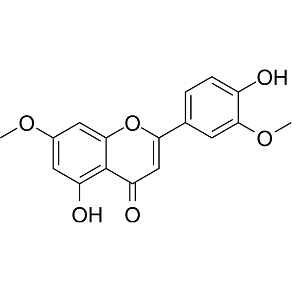
Velutin is a flavonoid. Velutin can be extracted from mistletoe. Velutin inhibits mushroom Tyrosinase activity with an IC50 of 910.1 μM. Velutin inhibits p38 phosphorylation, the NF-κB pathway and the MAPK pathway. Velutin prevents articular cartilage degeneration and subchondral bone loss. Velutin slows down the progression of intervertebral disc degeneration. Velutin exhibits inhibitory effects on melanogenesis, skin whitening, anti-inflammatory, anti-allergic, anti-oxidant and antibacterial activities. Velutin can be used in studies related to pigmented diseases, osteoarthritis and intervertebral disc degeneration.
For research use only. We do not sell to patients.
- Purity : 98.60%
- CAS No.: 25739-41-7
- Formula: C17H14O6
- Molecular Weight:314.29
-
Storage:
4°C, protect from light
* In solvent : -80°C, 6 months; -20°C, 1 month (protect from light)

To place orders, for customer services and technical support, please contact: MedChemExpress USA
Tel: 609-228-6898 E-mail: [email protected] [email protected]


-
 Biological Activity
-
Chemical Information
-
Solvent & Solubility
- Protocols
- Purity & Documentation
- References
-
 Help & FAQs


-
HY-101445
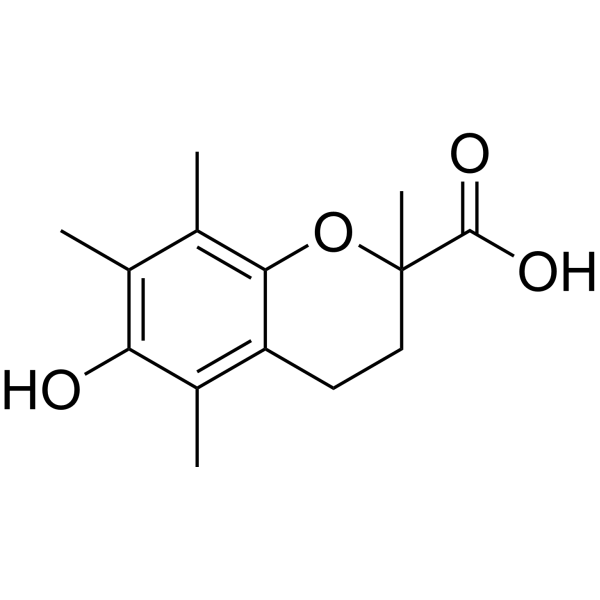
TroloxAntioxidant -

HY-N1098R
Velutin (Standard)Melanin Biosynthesis Inhibitor -

HY-101445R
Trolox (Standard)Membrane Damage Inhibitor -
HY-N5112B
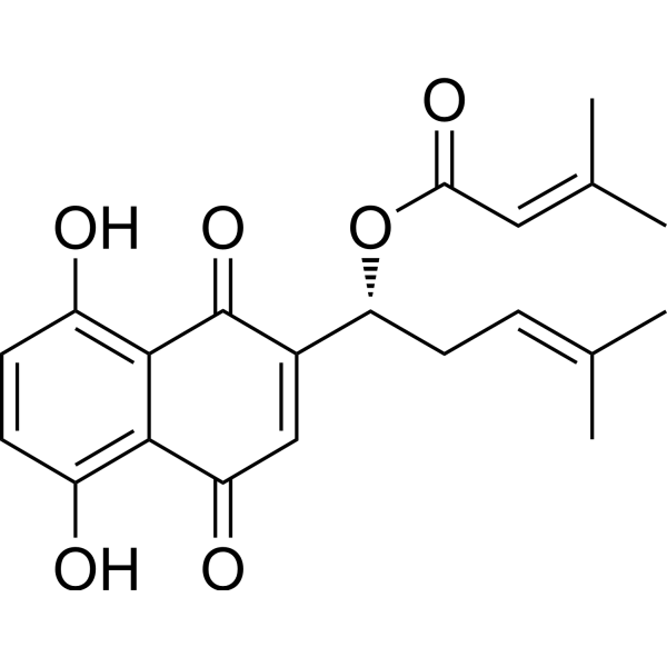
β,β-DimethylacrylshikoninAnticancer Compound -
HY-12867
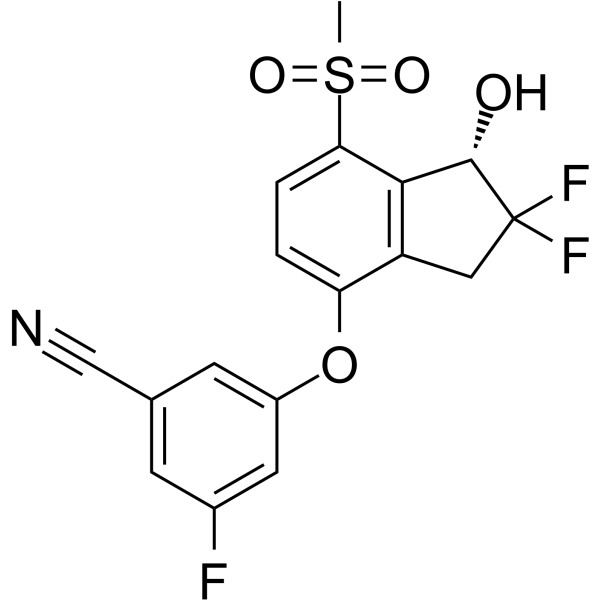
PT-2385HIF-2α Inhibitor -
HY-111547
M1001HIF-2α Agonist -

HY-101445G
Trolox (GMP)Antioxidant


Biological Activity
Description
Cellular Effect
|
Cell Line

|
Type | Value | Description | References |
|---|---|---|---|---|
| A2780 | IC50 |
>20 μg/mL
Compound: 4',5-dihydroxy-3',7-dimethoxyflavone
|
Cytotoxicity against human A2780 cells
Cytotoxicity against human A2780 cells
|
[PMID: 15217279] |
| KB | IC50 |
1.5 μg/mL
Compound: Velutin
|
Cytotoxicity against human KB cells after 72 hrs by neutral red assay
Cytotoxicity against human KB cells after 72 hrs by neutral red assay
|
[PMID: 10075750] |
| KB | IC50 |
1.5 μg/mL
Compound: 4, Velutin
|
Cytotoxicity against human KB cells
Cytotoxicity against human KB cells
|
[PMID: 8759170] |
| Neutrophil | IC50 |
<=12.51 μM
Compound: 59
|
Inhibition of fMLF/CB-induced human Neutrophil activation assessed as reduction in superoxide anion generation pre-incubated for 2 mins prior to CB induction for 3 mins followed by fMLF addition by spectrophotometric analysis
Inhibition of fMLF/CB-induced human Neutrophil activation assessed as reduction in superoxide anion generation pre-incubated for 2 mins prior to CB induction for 3 mins followed by fMLF addition by spectrophotometric analysis
|
[PMID: 37683361] |
| Neutrophil | IC50 |
<=15.25 μM
Compound: 59
|
Inhibition of fMLF/CB-induced human Neutrophil activation assessed as reduction in elastase release using MeO-Suc-Ala-Ala-Pro-Val-pnitroanilide as substrate pre-incubated for 2 mins followed by fMLF/CB-stimulation and measured for 5 mins
Inhibition of fMLF/CB-induced human Neutrophil activation assessed as reduction in elastase release using MeO-Suc-Ala-Ala-Pro-Val-pnitroanilide as substrate pre-incubated for 2 mins followed by fMLF/CB-stimulation and measured for 5 mins
|
[PMID: 37683361] |
| P388 | IC50 |
10 μg/mL
Compound: 4, Velutin
|
Cytotoxicity against mouse P388 cells
Cytotoxicity against mouse P388 cells
|
[PMID: 8759170] |
In Vitro
Velutin (2-200 μM; 10 min) potently scavenges ABTS free radicals in cell-free assays, with an EC50 value of 23.13 μM[1].
Velutin (0.01-1 mM; pre-incubated with tyrosinase for 30 min, followed by incubation with L-tyrosine for 10 min) inhibits mushroom tyrosinase activity in cell-free assays, with an IC50 of 910.1 μM[1].
Velutin (10 μM; 48 h) completely inhibits α-MSH-induced melanogenesis in B16F10 mouse melanoma cells[1].
At a concentration of 10 μM with 48 h incubation, velutin inhibits α-MSH-induced intracellular tyrosinase activity by 87% in B16F10 mouse melanoma cells[1].
Velutin (0-32 μM; 24-48 h) exhibits no cytotoxicity toward primary mouse chondrocytes, with the maximum concentration reaching 32 μM after 24 h or 48 h of incubation[2].
Velutin (32 μM; 7 days) protects primary mouse chondrocytes against IL-1β-induced inflammation and extracellular matrix degradation after 7 days of incubation[2].
Velutin (4 μM; 2 h) inhibits RANKL-induced activation of p38 MAPK in mouse bone marrow-derived macrophages[2].
MedChemExpress (MCE) has not independently confirmed the accuracy of these methods. They are for reference only. Further protocols information, click here.
-
Cell Line:B16F10 murine melanoma cells
-
Concentration:20 μM
-
Incubation Time:24 h
-
Result:Showed no significant cytotoxicity, with cell viability remaining near 100% at 20 μM.
In Vivo
Velutin (i.p.; once daily; for 4 consecutive weeks; 5-10 mg/kg) dose-dependently alleviates intervertebral disc degeneration in a mouse model of lumbar instability[3].
MedChemExpress (MCE) has not independently confirmed the accuracy of these methods. They are for reference only.
-
Animal Model:C57BL/6 (7-week-old male, DMM-induced osteoarthritis)[2]
-
Dosage:32 μM
-
Administration:intra-articular injection; once weekly; 8 weeks
-
Result:Showed significantly more regular articular cartilage surfaces, a thicker AC layer, a thinner calcified cartilage layer, and a lower osteophyte score.
Increased AC thickness, reduced CC thickness, and increased the AC/CC ratio.
Increased subchondral bone thickness, and significantly reduced the number of TRAP (+) multinucleated osteoclasts in SCB.
Increased aggrecan and type II collagen expression.
Reduced AC destruction and deeper Safranin O staining, with lower Osteoarthritis Research Society International scores.
Increased trabecular bone volume/tissue volume and trabecular number, and reduced trabecular separation in SCB, reducing SCB loss.
Significantly reduced p-p38 MAPK expression.


Chemical Information
-
CAS No. 25739-41-7
-
Appearance Solid
-
Molecular Weight 314.29
-
Formula C17H14O6
-
Color White to yellow
-
SMILES
O=C1C=C(C2=CC=C(O)C(OC)=C2)OC3=CC(OC)=CC(O)=C13
-
Structure Classification
-
Initial Source
-
Shipping
Room temperature in continental US; may vary elsewhere.
-
Storage
4°C, protect from light
* In solvent : -80°C, 6 months; -20°C, 1 month (protect from light)


Solvent & Solubility
In Vitro:
DMSO : 50 mg/mL (159.09 mM; Need ultrasonic; Hygroscopic DMSO has a significant impact on the solubility of product, please use newly opened DMSO)
Please refer to the solubility information to select the appropriate solvent. Once prepared, please aliquot and store the solution to prevent product inactivation from repeated freeze-thaw cycles.
Storage method and period of stock solution: -80°C, 6 months; -20°C, 1 month (protect from light). When stored at -80°C, please use it within 6 months. When stored at -20°C, please use it within 1 month.
Please refer to the solubility information to select the appropriate solvent. Once prepared, please aliquot and store the solution to prevent product inactivation from repeated freeze-thaw cycles.
Storage method and period of stock solution: -80°C, 6 months; -20°C, 1 month (protect from light). When stored at -80°C, please use it within 6 months. When stored at -20°C, please use it within 1 month.
Concentration (start) × Volume (start) = Concentration (final) × Volume (final)

Protocols
-
Kinase activity and phosphorylation assays
Kinase activity assays measure the ability of kinases to transfer phosphate groups from ATP to specific substrates, while phosphorylation assays detect the presence and levels of phosphorylated proteins. Common methods include radiolabeled ATP incorporation (e. g. ,), ADP release detection via bioluminescence (e. g. ,[3]), enzyme-linked immunosorbent assays (ELISA) for phospho-specific epitopes (e. g. ,[6]), and microtiter-based formats for high-throughput screening (e. g. ,[8]). The ADP-Glo assay quantifies kinase activity by measuring ADP produced during phosphorylation using a luciferase-based system. Radiometric assays involve autoradiography or scintillation counting after incorporation of 32P-labeled ATP into substrate proteins. ELISA-based approaches rely on phospho-specific antibodies to detect activated kinases in cell lysates or purified samples.
-
Western Blot
Western blotting (WB) is a commonly used experimental method in molecular biology, biochemistry, and immunogenetics for identifying and quantifying target proteins. It combines gel electrophoresis with immunoassay, enabling researchers to analyze protein expression, post-translational modifications, and molecular weight.
-
Research Protocol for Infectious Diseases
Infectious-disease experiments test how pathogens interact with host barriers, innate immune receptors, inflammatory signaling, pathogen replication, and tissue injury; pattern-recognition receptors such as TLRs, RIG-I-like receptors, NOD-like receptors, and inflammasomes detect microbial molecules and activate NF-κB, interferon, and cytokine responses. The central hypothesis is that infection severity reflects the balance between pathogen burden and host response: protective inflammation restricts pathogen growth, whereas excessive or mislocalized inflammation contributes to tissue damage and disease phenotype. Unresolved questions include which host pathways are protective versus pathogenic, why some infection models fail to translate to human disease, and which combined readouts best predict clinically relevant infection outcomes.
-
Cell Cytotoxicity Assay
Cytotoxicity assays are usually based on the assessment of cell membrane damage, which can also be indirectly detected by measuring cell viability. Detection methods include MTT assay, CKK-8 assay, LDH assay and ATP assay, etc.
-
Protocol for Kinase activity and phosphorylation assays
Kinase activity assays measure transfer of phosphate from ATP to a protein or peptide substrate, generating phosphorylated substrate, ADP, or incorporated radiolabeled phosphate as the readout; phosphorylation assays measure site-specific phosphorylation in cells or tissues as a proxy for kinase-pathway activation, inhibition, or substrate regulation. Phosphorylation can be detected by phospho-specific Western blot, immunoprecipitation kinase assay, phospho-immunofluorescence, phospho-flow cytometry, luminescent ADP detection, radiolabeled ATP incorporation, or reporter-based pathway assays, and these readouts can be applied to cancer cells, primary neurons, mouse tumors, organoids, inflammatory macrophages, ferroptosis studies, and mitophagy studies when the kinase target is biologically relevant.
-
Research Protocol for Inflammation-related Diseases
The NLRP3 inflammasome is a cytosolic innate immune signaling platform that integrates priming signals and danger-signal activation to promote caspase-1 activation, maturation of IL-1β and IL-18, and gasdermin D-mediated pyroptotic cell death. The core experimental logic is to determine whether inflammatory disease phenotypes are driven by increased NLRP3 expression, ASC-containing inflammasome assembly, caspase-1 cleavage, GSDMD cleavage, and extracellular release of IL-1β/IL-18 rather than by nonspecific cell injury alone. The pathway is strongly linked to inflammation-related disease phenotypes because monosodium urate crystals activate NALP3/NLRP3 inflammasome signaling in gout-like crystal inflammation, cholesterol crystals activate NLRP3 inflammasomes in atherogenesis models, and DSS-induced intestinal inflammation has been reported to involve NLRP3 inflammasome activity. However, experimental colitis studies also show context-dependent protective effects of NLRP3 inflammasome co


Purity & Documentation
-
Data Sheet (277 KB)
-
SDS (393 KB)
- English - EN (393 KB)
- Français - FR (393 KB)
- Deutsch - DE (393 KB)
- Norwegian - NO (393 KB)
- Español - ES (393 KB)
- Swedish - SV (393 KB)
- Italian - IT (393 KB)
- Korean - KR (393 KB)
- Portuguese - PT (393 KB)
-
Handling Instructions (2659 KB)
References
[1]. Jung SH, et al. Anti-Melanogenic Properties of Velutin and Its Analogs. Molecules. 2021;26(10):3033. Published 2021 May 19. [Content Brief]
[2]. Wang K, et al. Dual protective role of velutin against articular cartilage degeneration and subchondral bone loss via the p38 signaling pathway in murine osteoarthritis. Front Endocrinol (Lausanne). 2022;13:926934. Published 2022 Jul 22. [Content Brief]
[3]. Zhang Y, et al. Velutin Inhibits IL-1β-Induced Nucleus Pulposus Inflammatory and Extracellular Matrix Degradation Attenuating Mouse Intervertebral Disc Degeneration via the NF-κB and MAPK Pathways. Mediators Inflamm. 2025 Nov 20;2025:9625485. [Content Brief]


Complete Stock Solution Preparation Table
Please refer to the solubility information to select the appropriate solvent. Once prepared, please aliquot and store the solution to prevent product inactivation from repeated freeze-thaw cycles.
Storage method and period of stock solution: -80°C, 6 months; -20°C, 1 month (protect from light). When stored at -80°C, please use it within 6 months. When stored at -20°C, please use it within 1 month.
| Optional Solvent | Concentration Solvent Mass | 1 mg | 5 mg | 10 mg | 25 mg |
|---|---|---|---|---|---|
| DMSO | 1 mM | 3.1818 mL | 15.9089 mL | 31.8177 mL | 79.5444 mL |
| 5 mM | 0.6364 mL | 3.1818 mL | 6.3635 mL | 15.9089 mL | |
| 10 mM | 0.3182 mL | 1.5909 mL | 3.1818 mL | 7.9544 mL | |
| 15 mM | 0.2121 mL | 1.0606 mL | 2.1212 mL | 5.3030 mL | |
| 20 mM | 0.1591 mL | 0.7954 mL | 1.5909 mL | 3.9772 mL | |
| 25 mM | 0.1273 mL | 0.6364 mL | 1.2727 mL | 3.1818 mL | |
| 30 mM | 0.1061 mL | 0.5303 mL | 1.0606 mL | 2.6515 mL | |
| 40 mM | 0.0795 mL | 0.3977 mL | 0.7954 mL | 1.9886 mL | |
| 50 mM | 0.0636 mL | 0.3182 mL | 0.6364 mL | 1.5909 mL | |
| 60 mM | 0.0530 mL | 0.2651 mL | 0.5303 mL | 1.3257 mL | |
| 80 mM | 0.0398 mL | 0.1989 mL | 0.3977 mL | 0.9943 mL | |
| 100 mM | 0.0318 mL | 0.1591 mL | 0.3182 mL | 0.7954 mL |


Velutin Related Classifications
HY-N1098 Related Classifications
 Powered by Bioz