



Valnemulin
Based on 2 publication(s) in Google Scholar
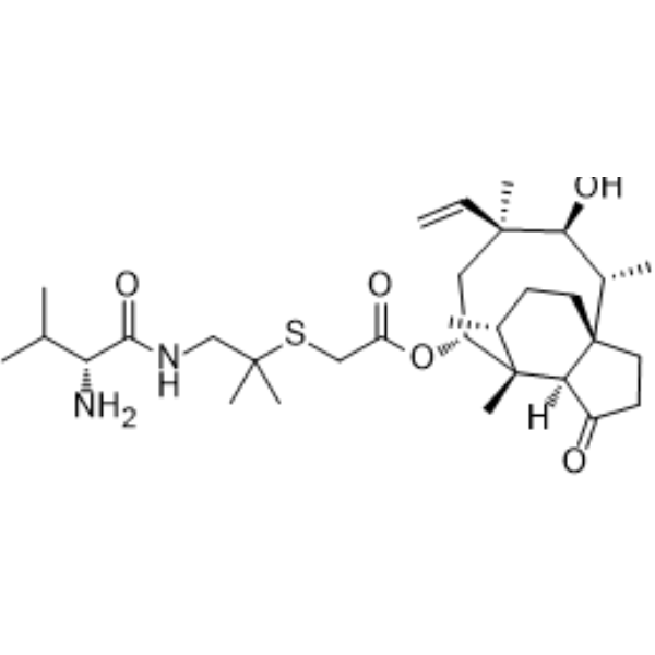
Valnemulin is an orally active broad-spectrum antibiotic against Gram-negative and Gram-positive bacteria, anaerobic bacteria, Mycoplasma, and Spirochetes. Valnemulin ameliorates enteric diseases, acute polyarthritis and enzootic pneumonia in pigs. Valnemulin exhibits anti-inflammatory efficacy against lipopolysaccharide (HY-D1056)-induced lung injury.
For research use only. We do not sell to patients.
- Purity : 99.0%
- CAS No.: 101312-92-9
- Formula: C31H52N2O5S
- Molecular Weight:564.82
-
Storage:Powder -20°C, 3 years ; In solvent -80°C, 6 months , -20°C, 1 month

To place orders, for customer services and technical support, please contact: MedChemExpress USA
Tel: 609-228-6898 E-mail: [email protected] [email protected]


-
 Biological Activity
-
Chemical Information
- Protocols
- Purity & Documentation
- References
-
 Help & FAQs


-
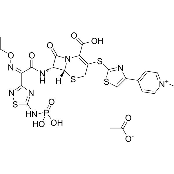
HY-14737
Ceftaroline fosamilCephalosporin Antibiotic -
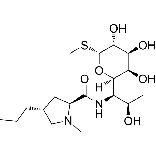
HY-117660
LincomycinRibosome Inhibitor -

HY-115693
CAP 3Antibacterial Agents -
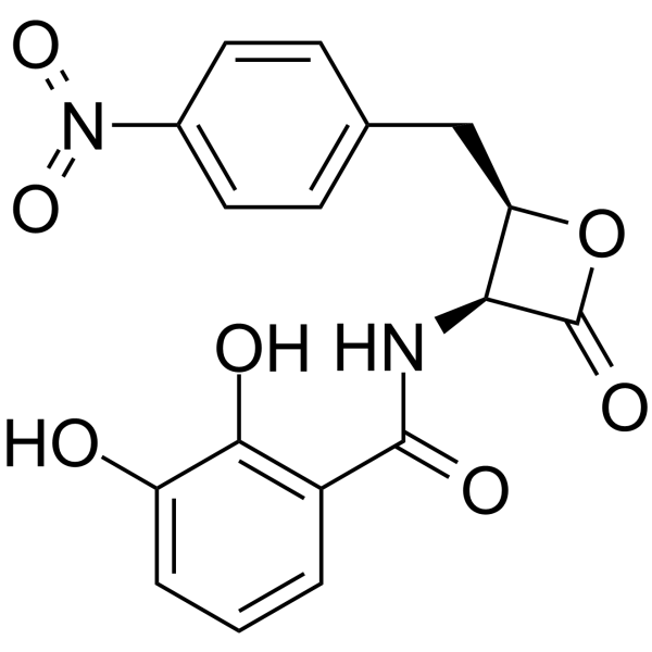
HY-138075
ObafluorinAntibiotic -

HY-14737R
Ceftaroline fosamil (Standard)Cephalosporin Antibiotic -

HY-117660R
Lincomycin (Standard)Standard of Lincomycin -

HY-120256
Corynecin II -

HY-Z7555
Cefazolin impurity HCefazolin Impurity -

HY-P3119
DesotamideAntibiotic -

HY-126746
Corynecin IAntibiotic
Publications Citing Use of MedChemExpress (MCE) Valnemulin
MoreAll Antibiotic Isoforms
More

Biological Activity
Description
Cellular Effect
|
Cell Line

|
Type | Value | Description | References |
|---|---|---|---|---|
| A549 | CC50 |
46.21 μM
Compound: Valnemulin
|
Cytotoxicity against human A549 cells incubated for 24 hrs by CCK8 assay
Cytotoxicity against human A549 cells incubated for 24 hrs by CCK8 assay
|
[PMID: 37531743] |
| A549 | CC50 |
88.01 μM
Compound: Valnemulin
|
Cytotoxicity against human A549 cells assessed as reduction in cell viability measured after 48 hrs by CCK8 assay
Cytotoxicity against human A549 cells assessed as reduction in cell viability measured after 48 hrs by CCK8 assay
|
[PMID: 37051724] |
| HEK293 | CC50 |
31.1 μM
Compound: Valnemulin
|
Cytotoxicity against HEK293 cells incubated for 24 hrs by CCK8 assay
Cytotoxicity against HEK293 cells incubated for 24 hrs by CCK8 assay
|
[PMID: 37531743] |
| HEK293 | CC50 |
65.44 μM
Compound: Valnemulin
|
Cytotoxicity against human HEK293 cells assessed as reduction in cell viability measured after 48 hrs by CCK8 assay
Cytotoxicity against human HEK293 cells assessed as reduction in cell viability measured after 48 hrs by CCK8 assay
|
[PMID: 37051724] |
| HepG2 | CC50 |
37.26 μM
Compound: Valnemulin
|
Cytotoxicity against human HepG2 cells incubated for 24 hrs by CCK8 assay
Cytotoxicity against human HepG2 cells incubated for 24 hrs by CCK8 assay
|
[PMID: 37531743] |
| HepG2 | CC50 |
87.31 μM
Compound: Valnemulin
|
Cytotoxicity against human HepG2 cells assessed as reduction in cell viability measured after 48 hrs by CCK8 assay
Cytotoxicity against human HepG2 cells assessed as reduction in cell viability measured after 48 hrs by CCK8 assay
|
[PMID: 37051724] |
| Vero | IC50 |
14.9 μg/mL
Compound: Valnemulin
|
Cytotoxicity against African green monkey Vero cells after 72 hrs by MTT assay
Cytotoxicity against African green monkey Vero cells after 72 hrs by MTT assay
|
[PMID: 28291943] |
| Vero | IC50 |
15 μg/mL
Compound: Valnemulin
|
Cytotoxicity against African green monkey Vero cells assessed as reduction in cell viability after 72 hrs by MTT assay
Cytotoxicity against African green monkey Vero cells assessed as reduction in cell viability after 72 hrs by MTT assay
|
[PMID: 30145051] |
In Vitro
Valnemulin exhibits antimicrobial efficacy against Mycoplasma hyopneumoniae and Mycoplasma hyosynoviae, with MIC90s of 0.0005 μg/mL and 0.0001 to 0.00025 μg/mL, respectively[1].
MedChemExpress (MCE) has not independently confirmed the accuracy of these methods. They are for reference only. Further protocols information, click here.
In Vivo
Valnemulin hydrochloride (10 mg/kg, p.o.; 10 days) has a control effect in experimental infection of calves with Mycoplasma bovis, and can effectively eliminate Mycobacterium bovis in the lungs [3].
MedChemExpress (MCE) has not independently confirmed the accuracy of these methods. They are for reference only.
-
Animal Model:Lipopolysaccharide induced acute lung injury in BALB/c mice[2].
-
Dosage:100 mg/kg
-
Administration:i.g., single dose
-
Result:Reduced levels of neutrophils, lymphocytes and macrophages, and expressions of TNF-α, IL-6, and IL-1β in bronchoalveolar lavage fluid (BALF).
Increased superoxidase dismutase (SOD) activity in BALF, decreased myeloperoxidase (MAO) activity in lung.
-
Animal Model:Mycoplasma bovis infection treated male calves aged 10-35 days[3]
-
Dosage:10 mg/kg
-
Administration:p.o., 10 days
-
Result:Resulted in a rapid diminution of clinical signs, restoration of appetite and reversal of weight loss.
Effectively reduced the isolation of Pasteurella multocida from the calves, lungs.


Chemical Information
-
CAS No. 101312-92-9
-
Appearance Solid
-
Molecular Weight 564.82
-
Formula C31H52N2O5S
-
Color White to off-white
-
SMILES
O=C(O[C@H]1[C@@]([C@H](C)CC2)(C)[C@@](C(CC3)=O)([H])[C@]32[C@@H](C)[C@H](O)[C@](C)(C=C)C1)CSC(C)(C)CNC([C@H](N)C(C)C)=O
-
Structure Classification
-
Initial Source
pig
-
Shipping
Room temperature in continental US; may vary elsewhere.
-
Storage
Powder -20°C 3 years In solvent -80°C 6 months -20°C 1 month


Publications (2)
-
Journal Impact Factor
-
Most Recent
-
Cell Rep
Pharmacological boosting of cGAS activation sensitizes chemotherapy by enhancing antitumor immunity. [Abstract]2023 Mar 20;42(3):112275. PMID: 36943864 -
J Biol Chem
2021 Jan-Jun:296:100525. PMID: 33689695

Protocols
-
Anaerobic Bacterial Culture
Anaerobic bacterial culture detects viable bacteria that can grow under oxygen-depleted conditions; the readout is visible colony formation or broth turbidity after incubation in a chamber, jar, pouch, bag, or roll-tube system that maintains anaerobiosis. Oxygen control is central to the method because recovery depends on limiting oxygen exposure during collection, transport, inoculation, and incubation.
-
Gram Staining of Tissue Sections
Gram staining of tissue sections is a histochemical technique used to differentiate Gram-positive and Gram-negative bacteria within histological specimens based on differences in bacterial cell wall structure and dye retention, adapted from classical bacteriological Gram staining into tissue-compatible “histological Gram stain” variants. In tissue applications, modifications of the Brown-Hopps and Brown-Brenn methods are commonly used to improve differentiation of microorganisms embedded within host connective tissue and to reduce overstaining or loss of Gram-negative signal, which are known limitations of earlier approaches. The principle relies on crystal violet-iodine complex retention in Gram-positive organisms and subsequent decolorization and counterstaining steps that allow contrast visualization of Gram-negative organisms against tissue background.
-
Research Protocol for Inflammation-related Diseases
The NLRP3 inflammasome is a cytosolic innate immune signaling platform that integrates priming signals and danger-signal activation to promote caspase-1 activation, maturation of IL-1β and IL-18, and gasdermin D-mediated pyroptotic cell death. The core experimental logic is to determine whether inflammatory disease phenotypes are driven by increased NLRP3 expression, ASC-containing inflammasome assembly, caspase-1 cleavage, GSDMD cleavage, and extracellular release of IL-1β/IL-18 rather than by nonspecific cell injury alone. The pathway is strongly linked to inflammation-related disease phenotypes because monosodium urate crystals activate NALP3/NLRP3 inflammasome signaling in gout-like crystal inflammation, cholesterol crystals activate NLRP3 inflammasomes in atherogenesis models, and DSS-induced intestinal inflammation has been reported to involve NLRP3 inflammasome activity. However, experimental colitis studies also show context-dependent protective effects of NLRP3 inflammasome co
-
Inhalation Toxicity Study
Inhalation toxicity studies expose rodents to a controlled aerosol, vapor, gas, or smoke atmosphere and assess respiratory and systemic toxicity using exposure-atmosphere characterization, clinical observations, body and organ weights, bronchoalveolar lavage fluid, histopathology, blood chemistry, hematology, and, when included, molecular endpoints such as transcriptomics, proteomics, lipidomics, or tissue burden analysis. The primary biological readouts are airway irritation, pulmonary inflammation, cytotoxicity, altered surfactant or lipid homeostasis, impaired particle clearance, and tissue remodeling, reflected by BALF cell differentials, BALF protein, LDH, phosphatase activities, cytokines, lung weight, microscopic respiratory-tract lesions, and retained lung burden.


Purity & Documentation
-
Data Sheet (275 KB)
-
SDS (393 KB)
- English - EN (393 KB)
- Français - FR (393 KB)
- Deutsch - DE (393 KB)
- Norwegian - NO (393 KB)
- Español - ES (393 KB)
- Swedish - SV (393 KB)
- Italian - IT (393 KB)
- Korean - KR (393 KB)
- Portuguese - PT (393 KB)
-
Handling Instructions (2659 KB)
References
[1]. Hannan PC, et al., In vitro susceptibilities of recent field isolates of Mycoplasma hyopneumoniae and Mycoplasma hyosynoviae to valnemulin (Econor), tiamulin and enrofloxacin and the in vitro development of resistance to certain antimicrobial agents in Mycoplasma hyopneumoniae. Res Vet Sci. 1997 Sep-Oct;63(2):157-60. [Content Brief]
[2]. Chen Z, et al., Preventive effects of valnemulin on lipopolysaccharide-induced acute lung injury in mice. Inflammation. 2010 Oct;33(5):306-14. [Content Brief]

Calculators
Concentration (start) × Volume (start) = Concentration (final) × Volume (final)


Valnemulin Related Classifications
HY-113829 Related Classifications
 Powered by Bioz