



Vidarabine phosphate

Based on 1 Customer Validation
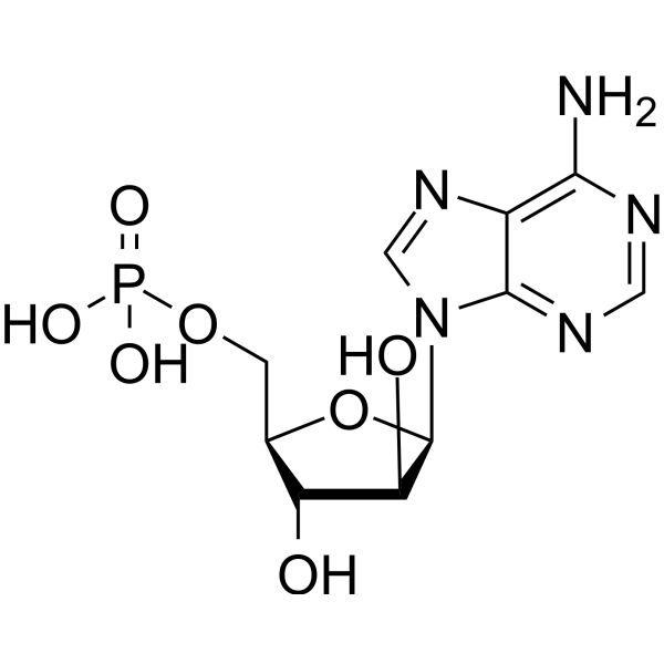
Vidarabine phosphate (ara-AMP; ara-A 5'-monophosphate) is a purine nucleoside antiviral agent and a prodrug of Vidarabine (HY-B0277). Vidarabine phosphate is rapidly converted into the antiviral active Vidarabine in vivo, which selectively inhibits viral DNA polymerase and cellular ribonucleotide reductase, thereby blocking viral replication. Vidarabine phosphate also exhibits antifungal activity, induces late-stage cellular apoptosis, and causes cell cycle arrest. Vidarabine phosphate can be used in research related to severe chronic active Epstein-Barr virus (EBV) infection, herpes infection, and candidiasis.
For research use only. We do not sell to patients.
- Purity : 99.44%
- CAS No.: 29984-33-6
- Formula: C10H14N5O7P
- Molecular Weight:347.22
-
Storage:
4°C, sealed storage, away from moisture
* In solvent : -80°C, 6 months; -20°C, 1 month (sealed storage, away from moisture)

To place orders, for customer services and technical support, please contact: MedChemExpress USA
Tel: 609-228-6898 E-mail: [email protected] [email protected]


-
 Biological Activity
-
Chemical Information
-
Solvent & Solubility
- Protocols
- Purity & Documentation
- References
-
 Help & FAQs


-
HY-N7138
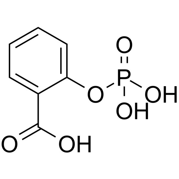
2-(Phosphonooxy)benzoic acidAnti-inflammatory Agent -

HY-B0277AR
Vidarabine phosphate (Standard)Standard of Vidarabine phosphate -
HY-13623A
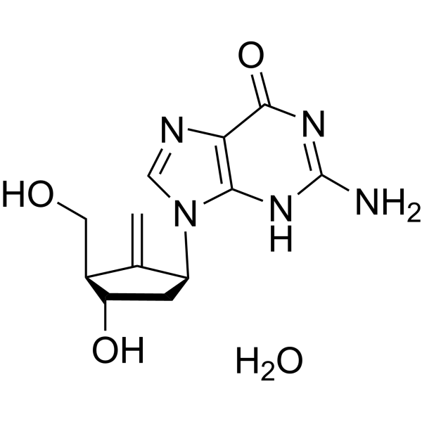
Entecavir monohydrateHBV Inhibitor -
HY-N0058
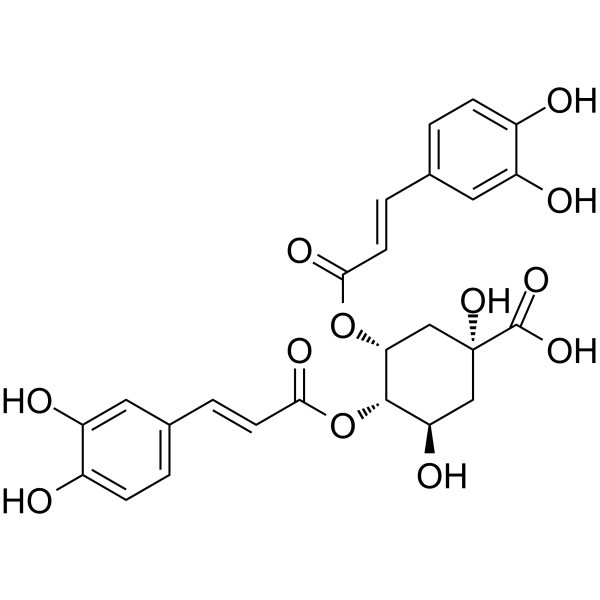
4,5-Dicaffeoylquinic acidAntioxidant -
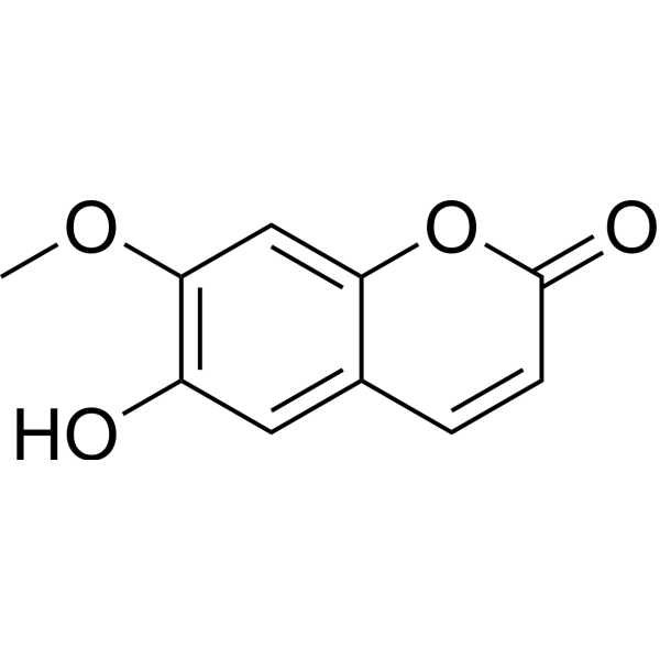
HY-N1365
IsoscopoletinHBV Inhibitor -
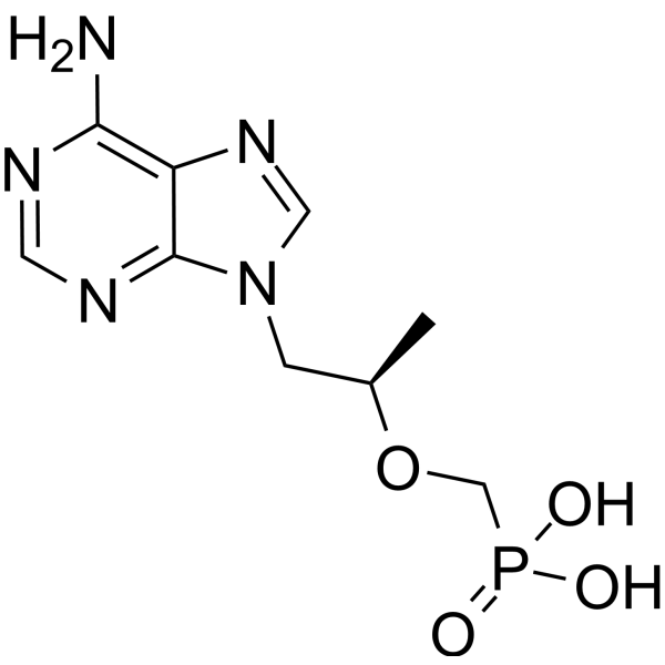
HY-13910
TenofovirNucleotide Reverse Transcriptase Inhibitor -
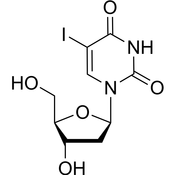
HY-B0307
IdoxuridineAntiviral Agent -
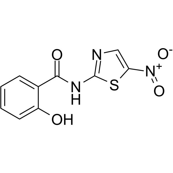
HY-12687
TizoxanideAnti-HIV Activity -
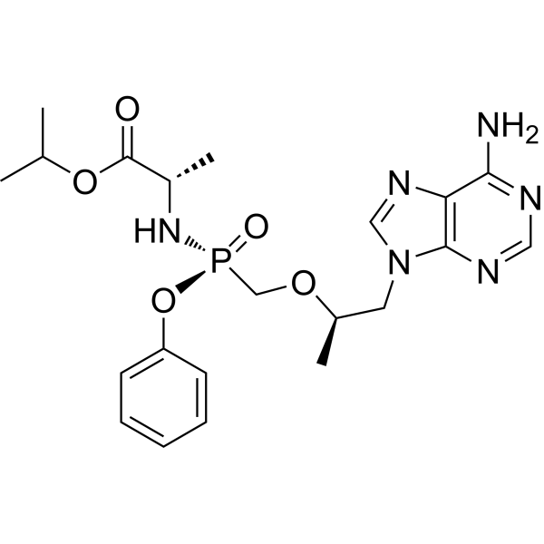
HY-15232
Tenofovir alafenamideReverse Transcriptase Inhibitor -
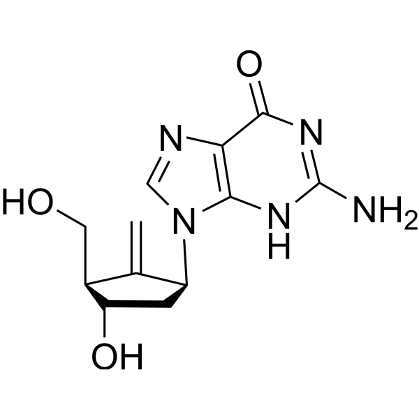
HY-13623
EntecavirHBV Inhibitor -
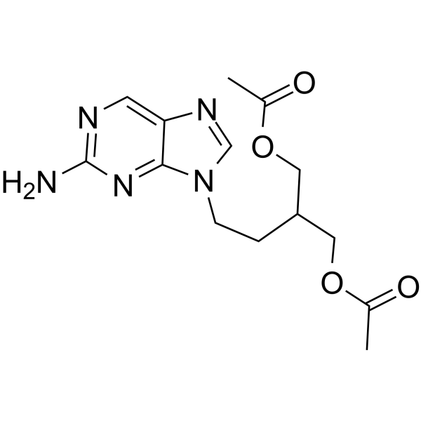
HY-17426
FamciclovirAntiviral Agent -
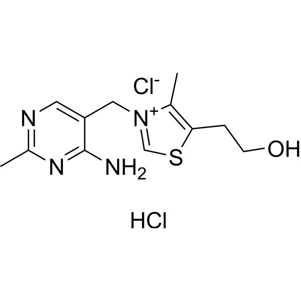
HY-N0680
Thiamine hydrochlorideVitamin B1
All DNA/RNA Synthesis Isoforms
More

Biological Activity
Description
In Vitro
Vidarabine (18-1200 μg/mL; 48 h) exhibits bactericidal activity against Candida albicans, with an MIC50 and MFC of 150 μg/mL and 300 μg/mL, respectively[3].
Vidarabine (150-300 μg/mL; 4-24 h) induces intracellular ROS accumulation, nuclear pyknosis, late-stage apoptosis and cell cycle arrest in Candida albicans cells, and reduces their ergosterol content[3].
MedChemExpress (MCE) has not independently confirmed the accuracy of these methods. They are for reference only.
-
Cell Line:Candida albicans cells
-
Concentration:150 μg/mL
-
Incubation Time:24 h
-
Result:Induced programmed cell death (apoptosis) in Candida albicans.
-
Cell Line:Candida albicans cells
-
Concentration:150 μg/mL
-
Incubation Time:4 h
-
Result:Caused the cells to undergo arrest (stall) at the G2/M phase, thereby preventing cell division.
In Vivo
MedChemExpress (MCE) has not independently confirmed the accuracy of these methods. They are for reference only.
-
Animal Model:CDR-CRL (male and female, 2 days old at study initiation)[2]
-
Dosage:25-400 mg/kg
-
Administration:i.p.; daily; 5 days
-
Result:Exhibited body weight gains comparable to untreated controls over the study period.
Showed delayed ventral injection-site hair growth measuring 2 cm2 (400 mg/kg group), 0.5 cm2 (100 mg/kg group), and an intermediate size (200 mg/kg group), first evident at 10 days and resolved by 16 days.
Displayed poorly developed hair follicles, adnexal glands, and mild dermal hypoplasia in injection-site skin 7 days after observation initiation, with no such changes at 5, 19, or 33 days.
Had no visceral lesions detected at necropsy at any interval, and no significant changes in absolute liver, brain, cerebellum, or kidney weights.
Showed mild cerebellar folia hypoplasia with an indistinct Purkinje cell layer in one 25 mg/kg female rat at 5 days, with no other histopathologic changes in brain, eye, liver, or kidney across all groups.


Chemical Information
-
CAS No. 29984-33-6
-
Appearance Solid
-
Molecular Weight 347.22
-
Formula C10H14N5O7P
-
Color White to off-white
-
SMILES
NC1=C(N=CN2[C@H]3[C@@H](O)[C@H](O)[C@@H](COP(O)(O)=O)O3)C2=NC=N1
-
Synonyms
ara-AMP; ara-A 5'-monophosphate
-
Shipping
Room temperature in continental US; may vary elsewhere.
-
Storage
4°C, sealed storage, away from moisture
* In solvent : -80°C, 6 months; -20°C, 1 month (sealed storage, away from moisture)


Solvent & Solubility
In Vitro:
DMSO : 16.67 mg/mL (48.01 mM; ultrasonic and warming and heat to 60°C; Hygroscopic DMSO has a significant impact on the solubility of product, please use newly opened DMSO)
H2O : 4.9 mg/mL (14.11 mM; ultrasonic and warming and adjust pH to 7 with NaOH and heat to 60°C)
Please refer to the solubility information to select the appropriate solvent. Once prepared, please aliquot and store the solution to prevent product inactivation from repeated freeze-thaw cycles.
Storage method and period of stock solution: -80°C, 6 months; -20°C, 1 month (sealed storage, away from moisture). When stored at -80°C, please use it within 6 months. When stored at -20°C, please use it within 1 month.
* Note: If you choose water as the stock solution, please dilute it to the working solution, then filter and sterilize it with a 0.22 μm filter before use.
Please refer to the solubility information to select the appropriate solvent. Once prepared, please aliquot and store the solution to prevent product inactivation from repeated freeze-thaw cycles.
Storage method and period of stock solution: -80°C, 6 months; -20°C, 1 month (sealed storage, away from moisture). When stored at -80°C, please use it within 6 months. When stored at -20°C, please use it within 1 month.
* Note: If you choose water as the stock solution, please dilute it to the working solution, then filter and sterilize it with a 0.22 μm filter before use.
Concentration (start) × Volume (start) = Concentration (final) × Volume (final)
In Vivo:
Select the appropriate dissolution method based on your experimental animal and administration route.
- For the following dissolution methods, please ensure to first prepare a clear stock solution using an In Vitro approach and then sequentially add co-solvents:
- To ensure reliable experimental results, the clarified stock solution can be appropriately stored based on storage conditions. As for the working solution for In Vivo experiments, it is recommended to prepare freshly and use it on the same day.
- The percentages shown for the solvents indicate their volumetric ratio in the final prepared solution. If precipitation or phase separation occurs during preparation, heat and/or sonication can be used to aid dissolution.
Add each solvent one by one: 10% DMSO 40% PEG300 5% Tween-80 45% Saline
Solubility: ≥ 1.67 mg/mL (4.81 mM); Clear solution
This protocol yields a clear solution of ≥ 1.67 mg/mL (saturation unknown).
Taking 1 mL working solution as an example, add 100 μL DMSO stock solution (16.7 mg/mL) to 400 μL PEG300, and mix evenly; then add 50 μL Tween-80 and mix evenly; then add 450 μL Saline to adjust the volume to 1 mL.
Preparation of Saline: Dissolve 0.9 g sodium chloride in ddH₂O and dilute to 100 mL to obtain a clear Saline solution.
Add each solvent one by one: 10% DMSO 90% (20% SBE-β-CD in Saline)
Solubility: ≥ 1.67 mg/mL (4.81 mM); Clear solution
This protocol yields a clear solution of ≥ 1.67 mg/mL (saturation unknown).
Taking 1 mL working solution as an example, add 100 μL DMSO stock solution (16.7 mg/mL) to 900 μL 20% SBE-β-CD in Saline, and mix evenly.
Preparation of 20% SBE-β-CD in Saline (4°C, storage for one week): 2 g SBE-β-CD powder is dissolved in 10 mL Saline, completely dissolve until clear.
In Vivo Dissolution Calculator
Please enter the basic information of animal experiments:
-
-
-
-
Recommended: Prepare an additional quantity of animals to account for potential losses during experiments.
Please enter your animal formula composition:
-
%DMSO +
Recommended: Keep the proportion of DMSO in working solution below 2% if your animal is weak.
-
%+
-
+%Tween-80 + +
-
%Saline +
The co-solvents required include: DMSO, . All of co-solvents are available by MedChemExpress (MCE). , Tween 80. All of co-solvents are available by MedChemExpress (MCE).
Working solution concentration: 0.22 mg/mL
Method for preparing stock solution: mg drug dissolved in μL DMSO. Stock solution concentration: mg/mL. * In solvent : -80°C, 6 months; -20°C, 1 month (sealed storage, away from moisture)
1. Take μL DMSO stock solution;
2. Add μL .
μL , mix evenly;
3. Then add μL Tween 80, mix evenly;
4. Then add μL
Please ensure that the stock solution in the first step is dissolved to a clear state, and add co-solvents in sequence. You can use ultrasonic heating (ultrasonic cleaner, recommended frequency 20-40 kHz), vortexing, etc. to assist dissolution.


Purity & Documentation
-
Data Sheet (282 KB)
-
SDS (419 KB)
- English - EN (419 KB)
- Français - FR (419 KB)
- Deutsch - DE (419 KB)
- Norwegian - NO (419 KB)
- Español - ES (419 KB)
- Swedish - SV (419 KB)
- Italian - IT (419 KB)
- Korean - KR (419 KB)
- Portuguese - PT (419 KB)
-
Handling Instructions (2659 KB)
References
[1]. Kimura H, et al. Vidarabine therapy for severe chronic active Epstein-Barr virus infection. J Pediatr Hematol Oncol. 2001;23(5):294-299. [Content Brief]
[3]. Gavandi TC, et al. Vidarabine as a novel antifungal agent against Candida albicans: insights on mechanism of action. Int Microbiol. 2025;28(3):589-602. [Content Brief]


Complete Stock Solution Preparation Table
Please refer to the solubility information to select the appropriate solvent. Once prepared, please aliquot and store the solution to prevent product inactivation from repeated freeze-thaw cycles.
Storage method and period of stock solution: -80°C, 6 months; -20°C, 1 month (sealed storage, away from moisture). When stored at -80°C, please use it within 6 months. When stored at -20°C, please use it within 1 month.
| Optional Solvent | Concentration Solvent Mass | 1 mg | 5 mg | 10 mg | 25 mg |
|---|---|---|---|---|---|
| H2O / DMSO | 1 mM | 2.8800 mL | 14.4001 mL | 28.8002 mL | 72.0005 mL |
| 5 mM | 0.5760 mL | 2.8800 mL | 5.7600 mL | 14.4001 mL | |
| 10 mM | 0.2880 mL | 1.4400 mL | 2.8800 mL | 7.2000 mL | |
| DMSO | 15 mM | 0.1920 mL | 0.9600 mL | 1.9200 mL | 4.8000 mL |
| 20 mM | 0.1440 mL | 0.7200 mL | 1.4400 mL | 3.6000 mL | |
| 25 mM | 0.1152 mL | 0.5760 mL | 1.1520 mL | 2.8800 mL | |
| 30 mM | 0.0960 mL | 0.4800 mL | 0.9600 mL | 2.4000 mL | |
| 40 mM | 0.0720 mL | 0.3600 mL | 0.7200 mL | 1.8000 mL |
* Note: If you choose water as the stock solution, please dilute it to the working solution, then filter and sterilize it with a 0.22 μm filter before use.


Vidarabine phosphate Related Classifications
HY-B0277A Related Classifications
 Powered by Bioz
Keywords
- Vidarabine
- 29984-33-6
- ara-AMP
- ara-A 5'-monophosphate
- EBV
- HSV
- Fungal
- DNA/RNA Synthesis
- Apoptosis
- Drug Intermediate
- Reactive Oxygen Species (ROS)
- viral DNA polymerase
- caspofungin
- fluconazole
- Candida albicans
- candidiasis
- natural killer cells
- Epstein-Barr virus
- neonatal rats
- NK cell-type severe chronic active EBV infection
- cellular ribonucleotide reductase
- Inhibitor
- inhibitor
- inhibit