



WP9QY

Based on 1 Customer Validation
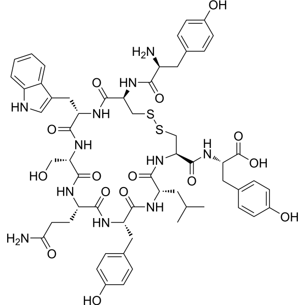
WP9QY is an inhibitor targeting TNFα and RANKL, which blocks the TNFα-TNFR1 interaction and inhibits TNFα-mediated apoptosis, cytotoxicity and bone destruction. WP9QY inhibits osteoclastogenesis and promotes osteoblast differentiation, induces chondrocyte proliferation and glycosaminoglycan production, and synergizes with TGF-β3 to promote chondrogenesis. WP9QY effectively repairs full-thickness articular cartilage defects in rabbits via intra-articular injection, and inhibits methylmercury-induced reduction of NeuN-positive cells in mouse brain slices. WP9QY can be applied to the research of diseases related to methylmercury-induced neuronal death, cartilage injury, osteoarthritis and bone loss.
For research use only. We do not sell to patients.
- Purity : 99.79%
- CAS No.: 199999-60-5
- Formula: C58H71N11O15S2
- Molecular Weight:1226.38
-
Storage:
Sealed storage, away from moisture.
Powder -80°C, 2 years , -20°C, 1 year* In solvent : -80°C, 6 months; -20°C, 1 month (sealed storage, away from moisture)

To place orders, for customer services and technical support, please contact: MedChemExpress USA
Tel: 609-228-6898 E-mail: [email protected] [email protected]


-
 Biological Activity
-
Chemical Information
-
Solvent & Solubility
- Protocols
- Purity & Documentation
- References
-
 Help & FAQs


-

HY-P9908
AdalimumabTNF-α Inhibitor -

HY-P9970
InfliximabTNF-α Inhibitor -
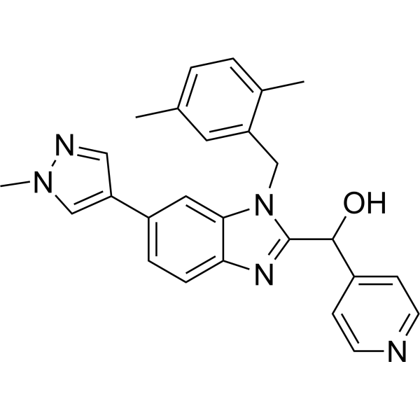
HY-133122
UCB-9260TNF Signaling Inhibitor -
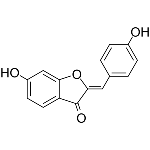
HY-102040
HispidolTNF-α Inhibitor -
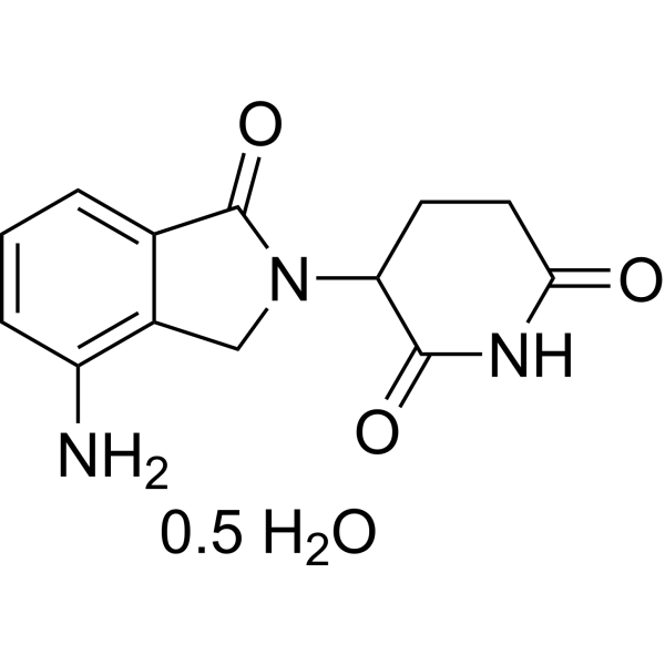
HY-A0003B
Lenalidomide hemihydrateImmunomodulator -
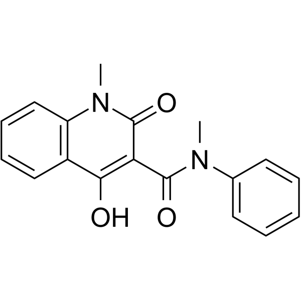
HY-13743
RoquinimexImmunomodulator -

HY-108847
EtanerceptTNF Inhibitor -
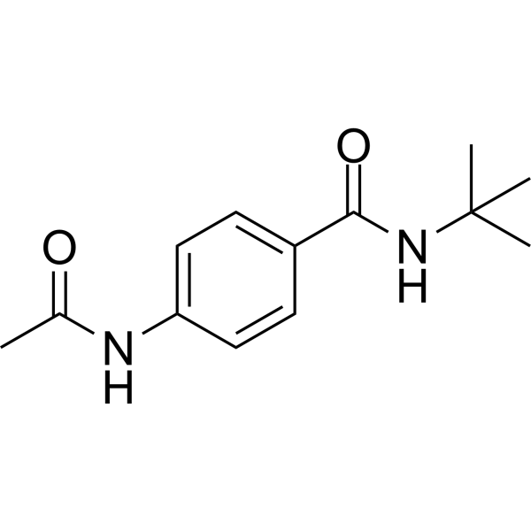
HY-100376
CPI-1189TNF-α Inhibitor -
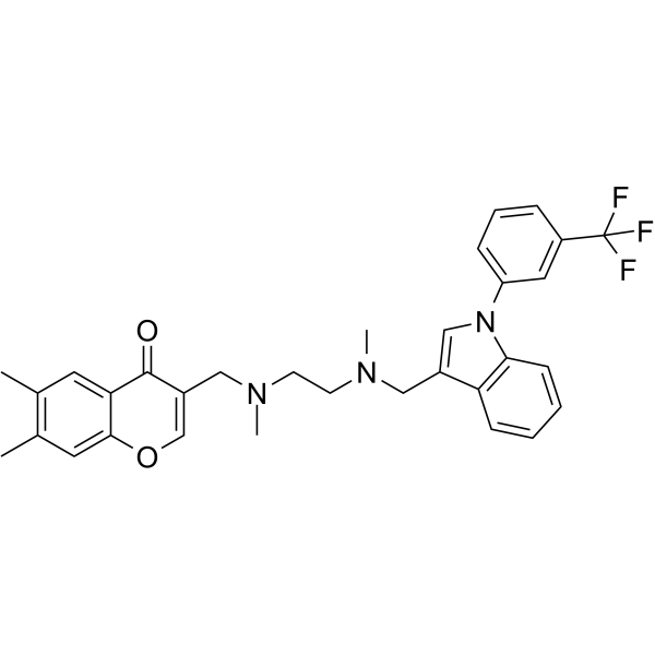
HY-111255
SPD304TNFα Inhibitor -
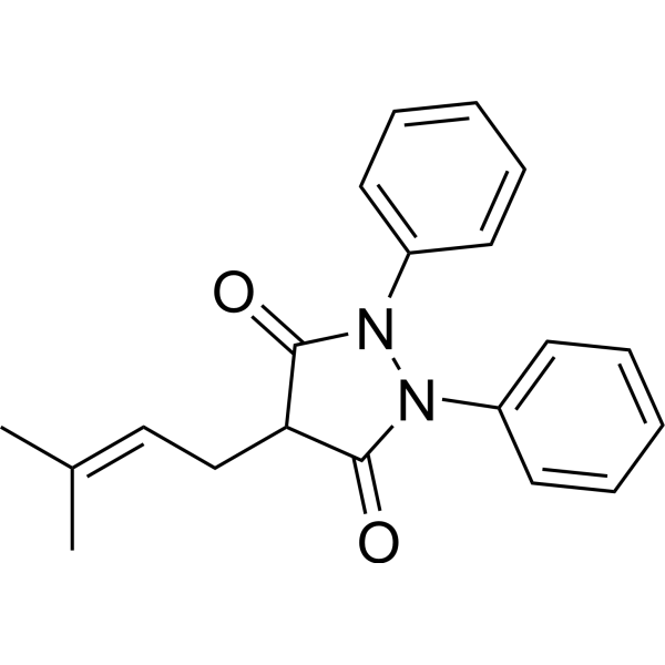
HY-114911
FeprazoneNonsteroidal Anti-inflammatory Agent -

HY-P9953
Certolizumab pegolTNF-α Monoclonal Antibody -

HY-P99111
GolimumabTNFα Antagonist


Biological Activity
Description
In Vitro
WP9QY-functionalized PEG hydrogels (100-250 μM; 24 h TNFα exposure, 6 d NGF differentiation culture) protect differentiated PC12 cells from apoptosis induced by 50 ng/mL human TNFα, maintain cell viability, and prevent the elevation of caspase 3/7 activation levels[1].
WP9QY-functionalized PEG hydrogel (100 μM; 24 h) protects encapsulated mouse islets from 50 ng/mL human TNFα-induced apoptosis and impaired insulin secretion, and maintains cell viability and function[1].
WP9QY (10 μM; 72 h) inhibits neuronal cell death induced by 50 μM methylmercury in cultured mouse organotypic brain slices in vitro[2].
WP9QY (100 μM; 14 d) significantly enhances chondrogenic differentiation of hMSCs in a two-dimensional culture system via accumulation of glycosaminoglycan matrix, as confirmed by Alcian Blue staining (HY-D0001) and absorbance measurement[3].
WP9QY (25-100 μM; 72 h) dose-dependently promotes the proliferation of hMSCs cultured in maintenance medium, chondrocyte differentiation medium, and osteoblast differentiation medium, and this result is confirmed by Hoechst 33342 (HY-15559) fluorescence assay[3].
WP9QY (100 μM; 14 d) induces chondrocyte differentiation in both control and RANKL-knockdown C3H10T1/2 cells in a RANKL signaling-independent manner[3].
WP9QY (5-50 μM; 20 min) dose-dependently inhibits RANKL-induced signaling pathways in RAW 264.7 cells, without affecting M-CSF-induced signaling pathways[4].
MedChemExpress (MCE) has not independently confirmed the accuracy of these methods. They are for reference only. Further protocols information, click here.
-
Cell Line:differentiated rat adrenal pheochromocytoma (PC12) cells
-
Concentration:100-250 μM WP9QY; 50 ng/mL human TNFα; 50 ng/mL 2.5S NGF
-
Incubation Time:24 hours (TNFα exposure); 6 days (NGF differentiation)
-
Result:Maintained viability comparable to unchallenged cells in PEG-RGD-WP9QY hydrogels (100 μM and 250 μM WP9QY) under TNFα challenge.
Showed no significant increase in caspase 3/7 activity.
Confirmed minimal cell death via Live/Dead imaging.
-
Cell Line:human mesenchymal stem cells (hMSCs)
-
Concentration:100 μM
-
Incubation Time:14 days
-
Result:Increased absorbance at 620 nm to ~0.27, which was significantly higher than control's ~0.17 with p < 0.01.
-
Cell Line:mouse chondrogenic ATDC5 cells
-
Concentration:25-100 μM
-
Incubation Time:18 days
-
Result:Induced dose-dependent chondrocytic differentiation, with more intense Alcian Blue staining at 100 μM compared to 25 μM, and no staining in untreated control cells.
-
Cell Line:human mesenchymal stem cells (hMSCs)
-
Concentration:50-150 μM (with 10 ng/mL TGF-β3 present throughout culture)
-
Incubation Time:14 days
-
Result:Induced dose-dependent enlargement of hMSC 3D pellets, with larger pellets observed at 150 μM compared to 50 μM.
Increased Alcian Blue-positive cartilage matrix production in treated pellets.
-
Cell Line:human mesenchymal stem cells (hMSCs)
-
Concentration:100 μM (tested alone and in combination with 20 ng/mL TGF-β3)
-
Incubation Time:15 days
-
Result:Strongly enhanced chondrogenesis alone, while TGF-β3 alone weakly enhanced chondrogenesis.
Combined treatment resulted in synergistically increased Alcian Blue staining intensity, showing greater chondrogenesis than either treatment alone.
-
Cell Line:human mesenchymal stem cells (hMSCs)
-
Concentration:100 μM (tested alone and in combination with 10 ng/mL TGF-β3)
-
Incubation Time:21 days
-
Result:Increased pellet size and induced cartilage matrix production (positive Alcian Blue and Safranin O staining) alone.
TGF-β3 alone increased chondrocytic differentiation but did not affect pellet size.
Combined treatment resulted in synergistically enhanced cartilage matrix production and larger pellet size compared to either treatment alone.
-
Cell Line:human mesenchymal stem cells (hMSCs)
-
Concentration:25-100 μM
-
Incubation Time:72 hours
-
Result:Promoted hMSC proliferation in maintenance, chondrocyte-differentiation, and osteoblast-differentiation media in a dose-dependent manner.
Elevated fluorescence intensity at 25 μM compared to untreated controls; further increased intensity at 100 μM, with p < 0.01 for all comparisons to untreated controls.
-
Cell Line:mouse mesenchymal C3H10T1/2 cells (control and RANKL-knockdown)
-
Concentration:100 μM
-
Incubation Time:14 days
-
Result:Enhanced chondrocytic differentiation (positive Alcian Blue staining) in both control and RANKL-knockdown C3H10T1/2 cells.
RANKL knockdown had no effect on WP9QY-induced chondrogenesis.
In Vivo
WP9QY (2.1 mg/kg/d; continuous infusion via osmotic minipump; 4 weeks) completely prevents ovariectomy-induced osteoporosis in female C57BL/6J mice, restoring cancellous bone parameters and reducing osteoclast activity to near sham-operated levels[4].
WP9QY (9 mg/kg; s.c.; every 3 hours; 48 hours) completely prevents low dietary calcium2+-induced osteoporosis in male CD-1 mice, restoring cancellous bone parameters and reducing osteoclast activity to near normal-calcium diet levels[4].
WP9QY (4 mg/kg/d; continuous infusion via osmotic minipump; 7 days) completely prevents low dietary calcium2+-induced osteoporosis in both TNFR(I)-/- TNFR(II)-/- and wild-type mice[4].
MedChemExpress (MCE) has not independently confirmed the accuracy of these methods. They are for reference only.
-
Animal Model:Japanese white (male, 19-20 weeks old)[3]
-
Dosage:5 mg
-
Administration:intraarticular; 2 doses (days 14 and 21)
-
Result:Caused partial filling of articular cartilage defects and expanded Safranin-O-positive regions in defects.
Reduced mean modified Wakitani score to ~7, versus ~12 for vehicle (P=0.04).
-
Animal Model:C57BL/6J Mice (female, 12 weeks old, bilateral ovariectomy-induced)[4]
-
Dosage:2.1 mg/kg/d
-
Administration:continuous infusion via osmotic minipump; 4 weeks
-
Result:Maintained bone mineral density of proximal tibiae at levels not significantly different from sham-operated mice.
Restored cancellous bone volume fraction from 9.10% to 14.79%.
Restored trabecular number from 6.10 no./mm to 8.01 no./mm.
Restored trabecular spacing from 150.12 μm to 109.998 μm.
Reduced osteoclast number per bone surface from 7.62 no./mm to 3.65 no./mm.
Reduced osteoclast surface per bone surface from 11.58% to 5.96%.
Blocked the ovariectomy-induced increase in urinary deoxypyridinoline cross-links.
Had no effect on body weight or uterine weight.
-
Animal Model:TNFR(I)-/- TNFR(II)-/- Mice (129/B6 background); wild-type (genetically matched, low dietary calcium-induced)[4]
-
Dosage:4 mg/kg/d
-
Administration:continuous infusion via osmotic minipump; 7 days
-
Result:Maintained bone mineral density at levels not significantly different from mice on a normal-calcium diet in both TNFR double-knockout and wild-type mice.
Blocked the low-calcium diet-induced increase in urinary deoxypyridinoline cross-links in both genotypes.


Chemical Information
-
CAS No. 199999-60-5
-
Appearance Solid
-
Molecular Weight 1226.38
-
Formula C58H71N11O15S2
-
Color White to off-white
-
Sequence
Tyr-Cys-Trp-Ser-Gln-Tyr-Leu-Cys-Tyr (Disulfide bridge:Cys2-Cys8)
-
Sequence Shortening
YCWSQYLCY (Disulfide bridge:Cys2-Cys8)
-
Shipping
Room temperature in continental US; may vary elsewhere.
-
Storage
Sealed storage, away from moisture
Powder -80°C 2 years -20°C 1 year * In solvent : -80°C, 6 months; -20°C, 1 month (sealed storage, away from moisture)


Solvent & Solubility
In Vitro:
DMSO : 100 mg/mL (81.54 mM; Need ultrasonic; Hygroscopic DMSO has a significant impact on the solubility of product, please use newly opened DMSO)
H2O : 1 mg/mL (0.82 mM; Need ultrasonic)
Please refer to the solubility information to select the appropriate solvent. Once prepared, please aliquot and store the solution to prevent product inactivation from repeated freeze-thaw cycles.
Storage method and period of stock solution: -80°C, 6 months; -20°C, 1 month (sealed storage, away from moisture). When stored at -80°C, please use it within 6 months. When stored at -20°C, please use it within 1 month.
* Note: If you choose water as the stock solution, please dilute it to the working solution, then filter and sterilize it with a 0.22 μm filter before use.
Please refer to the solubility information to select the appropriate solvent. Once prepared, please aliquot and store the solution to prevent product inactivation from repeated freeze-thaw cycles.
Storage method and period of stock solution: -80°C, 6 months; -20°C, 1 month (sealed storage, away from moisture). When stored at -80°C, please use it within 6 months. When stored at -20°C, please use it within 1 month.
* Note: If you choose water as the stock solution, please dilute it to the working solution, then filter and sterilize it with a 0.22 μm filter before use.
Concentration (start) × Volume (start) = Concentration (final) × Volume (final)
In Vivo:
Select the appropriate dissolution method based on your experimental animal and administration route.
- For the following dissolution methods, please ensure to first prepare a clear stock solution using an In Vitro approach and then sequentially add co-solvents:
- To ensure reliable experimental results, the clarified stock solution can be appropriately stored based on storage conditions. As for the working solution for In Vivo experiments, it is recommended to prepare freshly and use it on the same day.
- The percentages shown for the solvents indicate their volumetric ratio in the final prepared solution. If precipitation or phase separation occurs during preparation, heat and/or sonication can be used to aid dissolution.
In Vivo Dissolution Calculator
Please enter the basic information of animal experiments:
-
-
-
-
Recommended: Prepare an additional quantity of animals to account for potential losses during experiments.
Please enter your animal formula composition:
-
%DMSO +
Recommended: Keep the proportion of DMSO in working solution below 2% if your animal is weak.
-
%+
-
+%Tween-80 + +
-
%Saline +
The co-solvents required include: DMSO, . All of co-solvents are available by MedChemExpress (MCE). , Tween 80. All of co-solvents are available by MedChemExpress (MCE).
Working solution concentration: 0.22 mg/mL
Method for preparing stock solution: mg drug dissolved in μL DMSO. Stock solution concentration: mg/mL. * In solvent : -80°C, 6 months; -20°C, 1 month (sealed storage, away from moisture)
1. Take μL DMSO stock solution;
2. Add μL .
μL , mix evenly;
3. Then add μL Tween 80, mix evenly;
4. Then add μL
Please ensure that the stock solution in the first step is dissolved to a clear state, and add co-solvents in sequence. You can use ultrasonic heating (ultrasonic cleaner, recommended frequency 20-40 kHz), vortexing, etc. to assist dissolution.

Protocols
-
How to Select the Route of Administration for Mammals
Route-of-administration selection in mammals is a pharmacokinetic, pharmacodynamic, formulation, animal-welfare, and translational decision, not a default technical choice. The selected route should match the study goal: intravenous dosing is most useful when complete systemic exposure and rapid onset are required, oral dosing is most translational for orally intended medicines but is affected by absorption and first-pass metabolism, subcutaneous or intramuscular dosing can provide slower systemic exposure, and intraperitoneal dosing can be useful in rodent proof-of-concept studies but may have limited clinical translation. Published route-comparison studies show that the same compound can produce different exposure, onset, bioavailability, tissue distribution, and tolerability depending on route; therefore, route choice should be supported by pilot pharmacokinetic or pharmacodynamic evidence when the literature is insufficient. Unresolved questions include how to standardize route sel
-
Apoptosis
Apoptosis, also called programmed cell death, is generally characterized by distinct morphological characteristics.
-
TUNEL staining for apoptotic DNA fragmentation
TUNEL staining detects DNA strand breaks by using terminal deoxynucleotidyl transferase to add labeled nucleotides to exposed 3′-OH DNA termini, generating either microscopic staining in fixed cells or tissue sections, or fluorescence/cytometric signal in cell suspensions. TUNEL positivity reflects DNA fragmentation but should not be interpreted alone as definitive apoptosis, because TUNEL can also label necrotic, autolytic, mechanically damaged, or DNA-repair-associated DNA breaks.
-
Organotypic Brain Slice Culture
Organotypic brain slice culture is an ex vivo method in which CNS tissue slices are maintained on a stable support with culture medium and oxygen access, preserving tissue architecture, multiple resident brain cell types, and network organization better than dissociated cultures. The commonly used membrane-interface method places brain or hippocampal slices on a porous membrane insert at the air-liquid interface; culture medium reaches the tissue through the membrane while the slice remains oxygenated from the humidified incubator atmosphere. Readouts depend on the experimental aim: slice survival can be monitored by propidium iodide uptake or LDH release, tissue organization by immunostaining, live structural changes by repeated imaging, and neuronal/network function by electrophysiology or multi-electrode recordings.
-
Cell Cytotoxicity Assay
Cytotoxicity assays are usually based on the assessment of cell membrane damage, which can also be indirectly detected by measuring cell viability. Detection methods include MTT assay, CKK-8 assay, LDH assay and ATP assay, etc.
-
Mammalian live/dead viability and cytotoxicity staining
Live/dead viability and cytotoxicity staining assays are based on the simultaneous detection of intracellular esterase activity in metabolically active (viable) cells and membrane integrity loss in non-viable cells. In commonly used dual-staining approaches, membrane-permeant fluorogenic substrates are converted by intracellular esterases into fluorescent products in live cells, while impermeant DNA-binding dyes selectively enter cells with compromised plasma membranes and label nucleic acids in dead or dying cells, enabling discrimination between viable and non-viable populations by fluorescence microscopy or flow cytometry.
-
Mesenchymal stromal/stem cell chondrogenic differentiation
MSC chondrogenic differentiation is commonly induced by culturing bone marrow-derived mesenchymal stromal/stem cells as high-density three-dimensional pellets or micromass aggregates in defined chondrogenic medium containing TGF-β family stimulation; the readout is formation of cartilage-like extracellular matrix, especially sulfated proteoglycans, aggrecan, and type II collagen. The assay detects chondrogenesis by pellet enlargement, metachromatic or Alcian blue/Safranin O staining of proteoglycan-rich matrix, immunodetection of type II collagen and aggrecan, and gene-expression changes in cartilage matrix markers; hypertrophic or fibrocartilaginous drift can be assessed by collagen X and collagen I readouts when included.
-
Annexin V plus membrane-impermeant dye apoptosis staining
Annexin V-based apoptosis assays rely on the detection of phosphatidylserine (PS) externalization from the inner leaflet of the plasma membrane to the outer leaflet, an early biochemical hallmark of apoptosis. Fluorescently labeled Annexin V binds PS in a calcium-dependent manner, enabling identification of early apoptotic cells by flow cytometry or fluorescence microscopy. When combined with a membrane-impermeant DNA-binding dye (e. g. , propidium iodide), this approach allows discrimination between viable (Annexin V−/dye−), early apoptotic (Annexin V+/dye−), and late apoptotic or necrotic (Annexin V+/dye+) cell populations by assessing membrane integrity and PS exposure.
-
Apoptosis Solutions
Apoptosis is a regulated, generally non-lytic cell-death pathway that removes unwanted, damaged, infected, or abnormal cells through coordinated morphological changes, caspase activation, DNA fragmentation, and membrane remodeling. The intrinsic apoptosis pathway is controlled mainly by mitochondrial outer membrane permeabilization, BCL-2 family proteins, cytochrome c release, apoptosome formation, caspase-9 activation, and downstream executioner caspase-3/7 activation. The extrinsic apoptosis pathway is initiated by death receptors such as Fas, TNFR, and TRAIL receptors, which recruit adaptor proteins and activate caspase-8 before engaging executioner caspases or mitochondrial amplification through BID cleavage. Apoptosis is linked to many phenotypes, including cancer cell killing, tissue homeostasis, immune regulation, neurodegeneration, infection response, and treatment-induced cytotoxicity; unresolved questions include how apoptosis interacts with necroptosis, pyroptosis, ferroptos
-
Carbohydrates and Mucins: Alcian Blue/Alcian Blue-PAS Staining
Alcian Blue (AB) staining is a cationic copper phthalocyanine dye-based histochemical method that binds electrostatically to negatively charged acidic mucopolysaccharides (glycosaminoglycans and sialomucins), enabling visualization of acidic carbohydrate-rich structures such as epithelial mucins, cartilage matrix, and mast cell granules. Periodic Acid-Schiff (PAS) reaction detects neutral mucopolysaccharides and glycoconjugates by oxidizing vicinal diols to aldehydes, which subsequently react with Schiff reagent to produce a magenta signal. The combined Alcian Blue-PAS (AB-PAS) method allows simultaneous differentiation of acidic (blue) and neutral (magenta) mucins in the same tissue section, enabling mucin subtype discrimination in epithelial tissues and pathological lesions.
-
Cell Viability Determination by MTT Colorimetric Assay
The following protocol uses the MTT colorimetric assay as a classic literature-established method for assessing cell viability/metabolic activity in cultured mammalian cells. MTT[3-(4,5-dimethylthiazol-2-yl)-2,5-diphenyltetrazolium bromide] is reduced by metabolically active cells to a colored formazan product; the amount of formazan is quantified spectrophotometrically and provides an indirect measure of metabolically active viable cells. Importantly, MTT reduction reflects cellular oxidoreductase/metabolic activity rather than an absolute direct count of living cells, so changes in cellular metabolism can alter the signal independently of cell number.
-
Osteoclast differentiation from monocyte/macrophage precursors
Osteoclast differentiation is an in vitro induction assay in which monocyte/macrophage-lineage precursors are exposed to macrophage colony-stimulating factor (M-CSF) and receptor activator of NF-κB ligand (RANKL), generating multinucleated osteoclasts that are commonly identified by tartrate-resistant acid phosphatase (TRAP) staining and functionally confirmed by resorption pits on dentin, bone, or mineralized substrates. M-CSF supports survival and expansion of osteoclast precursors, while RANKL binding to RANK drives osteoclast commitment, fusion, maturation, and resorptive function; osteoprotegerin inhibits this pathway by binding RANKL and preventing RANK activation. The main readouts are the number of TRAP-positive multinucleated cells, formation of F-actin rings, and resorbed surface area; TRAP-positive multinucleated cells indicate osteoclast differentiation, whereas pit formation on dentin, bone, or mineralized coating indicates functional bone-resorbing activity.


Purity & Documentation
-
Data Sheet (299 KB)
-
SDS (254 KB)
- English - EN (254 KB)
- Français - FR (254 KB)
- Deutsch - DE (254 KB)
- Norwegian - NO (254 KB)
- Español - ES (254 KB)
- Swedish - SV (254 KB)
- Italian - IT (254 KB)
- Korean - KR (254 KB)
- Portuguese - PT (254 KB)
-
Handling Instructions (2659 KB)
References
[1]. Lin CC, et al. Functional PEG-peptide hydrogels to modulate local inflammation induced by the pro-inflammatory cytokine TNFalpha. Biomaterials. 2009;30(28):4907-4914. [Content Brief]
[2]. Toyama T, et al. Methylmercury induces neuronal cell death by inducing TNF-α expression through the ASK1/p38 signaling pathway in microglia. Sci Rep. 2021;11(1):9832. Published 2021 May 10. [Content Brief]
[3]. Furuya Y, et al. Induction of chondrogenesis with a RANKL-binding peptide, WP9QY, in vitro and in vivo in a rabbit model. Biochem Biophys Res Commun. 2022;602:98-104. [Content Brief]
[4]. Aoki K, et al. A TNF receptor loop peptide mimic blocks RANK ligand-induced signaling, bone resorption, and bone loss. J Clin Invest. 2006;116(6):1525-1534. [Content Brief]


Complete Stock Solution Preparation Table
Please refer to the solubility information to select the appropriate solvent. Once prepared, please aliquot and store the solution to prevent product inactivation from repeated freeze-thaw cycles.
Storage method and period of stock solution: -80°C, 6 months; -20°C, 1 month (sealed storage, away from moisture). When stored at -80°C, please use it within 6 months. When stored at -20°C, please use it within 1 month.
| Optional Solvent | Concentration Solvent Mass | 1 mg | 5 mg | 10 mg | 25 mg |
|---|---|---|---|---|---|
| DMSO | 1 mM | 0.8154 mL | 4.0770 mL | 8.1541 mL | 20.3852 mL |
| 5 mM | 0.1631 mL | 0.8154 mL | 1.6308 mL | 4.0770 mL | |
| 10 mM | 0.0815 mL | 0.4077 mL | 0.8154 mL | 2.0385 mL | |
| 15 mM | 0.0544 mL | 0.2718 mL | 0.5436 mL | 1.3590 mL | |
| 20 mM | 0.0408 mL | 0.2039 mL | 0.4077 mL | 1.0193 mL | |
| 25 mM | 0.0326 mL | 0.1631 mL | 0.3262 mL | 0.8154 mL | |
| 30 mM | 0.0272 mL | 0.1359 mL | 0.2718 mL | 0.6795 mL | |
| 40 mM | 0.0204 mL | 0.1019 mL | 0.2039 mL | 0.5096 mL | |
| 50 mM | 0.0163 mL | 0.0815 mL | 0.1631 mL | 0.4077 mL | |
| 60 mM | 0.0136 mL | 0.0680 mL | 0.1359 mL | 0.3398 mL | |
| 80 mM | 0.0102 mL | 0.0510 mL | 0.1019 mL | 0.2548 mL |
* Note: If you choose water as the stock solution, please dilute it to the working solution, then filter and sterilize it with a 0.22 μm filter before use.


WP9QY Related Classifications
HY-P2612 Related Classifications
 Powered by Bioz