



KW-2478
Based on 4 publication(s) in Google Scholar
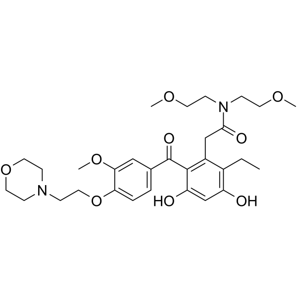
KW-2478 is an HSP90 inhibitor (IC50 = 3.8 nM). KW-2478 inhibits the growth and induces apoptosis in chronic myeloid leukemia (CML) cells and liver cancer cells. KW-2478 weakens the BCR/ABL and MAPK signaling pathways, leading to increased p27 and p21 expression and decreased cyclin B1 expression. KW-2478 downregulates STAT3 expression. KW-2478 may be used in research on cancers such as CML and liver cancer.
For research use only. We do not sell to patients.
- Purity : 99.53%
- CAS No.: 819812-04-9
- Formula: C30H42N2O9
- Molecular Weight:574.66
-
Storage:Powder -20°C, 3 years , 4°C, 2 years ; In solvent -80°C, 2 years , -20°C, 1 year

To place orders, for customer services and technical support, please contact: MedChemExpress USA
Tel: 609-228-6898 E-mail: [email protected] [email protected]


-
 Biological Activity
-
Chemical Information
-
Solvent & Solubility
- Protocols
- Purity & Documentation
- References
-
 Help & FAQs


-
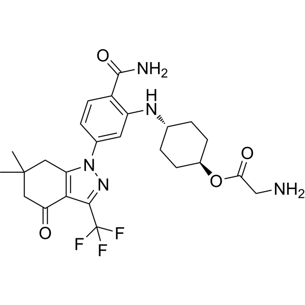
HY-10213
SNX-5422 -
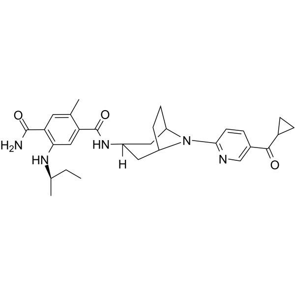
HY-13313
XL888HSP90 Inhibitor -
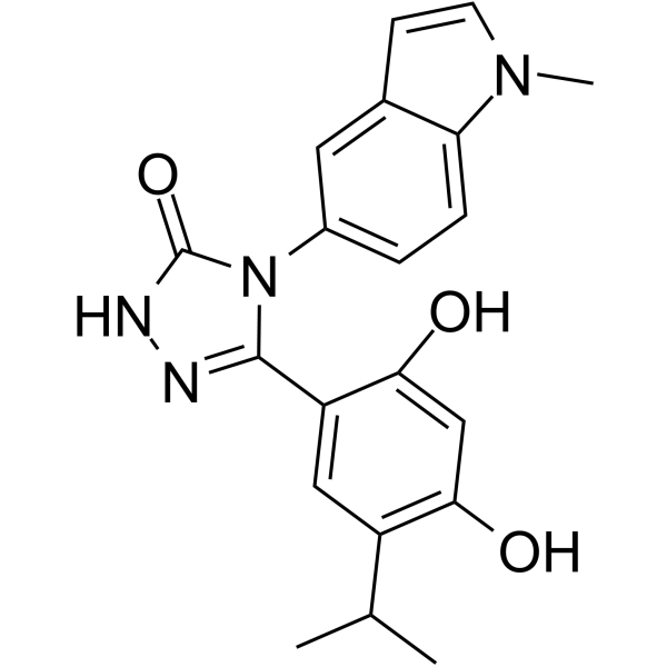
HY-15205
GanetespibHSP90 Inhibitor -
HY-10215
LuminespibHSP90 Inhibitor -
HY-10212
BIIB021HSP90 Inhibitor -
HY-14463
OnalespibHsp90 Inhibitor -
HY-10211
TanespimycinHSP90 Inhibitor -
HY-15230
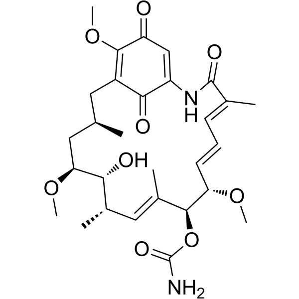
GeldanamycinHsp90 Inhibitor -
HY-11038
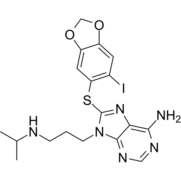
ZelavespibHsp90 Inhibitor -
HY-15785
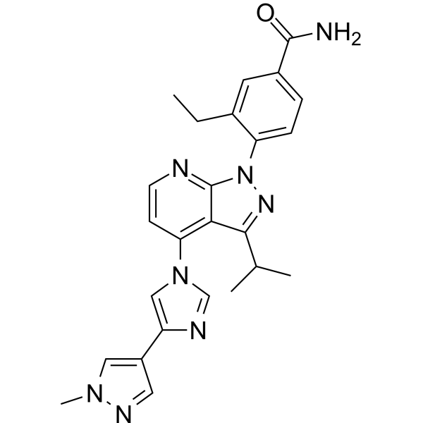
PimitespibHSP90α/HSP90β Inhibitor -
HY-10942
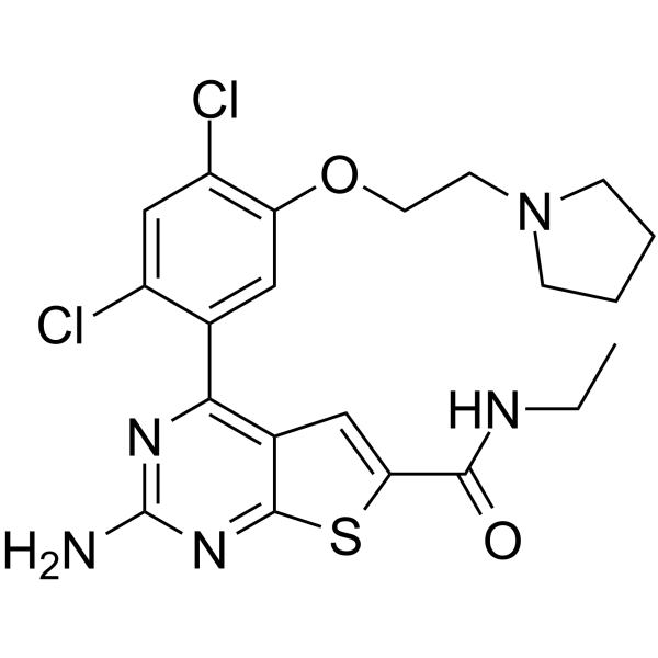
VER-82576HSP Inhibitor -
HY-10214
SNX-2112
Publications Citing Use of MedChemExpress (MCE) KW-2478
More Customer Validation & Images

-
RT-PCR

-
WB


Biological Activity
Description
IC50 & Target
[1]|
HSP90α 3.8 nM (IC50) |
In Vitro
KW-2478 is an inhibitor of Hsp90, with an IC50 of 3.8 nM for Hsp90α. KW-2478 shows anti-proliferative activity against multiple myeloma (MM) and non-Hodgkin’s lymphoma (NHL), with GI50s of 0.30 μM (OPM-2/GFP), 0.34 μM (KMS-11), 0.39 μM (RPMI 8226), 0.12 μM (NCI-H929), 0.36 μM (Raji), 0.098 μM (SR), and 0.33 μM μM (SC-1). KW-2478 also inhibits the transcription of c-Maf and Cyclin D1 genes by mainly suppressing the function of Cdk9[1].
MedChemExpress (MCE) has not independently confirmed the accuracy of these methods. They are for reference only.
In Vivo
MedChemExpress (MCE) has not independently confirmed the accuracy of these methods. They are for reference only.


Chemical Information
-
CAS No. 819812-04-9
-
Appearance Solid
-
Molecular Weight 574.66
-
Formula C30H42N2O9
-
Color Light yellow to yellow
-
SMILES
O=C(N(CCOC)CCOC)CC1=C(C(C2=CC=C(OCCN3CCOCC3)C(OC)=C2)=O)C(O)=CC(O)=C1CC
-
Shipping
Room temperature in continental US; may vary elsewhere.
-
Storage
Powder -20°C 3 years 4°C 2 years In solvent -80°C 2 years -20°C 1 year


Publications (4)
-
Journal Impact Factor
-
Most Recent
-
Exp Mol Med
2025 Jun;57(6):1308-1323. PMID: 40583061 -
Int J Mol Sci
2023 Nov 4;24(21):15971. PMID: 37958953
KW-2478 purchased from MedChemExpress. Usage Cited in: Int J Mol Sci. 2023 Nov 4;24(21):15971. [Abstract]
The relative mRNA level of TGEV M gene was determined by qRT-PCR treated with KW-2478 (250, 500, 1000 nM).
KW-2478 purchased from MedChemExpress. Usage Cited in: Int J Mol Sci. 2023 Nov 4;24(21):15971. [Abstract]
N protein level was determined by Western blot treated with KW-2478 (250, 500, 1000 nM).
-
J Biol Chem
2024 Jan;300(1):105536. PMID: 38092149 -
Vet Microbiol
HSP90 inhibitors 17-AAG and VER-82576 inhibit porcine deltacoronavirus replication in vitro. [Abstract]2022 Feb:265:109316. PMID: 34954542


Solvent & Solubility
In Vitro:
DMSO : 200 mg/mL (348.03 mM; Need ultrasonic; Hygroscopic DMSO has a significant impact on the solubility of product, please use newly opened DMSO)
Please refer to the solubility information to select the appropriate solvent. Once prepared, please aliquot and store the solution to prevent product inactivation from repeated freeze-thaw cycles.
Storage method and period of stock solution: -80°C, 2 years; -20°C, 1 year. When stored at -80°C, please use it within 2 years. When stored at -20°C, please use it within 1 year.
Please refer to the solubility information to select the appropriate solvent. Once prepared, please aliquot and store the solution to prevent product inactivation from repeated freeze-thaw cycles.
Storage method and period of stock solution: -80°C, 2 years; -20°C, 1 year. When stored at -80°C, please use it within 2 years. When stored at -20°C, please use it within 1 year.
Concentration (start) × Volume (start) = Concentration (final) × Volume (final)
In Vivo:
Select the appropriate dissolution method based on your experimental animal and administration route.
- For the following dissolution methods, please ensure to first prepare a clear stock solution using an In Vitro approach and then sequentially add co-solvents:
- To ensure reliable experimental results, the clarified stock solution can be appropriately stored based on storage conditions. As for the working solution for In Vivo experiments, it is recommended to prepare freshly and use it on the same day.
- The percentages shown for the solvents indicate their volumetric ratio in the final prepared solution. If precipitation or phase separation occurs during preparation, heat and/or sonication can be used to aid dissolution.
Add each solvent one by one: 10% DMSO 40% PEG300 5% Tween-80 45% Saline
Solubility: ≥ 5 mg/mL (8.70 mM); Clear solution
This protocol yields a clear solution of ≥ 5 mg/mL (saturation unknown).
Taking 1 mL working solution as an example, add 100 μL DMSO stock solution (50.0 mg/mL) to 400 μL PEG300, and mix evenly; then add 50 μL Tween-80 and mix evenly; then add 450 μL Saline to adjust the volume to 1 mL.
Preparation of Saline: Dissolve 0.9 g sodium chloride in ddH₂O and dilute to 100 mL to obtain a clear Saline solution.
Add each solvent one by one: 10% DMSO 90% (20% SBE-β-CD in Saline)
Solubility: ≥ 5 mg/mL (8.70 mM); Clear solution
This protocol yields a clear solution of ≥ 5 mg/mL (saturation unknown).
Taking 1 mL working solution as an example, add 100 μL DMSO stock solution (50.0 mg/mL) to 900 μL 20% SBE-β-CD in Saline, and mix evenly.
Preparation of 20% SBE-β-CD in Saline (4°C, storage for one week): 2 g SBE-β-CD powder is dissolved in 10 mL Saline, completely dissolve until clear.
In Vivo Dissolution Calculator
Please enter the basic information of animal experiments:
-
-
-
-
Recommended: Prepare an additional quantity of animals to account for potential losses during experiments.
Please enter your animal formula composition:
-
%DMSO +
Recommended: Keep the proportion of DMSO in working solution below 2% if your animal is weak.
-
%+
-
+%Tween-80 + +
-
%Saline +
The co-solvents required include: DMSO, . All of co-solvents are available by MedChemExpress (MCE). , Tween 80. All of co-solvents are available by MedChemExpress (MCE).
Working solution concentration: 0.22 mg/mL
Method for preparing stock solution: mg drug dissolved in μL DMSO. Stock solution concentration: mg/mL.
1. Take μL DMSO stock solution;
2. Add μL .
μL , mix evenly;
3. Then add μL Tween 80, mix evenly;
4. Then add μL
Please ensure that the stock solution in the first step is dissolved to a clear state, and add co-solvents in sequence. You can use ultrasonic heating (ultrasonic cleaner, recommended frequency 20-40 kHz), vortexing, etc. to assist dissolution.


Purity & Documentation
-
Data Sheet (280 KB)
-
SDS (393 KB)
- English - EN (393 KB)
- Français - FR (393 KB)
- Deutsch - DE (393 KB)
- Norwegian - NO (393 KB)
- Español - ES (393 KB)
- Swedish - SV (393 KB)
- Italian - IT (393 KB)
- Korean - KR (393 KB)
- Portuguese - PT (393 KB)
-
Handling Instructions (2659 KB)
References
[1]. Nakashima T, et al. New molecular and biological mechanism of antitumor activities of KW-2478, a novel nonansamycin heat shock protein 90 inhibitor, in multiple myeloma cells. Clin Cancer Res. 2010 May 15;16(10):2792-802. [Content Brief]
[2]. Zeng D, et al. The HSP90 inhibitor KW-2478 depletes the malignancy of BCR/ABL and overcomes the imatinib-resistance caused by BCR/ABL amplification. Exp Hematol Oncol. 2022 May 27;11(1):33. [Content Brief]
[3]. Chang X, et al. Effect of Hsp90 Inhibitor KW-2478 on HepG2 Cells. Anticancer Agents Med Chem. 2019;19(18):2231-2242. [Content Brief]


Complete Stock Solution Preparation Table
Please refer to the solubility information to select the appropriate solvent. Once prepared, please aliquot and store the solution to prevent product inactivation from repeated freeze-thaw cycles.
Storage method and period of stock solution: -80°C, 2 years; -20°C, 1 year. When stored at -80°C, please use it within 2 years. When stored at -20°C, please use it within 1 year.
| Optional Solvent | Concentration Solvent Mass | 1 mg | 5 mg | 10 mg | 25 mg |
|---|---|---|---|---|---|
| DMSO | 1 mM | 1.7402 mL | 8.7008 mL | 17.4016 mL | 43.5040 mL |
| 5 mM | 0.3480 mL | 1.7402 mL | 3.4803 mL | 8.7008 mL | |
| 10 mM | 0.1740 mL | 0.8701 mL | 1.7402 mL | 4.3504 mL | |
| 15 mM | 0.1160 mL | 0.5801 mL | 1.1601 mL | 2.9003 mL | |
| 20 mM | 0.0870 mL | 0.4350 mL | 0.8701 mL | 2.1752 mL | |
| 25 mM | 0.0696 mL | 0.3480 mL | 0.6961 mL | 1.7402 mL | |
| 30 mM | 0.0580 mL | 0.2900 mL | 0.5801 mL | 1.4501 mL | |
| 40 mM | 0.0435 mL | 0.2175 mL | 0.4350 mL | 1.0876 mL | |
| 50 mM | 0.0348 mL | 0.1740 mL | 0.3480 mL | 0.8701 mL | |
| 60 mM | 0.0290 mL | 0.1450 mL | 0.2900 mL | 0.7251 mL | |
| 80 mM | 0.0218 mL | 0.1088 mL | 0.2175 mL | 0.5438 mL | |
| 100 mM | 0.0174 mL | 0.0870 mL | 0.1740 mL | 0.4350 mL |


KW-2478 Related Classifications
HY-13468 Related Classifications
 Powered by Bioz