



Loganin
Based on 7 publication(s) in Google Scholar
Loganin is a type of iridoid glycoside compound that possesses anti-inflammatory, antioxidant, and antitumor properties, and offers protective effects against acute lung injury and pulmonary fibrosis. Loganin exerts its protective effects against LPS (HY-D1056)-mediated inflammation and oxidative stress by upregulating the Nrf2/HO-1 signaling pathway, and it reduces neuroinflammation caused by spinal cord injury (SCI).
Para uso exclusivo en investigación. No vendemos a pacientes.
- Pureza : 99.85%
- No. CAS: 18524-94-2
- Fòrmula: C17H26O10
- Peso molecular:390.38
-
Almacenamiento:Powder -20°C, 3 years , 4°C, 2 years ; In solvent -80°C, 2 years , -20°C, 1 year

To place orders, for customer services and technical support, please contact: MedChemExpress USA
Tel: 609-228-6898 E-mail: [email protected] [email protected]


-
 Actividad biológica
-
Chemical Information
-
Solvente y solubilidad
- Protocolo
- Pureza y Documentación
- Referencias
-
 Help & FAQs


-
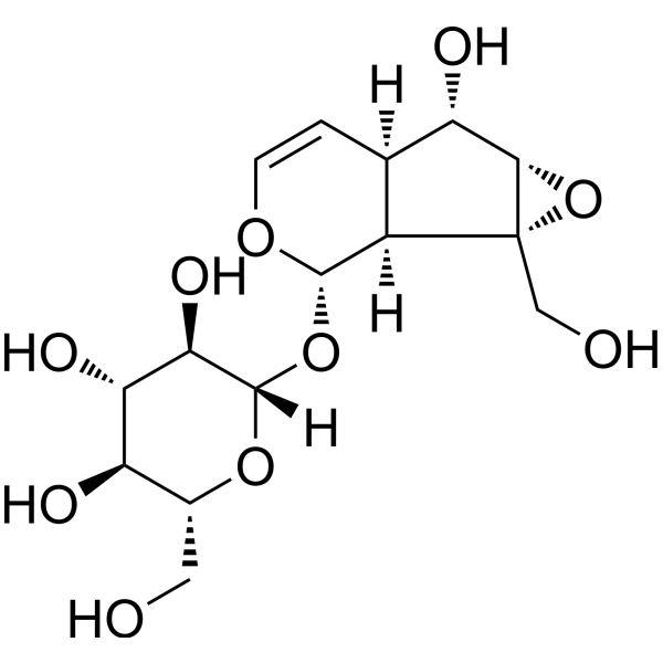
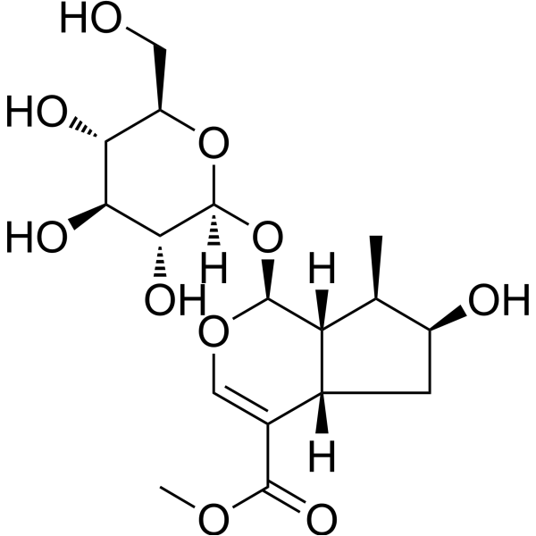
HY-N0820
CatalpolHBV Inhibitor -
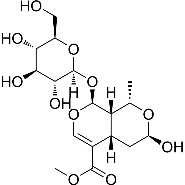
HY-N0532
MorronisidePyroptosis Inhibitor -
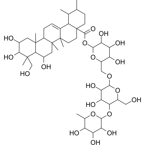
HY-N0568
MadecassosideEndogenous Metabolite -
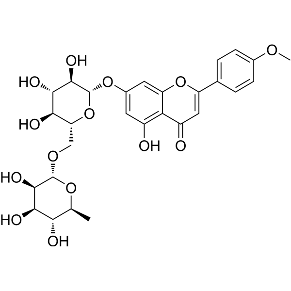
HY-N0528
LinarinAChE Inhibitor -

HY-N0512R
Loganin (Standard)Natural Product -
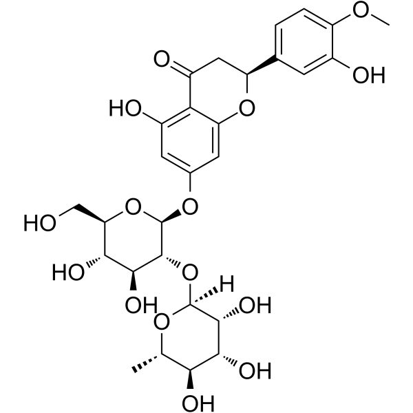
HY-N0101
NeohesperidinWnt/β-Catenin Activator -
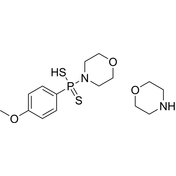
HY-107632
GYY4137H2S Donor -
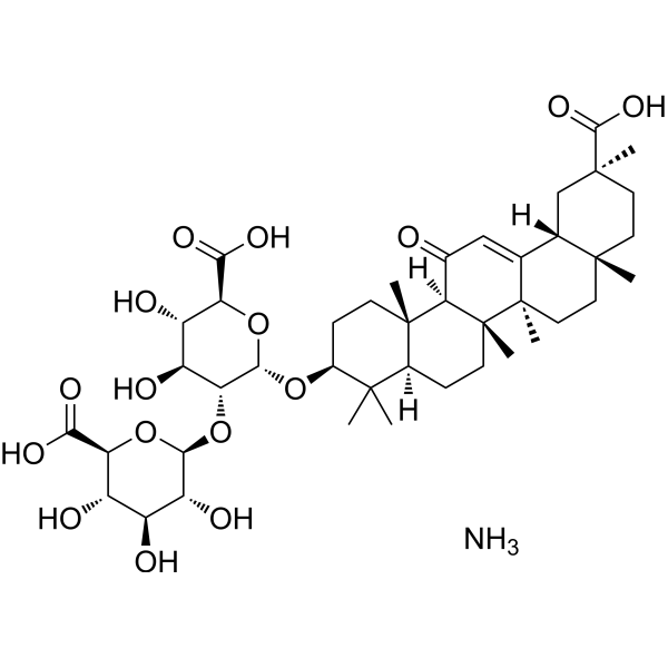
HY-76225
Ammonium glycyrrhizinateAnti-tumor Agent -
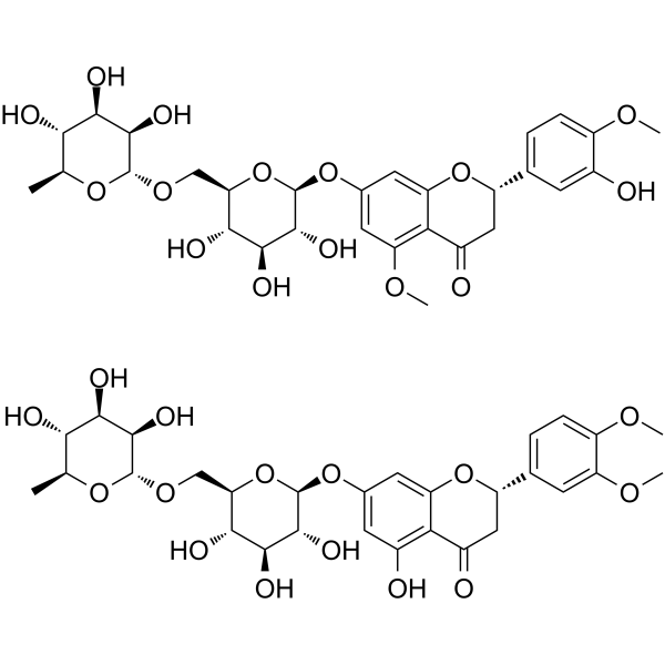
HY-N0165
Methyl-HesperidinVasodilating Agent -
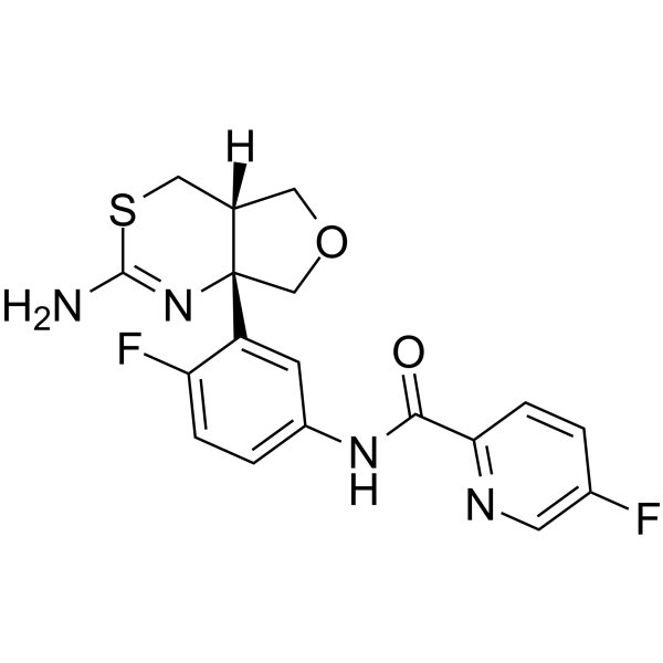
HY-13240
LY2886721BACE1 Inhibitor -
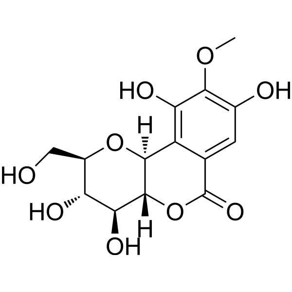
HY-N0017
BergeninBacterial Inhibitor -
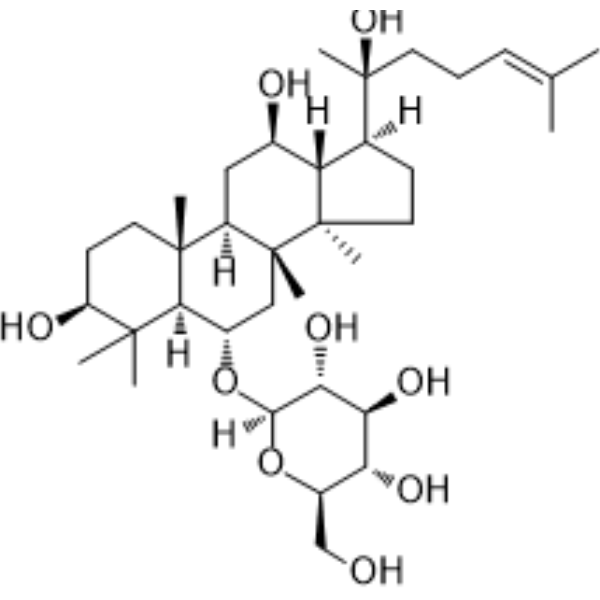
HY-N0604
Ginsenoside Rh1Endogenous Metabolite
Publications Citing Use of MedChemExpress (MCE) Loganin
More- PLoS Biol. 2024 Jun 27;22(6):e3002672. [Abstract]
- Biochem Pharmacol. 2026 Jun 25:252:118202. [Abstract]
- Bioorg Chem. 2025 Jan:154:108108. [Abstract]
- Sci Rep. 2026 May 28. [Abstract]
- Int J Cardiol. 2024 Jan 15:395:131426. [Abstract]
- J Stroke Cerebrovasc Dis. 2025 Jan;34(1):108114. [Abstract]
- bioRxiv. 2024 Apr 3:2023.06.02.542933. [Abstract]
 Customer Validation & Images

-
Histological Imaging/Staining
-
IF
-
WB
Ver todos los productos específicos de isoformas TNF Receptor
More

Actividad biológica
Descripciòn
Cellular Effect
|
Cell Line

|
Type | Value | Description | References |
|---|---|---|---|---|
| Caco-2 | IC50 |
1.28 μg/mL
Compound: 1
|
Cytotoxic activity against Homo sapiens (human) Caco2 cells incubated for 4 hr followed by compound wash-out measured after doubling time by MTT assay
Cytotoxic activity against Homo sapiens (human) Caco2 cells incubated for 4 hr followed by compound wash-out measured after doubling time by MTT assay
|
10.1007/s00044-011-9832-9 |
| Caco-2 | IC50 |
3.45 μg/mL
Compound: 1
|
Cytotoxic activity against Homo sapiens (human) Caco2 cells by clonogenic assay
Cytotoxic activity against Homo sapiens (human) Caco2 cells by clonogenic assay
|
10.1007/s00044-011-9832-9 |
| COLO 320 | IC50 |
1 μg/mL
Compound: 1
|
Cytotoxic activity against Homo sapiens (human) COLO320 cells incubated for 4 hr followed by compound wash-out measured after doubling time by MTT assay
Cytotoxic activity against Homo sapiens (human) COLO320 cells incubated for 4 hr followed by compound wash-out measured after doubling time by MTT assay
|
10.1007/s00044-011-9832-9 |
| COLO 320 | IC50 |
2.2 μg/mL
Compound: 1
|
Cytotoxic activity against Homo sapiens (human) COLO320 cells by clonogenic assay
Cytotoxic activity against Homo sapiens (human) COLO320 cells by clonogenic assay
|
10.1007/s00044-011-9832-9 |
| Huh-7 | IC50 |
16 μg/mL
Compound: 5, ASB-00012351-010
|
Antiviral activity against HCV infected in human Huh7 cells assessed as inhibition of viral replication after 72 hrs by NS5A staining
Antiviral activity against HCV infected in human Huh7 cells assessed as inhibition of viral replication after 72 hrs by NS5A staining
|
[PMID: 19904996] |
| MCF7 | IC50 |
4.85 μg/mL
Compound: 1
|
Cytotoxic activity against Homo sapiens (human) MCF7 cells incubated for 4 hr followed by compound wash-out measured after doubling time by MTT assay
Cytotoxic activity against Homo sapiens (human) MCF7 cells incubated for 4 hr followed by compound wash-out measured after doubling time by MTT assay
|
10.1007/s00044-011-9832-9 |
| MCF7 | IC50 |
6.45 μg/mL
Compound: 1
|
Cytotoxic activity against Homo sapiens (human) MCF7 cells by clonogenic assay
Cytotoxic activity against Homo sapiens (human) MCF7 cells by clonogenic assay
|
10.1007/s00044-011-9832-9 |
| PA-1 | IC50 |
1.45 μg/mL
Compound: 1
|
Cytotoxic activity against Homo sapiens (human) PA1 cells incubated for 4 hr followed by compound wash-out measured after doubling time by MTT assay
Cytotoxic activity against Homo sapiens (human) PA1 cells incubated for 4 hr followed by compound wash-out measured after doubling time by MTT assay
|
10.1007/s00044-011-9832-9 |
| PA-1 | IC50 |
4.85 μg/mL
Compound: 1
|
Cytotoxic activity against Homo sapiens (human) PA1 cells by clonogenic assay
Cytotoxic activity against Homo sapiens (human) PA1 cells by clonogenic assay
|
10.1007/s00044-011-9832-9 |
| WRL68 | IC50 |
1.86 μg/mL
Compound: 1
|
Cytotoxic activity against Homo sapiens (human) WRL68 cells incubated for 4 hr followed by compound wash-out measured after doubling time by MTT assay
Cytotoxic activity against Homo sapiens (human) WRL68 cells incubated for 4 hr followed by compound wash-out measured after doubling time by MTT assay
|
10.1007/s00044-011-9832-9 |
| WRL68 | IC50 |
3.2 μg/mL
Compound: 1
|
Cytotoxic activity against Homo sapiens (human) WRL68 cells by clonogenic assay
Cytotoxic activity against Homo sapiens (human) WRL68 cells by clonogenic assay
|
10.1007/s00044-011-9832-9 |
In Vitro
Loganin (0-50 µM; 24 h) shows significant cytotoxicity to RAW264.7 at high concentrations[3]. Loganin (0-30 µM; 1 h) significantly reduces the production of NO, PGE2, TNF-α, and IL-1β in RAW264.7 macrophages stimulated by LPS (100 ng/ml, 24 h) in a dose-dependent manner, and increases the expression of Nrf2 and HO-1 in a concentration-dependent manner[3]. Loganin (30 µM; 1 h) alleviates LPS-mediated ROS generation in RAW264.7 macrophages[3]. Loganin (0-150 μg/mL; 48 h) reduces the survival rate, migration and invasion ability of Huh7 cells[6].
MedChemExpress (MCE) has not independently confirmed the accuracy of these methods. They are for reference only. Further protocols information, click here.
-
Cell Line:RAW264.7 cells
-
Concentration:0,5,10, 30 and 50 µM
-
Incubation Time:24 h
-
Result:Did not affect cytotoxicity at concentrations below 30 µM, but significant cytotoxicity was observed at 50 µM.
-
Cell Line:Huh7 cells
-
Concentration:0、50、100 and 150 μg/ml
-
Incubation Time:48 h
-
Result:Increased E-cadherin levels and decreased N-cadherin and vimentin levels in cells.
-
Cell Line:RAW264.7 cells
-
Concentration:0,5,10 and 30 µM
-
Incubation Time:1 h
-
Result:Significantly inhibited LPS-induced upregulation of iNOS and COX-2.
Compared with cells treated with loganin or LPS alone, Nrf2 protein expression was significantly upregulated in the nucleus of loganin-treated cells, but HO-1 expression was only observed in the cytoplasm.
In Vivo
MedChemExpress (MCE) has not independently confirmed the accuracy of these methods. They are for reference only.
-
Animal Model:Spinal cord injury rat model[4]
-
Dosage:1, 5 and 10 mg/kg
-
Administration:i.p.; Administer once daily from 1 to 28 days after SCI
-
Result:Improved the results of the inclined board test at a dose of 5 mg/kg on the 28th day after surgery and significantly improved the results of the inclined board test at a dose of 10 mg/kg from the 14th day after surgery.
-
Animal Model:Bleomycin-induced pulmonary fibrosis model in mice[5]
-
Dosage:80、160 和 200 mg/kg
-
Administration:i.g.; Bleomycin induction for 8 weeks, twice a week from the fourth week (day 28)
-
Result:Improved alveolar structural destruction and interstitial inflammatory infiltration and reduced the hydroxyproline content in lung tissue.


Chemical Information
-
No. CAS 18524-94-2
-
Appearance Solid
-
Peso molecular 390.38
-
Fòrmula C17H26O10
-
Color White to off-white
-
SMILES
O[C@H]([C@@H](O)[C@@H]1O)[C@](O[C@@H]1CO)([H])O[C@H]2[C@@]([C@H]3C)([H])[C@@](C[C@@H]3O)([H])C(C(OC)=O)=CO2
-
Synonyms
Loganoside
-
Structure Classification
-
Initial Source
-
Envío
Room temperature in continental US; may vary elsewhere.
-
Almacenamiento
Powder -20°C 3 years 4°C 2 years In solvent -80°C 2 years -20°C 1 year


Publications (7)
-
Journal Impact Factor
-
Most Recent
-
PLoS Biol
2024 Jun 27;22(6):e3002672. PMID: 38935621 -
Biochem Pharmacol
Loganin triggers ferroptosis in renal cell carcinoma by targeting the IGF2BP2/HMOX1 axis in an m6A-dependent manner. [Abstract]2026 Jun 25:252:118202. PMID: 42349616 -
Bioorg Chem
Discovery of cyanidin-3-O-galactoside as a novel CNT2 inhibitor for the treatment of hyperuricemia. [Abstract]2025 Jan:154:108108. PMID: 39753042 -
Sci Rep
Loganin alleviates oxidative stress-induced apoptosis by modulating mitochondrial function and STAT3 signaling in DSS-induced colitis and H2O2-injured Caco-2 cells. [Abstract]2026 May 28. PMID: 42203917 -
Int J Cardiol
Loganin protects against myocardial ischemia-reperfusion injury by modulating oxidative stress and cellular apoptosis via activation of JAK2/STAT3 signaling. [Abstract]2024 Jan 15:395:131426. PMID: 37813285
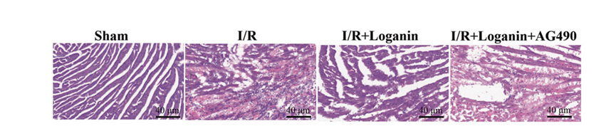
Loganin purchased from MedChemExpress. Usage Cited in: Int J Cardiol. 2024 Jan 15:395:131426. [Abstract]
Representative images of H&E staining showed that Loganin (20 mg/kg, i.p.)treatment prevented I/R-induced disorganization of cardiomyocytes, but administration of AG490 suppressed Loganin-induced enhancement of myocardial morphology.
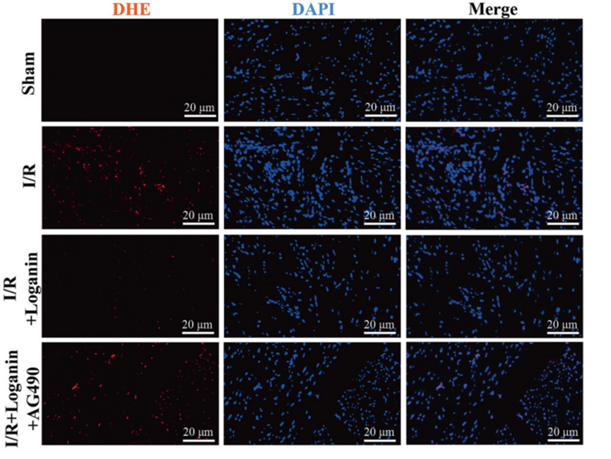
Loganin purchased from MedChemExpress. Usage Cited in: Int J Cardiol. 2024 Jan 15:395:131426. [Abstract]
DHE signaling in ischemic heart tissue treated with Loganin (20 mg/kg, i.p.).
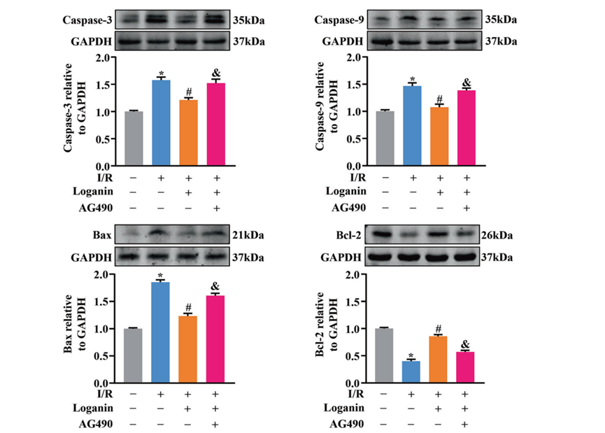
Loganin purchased from MedChemExpress. Usage Cited in: Int J Cardiol. 2024 Jan 15:395:131426. [Abstract]
Quantitative analysis showed that Loganin (20 mg/kg, i.p.)treatment prevented the I/Rinduced increase in the expression levels of caspase-3, caspase-9, and Bax in ischemic heart tissue and the I/R-induced decrease in the expression levels of Bcl-2.
-
J Stroke Cerebrovasc Dis
Loganin attenuates the inflammation, oxidative stress, and apoptosis through the JAK2/STAT3 pathway in cerebral ischemia-reperfusion injury. [Abstract]2025 Jan;34(1):108114. PMID: 39537043 -
bioRxiv
An efficient behavioral screening platform classifies natural products and other chemical cues according to their chemosensory valence in C. elegans. [Abstract]2024 Apr 3:2023.06.02.542933. PMID: 37333363


Solvente y solubilidad
In Vitro:
DMSO : 50 mg/mL (128.08 mM; ultrasonic and warming and heat to 60°C; Hygroscopic DMSO has a significant impact on the solubility of product, please use newly opened DMSO)
Please refer to the solubility information to select the appropriate solvent. Once prepared, please aliquot and store the solution to prevent product inactivation from repeated freeze-thaw cycles.
Storage method and period of stock solution: -80°C, 2 years; -20°C, 1 year. When stored at -80°C, please use it within 2 years. When stored at -20°C, please use it within 1 year.
Please refer to the solubility information to select the appropriate solvent. Once prepared, please aliquot and store the solution to prevent product inactivation from repeated freeze-thaw cycles.
Storage method and period of stock solution: -80°C, 2 years; -20°C, 1 year. When stored at -80°C, please use it within 2 years. When stored at -20°C, please use it within 1 year.
Concentration (start) × Volume (start) = Concentration (final) × Volume (final)
In Vivo:
Select the appropriate dissolution method based on your experimental animal and administration route.
- For the following dissolution methods, please ensure to first prepare a clear stock solution using an In Vitro approach and then sequentially add co-solvents:
- To ensure reliable experimental results, the clarified stock solution can be appropriately stored based on storage conditions. As for the working solution for In Vivo experiments, it is recommended to prepare freshly and use it on the same day.
- The percentages shown for the solvents indicate their volumetric ratio in the final prepared solution. If precipitation or phase separation occurs during preparation, heat and/or sonication can be used to aid dissolution.
Add each solvent one by one: 10% DMSO 40% PEG300 5% Tween-80 45% Saline
Solubility: ≥ 2.5 mg/mL (6.40 mM); Clear solution
This protocol yields a clear solution of ≥ 2.5 mg/mL (saturation unknown).
Taking 1 mL working solution as an example, add 100 μL DMSO stock solution (25.0 mg/mL) to 400 μL PEG300, and mix evenly; then add 50 μL Tween-80 and mix evenly; then add 450 μL Saline to adjust the volume to 1 mL.
Preparation of Saline: Dissolve 0.9 g sodium chloride in ddH₂O and dilute to 100 mL to obtain a clear Saline solution.
Add each solvent one by one: 10% DMSO 90% (20% SBE-β-CD in Saline)
Solubility: ≥ 2.5 mg/mL (6.40 mM); Clear solution
This protocol yields a clear solution of ≥ 2.5 mg/mL (saturation unknown).
Taking 1 mL working solution as an example, add 100 μL DMSO stock solution (25.0 mg/mL) to 900 μL 20% SBE-β-CD in Saline, and mix evenly.
Preparation of 20% SBE-β-CD in Saline (4°C, storage for one week): 2 g SBE-β-CD powder is dissolved in 10 mL Saline, completely dissolve until clear.
In Vivo Dissolution Calculator
Please enter the basic information of animal experiments:
-
-
-
-
Recommended: Prepare an additional quantity of animals to account for potential losses during experiments.
Please enter your animal formula composition:
-
%DMSO +
Recommended: Keep the proportion of DMSO in working solution below 2% if your animal is weak.
-
%+
-
+%Tween-80 + +
-
%Saline +
The co-solvents required include: DMSO, . All of co-solvents are available by MedChemExpress (MCE). , Tween 80. All of co-solvents are available by MedChemExpress (MCE).
Working solution concentration: 0.22 mg/mL
Method for preparing stock solution: mg drug dissolved in μL DMSO. Stock solution concentration: mg/mL.
1. Take μL DMSO stock solution;
2. Add μL .
μL , mix evenly;
3. Then add μL Tween 80, mix evenly;
4. Then add μL
Please ensure that the stock solution in the first step is dissolved to a clear state, and add co-solvents in sequence. You can use ultrasonic heating (ultrasonic cleaner, recommended frequency 20-40 kHz), vortexing, etc. to assist dissolution.

Protocolo
-
Fibrosis/Collagen Morphometry
Fibrosis and collagen morphometry is based on the quantitative visualization of fibrillar collagen deposition in tissue sections using histochemical stains such as Sirius Red (Picrosirius Red) or Masson's trichrome, followed by image-based or polarization-enhanced analysis to estimate collagen proportional area as a surrogate of extracellular matrix accumulation during fibrotic remodeling. Sirius Red combined with polarized light microscopy enhances detection of collagen fibers due to birefringence properties, enabling more specific visualization of collagen type I and III fibrils compared to conventional bright-field histology, while whole-section or region-restricted digital morphometry reduces field-selection bias in fibrosis assessment. Alternative quantitative approaches include second harmonic generation (SHG) and two-photon excited fluorescence microscopy, which enable label-free detection of fibrillar collagen and have been validated against histological staining and biochemica
-
Collagen: Sirius Red Staining
Sirius Red or picrosirius red staining is a histochemical method for visualizing collagen-rich extracellular matrix in tissue sections, and collagen fibers are detected as red-stained structures under bright-field microscopy with enhanced birefringence under polarized light. Picrosirius red is useful for assessing total collagen organization, distribution, and fibrosis burden, but polarized color should not be interpreted as a definitive collagen type I versus type III readout because color is affected by fiber orientation, thickness, and packing.
-
ROS/oxidative-stress fluorescent staining
ROS/oxidative-stress fluorescent staining uses cell-permeant fluorogenic probes that become fluorescent after oxidation inside cells or tissues; commonly used examples include DCFH-DA/DCFDA for broad cellular oxidant detection, DHE for superoxide-related signal detection, MitoSOX for mitochondrial superoxide-related signal detection, and CellROX probes for oxidative-stress-associated fluorescence readouts. The assay detects probe oxidation rather than a single ROS species unless the probe and analysis method have been chemically validated for that species. DCFH-DA enters cells, is deacetylated by intracellular esterases to DCFH, and produces fluorescent DCF after oxidation, so the readout is used as an operational measure of total cellular oxidative stress rather than a species-specific ROS measurement. DHE and MitoSOX can report superoxide-related oxidation, but red fluorescence alone can include non-specific ethidium-like oxidation products; HPLC or optimized spectral approaches are
-
Connective Tissue: Masson's Trichrome/Collagen Trichrome Staining
Masson’s Trichrome (collagen/trichrome staining) is a histological technique that differentially stains tissue compartments using sequential acidic dyes to distinguish collagen from muscle and cytoplasmic components based on dye affinity and tissue permeability differences, enabling visualization of fibrosis and connective tissue architecture in histological sections. The classical formulation typically uses Weigert's iron hematoxylin for nuclear staining, Biebrich scarlet-acid fuchsin for cytoplasm and muscle, and aniline blue (or light green variants) for collagen, producing a characteristic blue/green collagen signal contrasted against red cytoplasm and dark nuclei. The staining principle relies on selective displacement of smaller dye molecules by larger anionic dyes in collagen-rich regions under controlled acidified conditions, which enhances collagen-specific dye retention. This property makes the method widely used for fibrosis assessment in organs such as heart, liver, lung, a
-
Research Protocol for Inflammation-related Diseases
The NLRP3 inflammasome is a cytosolic innate immune signaling platform that integrates priming signals and danger-signal activation to promote caspase-1 activation, maturation of IL-1β and IL-18, and gasdermin D-mediated pyroptotic cell death. The core experimental logic is to determine whether inflammatory disease phenotypes are driven by increased NLRP3 expression, ASC-containing inflammasome assembly, caspase-1 cleavage, GSDMD cleavage, and extracellular release of IL-1β/IL-18 rather than by nonspecific cell injury alone. The pathway is strongly linked to inflammation-related disease phenotypes because monosodium urate crystals activate NALP3/NLRP3 inflammasome signaling in gout-like crystal inflammation, cholesterol crystals activate NLRP3 inflammasomes in atherogenesis models, and DSS-induced intestinal inflammation has been reported to involve NLRP3 inflammasome activity. However, experimental colitis studies also show context-dependent protective effects of NLRP3 inflammasome co
-
Inhalation Toxicity Study
Inhalation toxicity studies expose rodents to a controlled aerosol, vapor, gas, or smoke atmosphere and assess respiratory and systemic toxicity using exposure-atmosphere characterization, clinical observations, body and organ weights, bronchoalveolar lavage fluid, histopathology, blood chemistry, hematology, and, when included, molecular endpoints such as transcriptomics, proteomics, lipidomics, or tissue burden analysis. The primary biological readouts are airway irritation, pulmonary inflammation, cytotoxicity, altered surfactant or lipid homeostasis, impaired particle clearance, and tissue remodeling, reflected by BALF cell differentials, BALF protein, LDH, phosphatase activities, cytokines, lung weight, microscopic respiratory-tract lesions, and retained lung burden.
-
Research Protocol for Neurological Diseases
PINK1/Parkin-mediated mitophagy pathway is a mitochondrial quality-control signaling axis in which mitochondrial depolarization stabilizes PINK1 on damaged mitochondria, activates Parkin recruitment and E3 ubiquitin ligase activity, promotes ubiquitination of outer mitochondrial membrane proteins, recruits selective autophagy adaptors, and drives lysosomal degradation of damaged mitochondria. In neurological disease research, this pathway is experimentally important because neurons, especially dopaminergic neurons, are highly dependent on mitochondrial integrity, and defective mitochondrial turnover can lead to mitochondrial dysfunction, oxidative stress, impaired neuronal survival, α-synuclein accumulation, and neuroinflammatory damage-associated signals. The genetic disease link is strongest in Parkinson’s disease because mutations in PRKN/parkin cause autosomal recessive juvenile parkinsonism, mutations in PINK1 cause hereditary early-onset Parkinson’s disease, and Drosophila studie


Pureza y Documentación
-
Ficha de datos (287 KB)
-
SDS (393 KB)
- English - EN (393 KB)
- Français - FR (393 KB)
- Deutsch - DE (393 KB)
- Norwegian - NO (393 KB)
- Español - ES (393 KB)
- Swedish - SV (393 KB)
- Italian - IT (393 KB)
- Korean - KR (393 KB)
- Portuguese - PT (393 KB)
-
Instrucciones de manejo (2659 KB)
Referencias
[1]. Kim MJ, et al. Loganin protects against pancreatitis by inhibiting NF-κB activation. Eur J Pharmacol. 2015 Oct 15;765:541-50. [Content Brief]
[2]. Tsai WH, et al. Ba-Wei-Di-Huang-Wan through its active ingredient loganin counteracts substance P-enhanced NF-κB/ICAM-1 signaling in rats with bladder hyperactivity. Neurourol Urodyn. 2015 Aug 19. [Content Brief]
[3]. Park C, et al. Loganin Inhibits Lipopolysaccharide-Induced Inflammation and Oxidative Response through the Activation of the Nrf2/HO-1 Signaling Pathway in RAW264.7 Macrophages. Biol Pharm Bull. [Content Brief]
[4]. Huang X, et al. Loganin reduces neuroinflammation and promotes functional recovery after spinal cord injury through inhibiting NF-κB/NLRP3 signalling. Toxicon. 2023 Aug 15;232:107202. [Content Brief]
[5]. Chen L, et al. Protective effect and mechanism of loganin and morroniside on acute lung injury and pulmonary fibrosis. Phytomedicine. 2022 May;99:154030. [Content Brief]


Complete Stock Solution Preparation Table
Please refer to the solubility information to select the appropriate solvent. Once prepared, please aliquot and store the solution to prevent product inactivation from repeated freeze-thaw cycles.
Storage method and period of stock solution: -80°C, 2 years; -20°C, 1 year. When stored at -80°C, please use it within 2 years. When stored at -20°C, please use it within 1 year.
| Optional Solvent | Concentration Solvent Mass | 1 mg | 5 mg | 10 mg | 25 mg |
|---|---|---|---|---|---|
| DMSO | 1 mM | 2.5616 mL | 12.8080 mL | 25.6161 mL | 64.0402 mL |
| 5 mM | 0.5123 mL | 2.5616 mL | 5.1232 mL | 12.8080 mL | |
| 10 mM | 0.2562 mL | 1.2808 mL | 2.5616 mL | 6.4040 mL | |
| 15 mM | 0.1708 mL | 0.8539 mL | 1.7077 mL | 4.2693 mL | |
| 20 mM | 0.1281 mL | 0.6404 mL | 1.2808 mL | 3.2020 mL | |
| 25 mM | 0.1025 mL | 0.5123 mL | 1.0246 mL | 2.5616 mL | |
| 30 mM | 0.0854 mL | 0.4269 mL | 0.8539 mL | 2.1347 mL | |
| 40 mM | 0.0640 mL | 0.3202 mL | 0.6404 mL | 1.6010 mL | |
| 50 mM | 0.0512 mL | 0.2562 mL | 0.5123 mL | 1.2808 mL | |
| 60 mM | 0.0427 mL | 0.2135 mL | 0.4269 mL | 1.0673 mL | |
| 80 mM | 0.0320 mL | 0.1601 mL | 0.3202 mL | 0.8005 mL | |
| 100 mM | 0.0256 mL | 0.1281 mL | 0.2562 mL | 0.6404 mL |


Loganin Related Classifications
HY-N0512 Related Classifications
 Powered by Bioz