



Lomitapide mesylate
Based on 7 publication(s) in Google Scholar
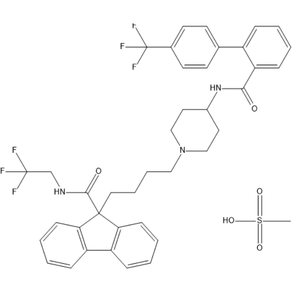
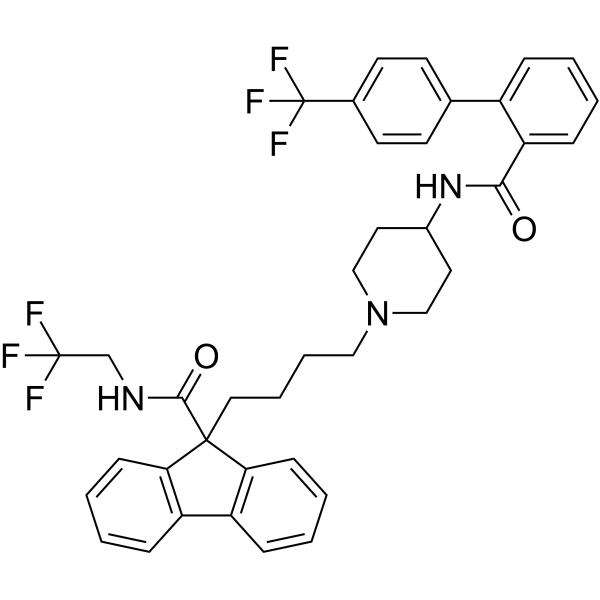
Lomitapide (AEGR-733; BMS-201038) mesylate is an orally active microsomal triglyceride transfer protein (MTP) inhibitor and a selective mTORC1 inhibitor with lipid-lowering activity and BBB permeability. Lomitapide mesylate significantly reduces plasma LDL levels by blocking the assembly and secretion of very-low-density lipoprotein (VLDL). Lomitapide mesylate inhibits mTORC1 in an ATP-dependent manner, thereby inducing AMPK-independent autophagic cell death and suppressing cancer cell growth and apoptosis. Lomitapide mesylate also enhances tumor infiltration of CD8+ T cells. In addition, Lomitapide mesylate inhibits HDAC, improves endothelial function, effectively alleviates vascular inflammation and oxidative stress, and exerts neuroprotective effects in a cerebral ischemia/reperfusion injury model. Lomitapide mesylate can be used in research on related diseases such as colorectal cancer, breast cancer, melanoma, ischemic stroke, and familial hypercholesterolemia.
For research use only. We do not sell to patients.
- Purity : 99.68%
- CAS No.: 202914-84-9
- Formula: C40H41F6N3O5S
- Molecular Weight:789.83
-
Storage:
4°C, sealed storage, away from moisture
* In solvent : -80°C, 6 months; -20°C, 1 month (sealed storage, away from moisture)

To place orders, for customer services and technical support, please contact: MedChemExpress USA
Tel: 609-228-6898 E-mail: [email protected] [email protected]


-
 Biological Activity
-
Chemical Information
-
Solvent & Solubility
- Protocols
- Purity & Documentation
- References
-
 Help & FAQs


-
HY-14667
LomitapideMTP Inhibitor -
HY-12005
Fingolimod hydrochlorideS1P Receptors Modulator -
HY-15656
CeritinibALK Inhibitor -
HY-13955
TelmisartanAngiotensin II Type 1 Receptor -
HY-14557
Pimavanserin5-HT2A Inverse Agonist -
HY-N0052A
Sanguinarine chlorideApoptosis Inducer -
HY-14153A
Tegaserod maleateSerotonin Receptor 4 Agonist -
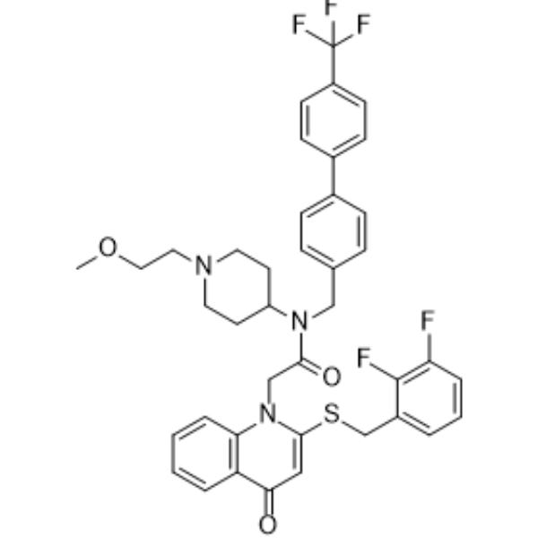
HY-102004
RilapladibLp-PLA2 Inhibitor -
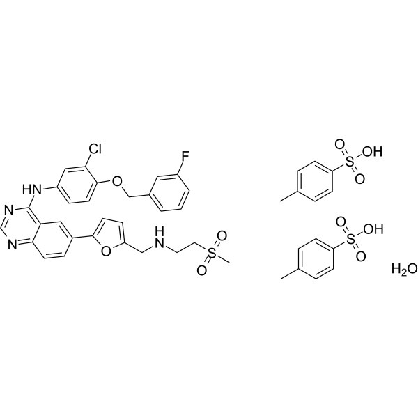
HY-50898B
Lapatinib ditosylate monohydrateErbB-2/EGFR Inhibitor -
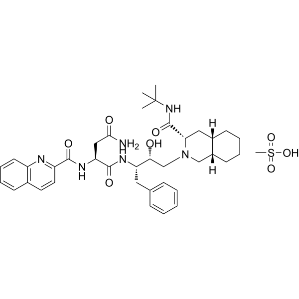
HY-17003
Saquinavir mesylateHIV Protease Inhibitor -

HY-14668R
Lomitapide mesylate (Standard)MTP Inhibitor -
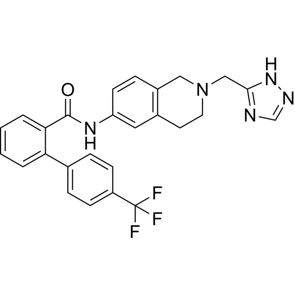
HY-113955
CP-346086MTP Inhibitor
Publications Citing Use of MedChemExpress (MCE) Lomitapide mesylate
More- Adv Mater. 2024 Jun;36(24):e2311760. [Abstract]
- Free Radic Biol Med. 2021 Aug 20:172:213-225. [Abstract]
- Cell Rep. 2026 Apr 21;45(5):117295. [Abstract]
- Liver Int. 2025 Apr;45(4):e70027. [Abstract]
- BMC Microbiol. 2022 Apr 26;22(1):114. [Abstract]
- J Lipid Res. 2024 Oct;65(10):100635. [Abstract]
- bioRxiv. 2025 Jul 21.
 Customer Validation & Images

-
Cell Proliferation/Viability Assay
-
Cell Imaging/Staining
-
Cell Proliferation/Viability Assay
-
Flow Cytometry
-
Cell Imaging/Staining


Biological Activity
Description
Cellular Effect
|
Cell Line

|
Type | Value | Description | References |
|---|---|---|---|---|
| HepG2 | ED50 |
0.8 nM
Compound: Juxtapid
|
Inhibition of ApoB lipoprotein secretion in human HepG2 cells
Inhibition of ApoB lipoprotein secretion in human HepG2 cells
|
[PMID: 38889609] |
In Vitro
Lomitapide mesylate (5 μM; 48 h) inhibits the proliferation of human HCT116, HT29 and SW480 colorectal cancer cells by reducing colony formation, and (2 μM; 96 h) inhibits the proliferation of HT29 cells overexpressing eIF4E[1].
Lomitapide mesylate (5 μM; 24 h) induces autophagic cell death in human HCT116 and HT29 colorectal cancer cells; autophagy inhibition restores cell viability, while no apoptosis-related caspase 3/7 activity is detected[1].
Lomitapide mesylate (5-20 μM; 48-72 h) potently inhibits the growth and induces autophagy in patient-derived human colorectal cancer organoids CRC-01 and CRC-02, and exhibits better efficacy than 5-FU (HY-90006) at equivalent concentrations[1].
Lomitapide mesylate (0.01-1 μM; 24 h) shows no toxicity to mouse Neuro-2a cells, and improves the survival rate of OGD-injured Neuro-2a cells in a concentration-dependent manner[2].
Lomitapide mesylate (0.01-1 μM; 24 h) shows no toxicity to primary mouse cortical neurons, and improves the cell viability of primary mouse cortical neurons injured by OGD in a concentration-dependent manner[2].
Lomitapide mesylate restores autophagic flux and inhibits apoptosis in OGD-injured mouse Neuro-2a cells co-cultured with LPS-treated BV2 microglia, which is evidenced by the normalization of autophagy protein expression and increased Bcl-2 levels[2].
Lomitapide mesylate significantly inhibits the migration of mouse BV2 microglial cells toward OGD-injured Neuro-2a cells[2].
MedChemExpress (MCE) has not independently confirmed the accuracy of these methods. They are for reference only. Further protocols information, click here.
-
Cell Line:HEK293T cells
-
Concentration:10-100 μM; 50 μM with 500-5000 μM ATP
-
Incubation Time:15 min at 37 °C
-
Result:Dose-dependently inhibited phosphorylation of the mTORC1 substrate T389 S6K1, with reduced phosphorylation observed at all tested concentrations.
Showed reversed inhibitory effect when ATP concentrations were increased, indicating competition with ATP for binding to mTORC1.
Exhibited no inhibition of mTORC2 activity (measured by S473 Akt phosphorylation).
-
Cell Line:Human colorectal cancer cell lines (HCT116, HT29)
-
Concentration:5 μM; 5 μM with 1 mM 3-MA; 5 μM with bafilomycin
-
Incubation Time:24 h
-
Result:Increased GFP-LC3 puncta formation in HT29 cells, indicating autophagosome accumulation.
Showed rescued cell viability reduction when treated with bafilomycin (in HT29 cells) or 3-MA (in HCT116 cells).
Showed inhibited LC3-II elevation when treated with 3-MA, and diminished LC3-II levels when ATG7 or Beclin-1 was knocked down.
Did not induce caspase 3/7 activity, indicating no apoptotic response.
-
Cell Line:murine Neuro-2a cells
-
Concentration:0.01, 0.1, 1 μM
-
Incubation Time:24 h; 4 h (OGD exposure prior to treatment)
-
Result:Showed no cytotoxicity at all tested concentrations compared to control.
Alleviated cell damage and improved cell viability in a concentration-dependent manner in OGD-injured cells, with statistically significant differences relative to the OGD model group.
In Vivo
Lomitapide (20 mg/kg; intraperitoneal injection; administered 5 times; initiated on day 10 post tumor implantation) mesylate inhibits the growth of MC38 colorectal cancer and B16-F10 melanoma in C57B6/N mice, respectively[1].
Lomitapide (0.5 mg/kg; p.o.; once daily; for 14 consecutive days) mesylate significantly improves neurological function, reduces neuronal tissue loss by 41.05%, enhances neuronal autophagy, inhibits the migration of pro-inflammatory microglia, and increases the survival rate of mice with ischemic stroke induced by middle cerebral artery occlusion (MCAO)[2].
Lomitapide (1 mg/kg/day; oral administration; daily dosing; for 2 consecutive weeks) mesylate improves cardiovascular function, reduces body weight and fat mass, downregulates blood glucose and lipid levels, decreases atherosclerotic plaque area, and alleviates vascular stress and inflammatory responses in obese LDLr−/− mice[3].
MedChemExpress (MCE) has not independently confirmed the accuracy of these methods. They are for reference only.
-
Animal Model:C57B6/N (6-week-old wild-type female; 6-week-old wild-type male; subcutaneous implantation of MC38 colorectal cancer cells)[1]
-
Dosage:20 mg/kg
-
Administration:i.p.; 5 doses; starting 10 days post-tumor injection
-
Result:Achieved a tumor growth inhibition (TGI) of 66%.
Did not cause changes in mouse body weight.
Showed no toxicity in liver, kidney, or lung tissues.
Increased infiltration of CD8+ T cells into tumor tissues.
-
Animal Model:C57B6/N (6-week-old wild-type female; 6-week-old wild-type male; subcutaneous implantation of B16-F10 melanoma cells)[1]
-
Dosage:20 mg/kg
-
Administration:i.p.; 5 doses every other day; starting 10 days post-tumor injection
-
Result:Reduced B16-F10 melanoma tumor growth.
Did not cause changes in mouse body weight.
Showed no toxicity in liver, kidney, or lung tissues.
Clinical Trial
| NCT Number | Sponsor | Condition | Start Date |
Phase
|
|---|---|---|---|---|
| NCT01329991 | Plexxikon| | 2011-05 | PHASE1 |


Chemical Information
-
CAS No. 202914-84-9
-
Appearance Solid
-
Molecular Weight 789.83
-
Formula C40H41F6N3O5S
-
Color White to off-white
-
SMILES
O=C(C1(CCCCN2CCC(NC(C3=CC=CC=C3C4=CC=C(C(F)(F)F)C=C4)=O)CC2)C5=C(C6=C1C=CC=C6)C=CC=C5)NCC(F)(F)F.O=S(C)(O)=O
-
Synonyms
AEGR-733 mesylate; BMS-201038 mesylate
-
Shipping
Room temperature in continental US; may vary elsewhere.
-
Storage
4°C, sealed storage, away from moisture
* In solvent : -80°C, 6 months; -20°C, 1 month (sealed storage, away from moisture)


Publications (7)
-
Journal Impact Factor
-
Most Recent
-
Adv Mater
Efficient Delivery of Lomitapide using Hybrid Membrane-Coated Tetrahedral DNA Nanostructures for Glioblastoma Therapy. [Abstract]2024 Jun;36(24):e2311760. PMID: 38569065
Lomitapide mesylate purchased from MedChemExpress. Usage Cited in: Adv Mater. 2024 Jun;36(24):e2311760. [Abstract]
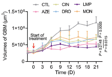
Lomitapide (LMP) (10 μM; 21 d) significantly inhibited the growth of 3D U251 cells.
Lomitapide mesylate purchased from MedChemExpress. Usage Cited in: Adv Mater. 2024 Jun;36(24):e2311760. [Abstract]
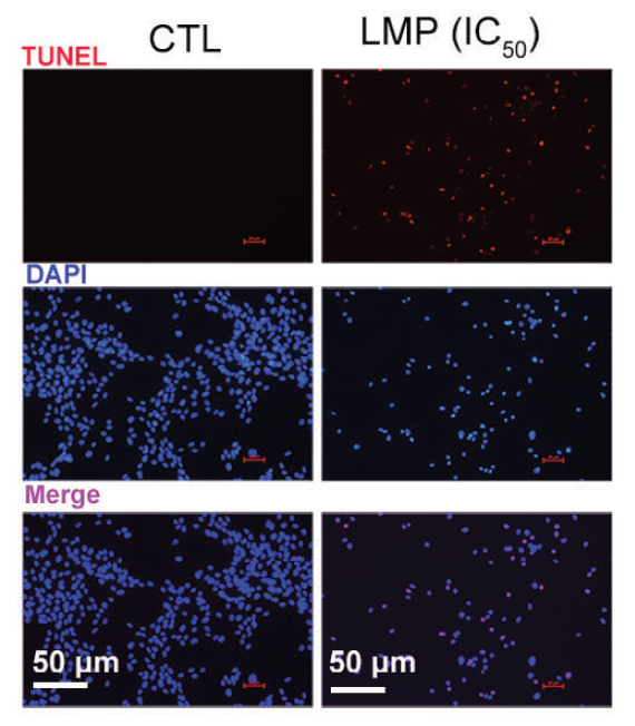
Lomitapide (LMP) (5.8 μM) induced apoptosis in GBM cells.
Lomitapide mesylate purchased from MedChemExpress. Usage Cited in: Adv Mater. 2024 Jun;36(24):e2311760. [Abstract]
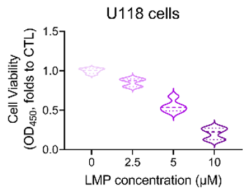
Lomitapide (LMP) (2.5-10 μM; 24 h) exhibited a concentration-dependent increase in cytotoxicity against U118 cells.
Lomitapide mesylate purchased from MedChemExpress. Usage Cited in: Adv Mater. 2024 Jun;36(24):e2311760. [Abstract]
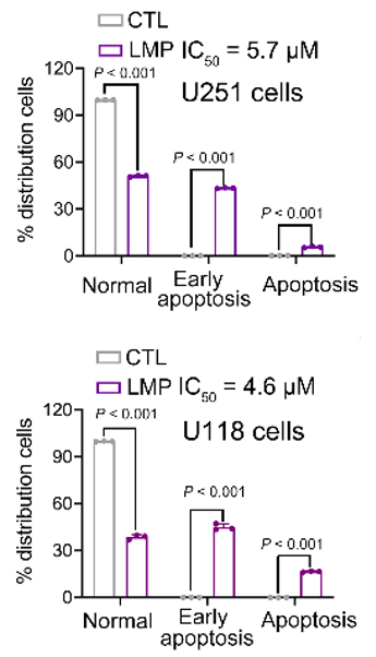
Lomitapide (LMP) (5.8 μM) mainly induced GBM cells to undergo early apoptosis.
Lomitapide mesylate purchased from MedChemExpress. Usage Cited in: Adv Mater. 2024 Jun;36(24):e2311760. [Abstract]
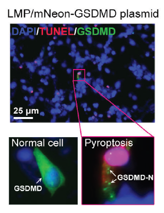
Lomitapide (LMP) (5.8 μM) induced the oligomeric GSDMD-N-terminal structural domains to aggregate on the membrane, thereby expediting pyroptosis of U251 cells.
-
Free Radic Biol Med
Lipid overload impairs hepatic VLDL secretion via oxidative stress-mediated PKCδ-HNF4α-MTP pathway in large yellow croaker (Larimichthys crocea). [Abstract]2021 Aug 20:172:213-225. PMID: 34116177 -
Cell Rep
Increased yolk lipid mobilization promotes zebrafish post-segmentation growth via an Hnf4-lipoprotein axis. [Abstract]2026 Apr 21;45(5):117295. PMID: 42018437 -
Liver Int
Disentangling Organ-Specific Roles of Farnesoid X Receptor in Bile Acid and Glucolipid Metabolism. [Abstract]2025 Apr;45(4):e70027. PMID: 40052709 -
BMC Microbiol
Antibacterial activities and action mode of anti-hyperlipidemic lomitapide against Staphylococcus aureus. [Abstract]2022 Apr 26;22(1):114. PMID: 35473561 -
J Lipid Res
Therapeutic siRNA targeting PLIN2 ameliorates steatosis, inflammation, and fibrosis in steatotic liver disease models. [Abstract]2024 Oct;65(10):100635. PMID: 39187042 -


Solvent & Solubility
In Vitro:
DMSO : 100 mg/mL (126.61 mM; Need ultrasonic; Hygroscopic DMSO has a significant impact on the solubility of product, please use newly opened DMSO)
Please refer to the solubility information to select the appropriate solvent. Once prepared, please aliquot and store the solution to prevent product inactivation from repeated freeze-thaw cycles.
Storage method and period of stock solution: -80°C, 6 months; -20°C, 1 month (sealed storage, away from moisture). When stored at -80°C, please use it within 6 months. When stored at -20°C, please use it within 1 month.
Please refer to the solubility information to select the appropriate solvent. Once prepared, please aliquot and store the solution to prevent product inactivation from repeated freeze-thaw cycles.
Storage method and period of stock solution: -80°C, 6 months; -20°C, 1 month (sealed storage, away from moisture). When stored at -80°C, please use it within 6 months. When stored at -20°C, please use it within 1 month.
Concentration (start) × Volume (start) = Concentration (final) × Volume (final)
In Vivo:
Select the appropriate dissolution method based on your experimental animal and administration route.
- For the following dissolution methods, please ensure to first prepare a clear stock solution using an In Vitro approach and then sequentially add co-solvents:
- To ensure reliable experimental results, the clarified stock solution can be appropriately stored based on storage conditions. As for the working solution for In Vivo experiments, it is recommended to prepare freshly and use it on the same day.
- The percentages shown for the solvents indicate their volumetric ratio in the final prepared solution. If precipitation or phase separation occurs during preparation, heat and/or sonication can be used to aid dissolution.
Add each solvent one by one: 10% DMSO 40% PEG300 5% Tween-80 45% Saline
Solubility: ≥ 2.5 mg/mL (3.17 mM); Clear solution
This protocol yields a clear solution of ≥ 2.5 mg/mL (saturation unknown).
Taking 1 mL working solution as an example, add 100 μL DMSO stock solution (25.0 mg/mL) to 400 μL PEG300, and mix evenly; then add 50 μL Tween-80 and mix evenly; then add 450 μL Saline to adjust the volume to 1 mL.
Preparation of Saline: Dissolve 0.9 g sodium chloride in ddH₂O and dilute to 100 mL to obtain a clear Saline solution.
Add each solvent one by one: 10% DMSO 90% (20% SBE-β-CD in Saline)
Solubility: ≥ 2.5 mg/mL (3.17 mM); Clear solution
This protocol yields a clear solution of ≥ 2.5 mg/mL (saturation unknown).
Taking 1 mL working solution as an example, add 100 μL DMSO stock solution (25.0 mg/mL) to 900 μL 20% SBE-β-CD in Saline, and mix evenly.
Preparation of 20% SBE-β-CD in Saline (4°C, storage for one week): 2 g SBE-β-CD powder is dissolved in 10 mL Saline, completely dissolve until clear.
In Vivo Dissolution Calculator
Please enter the basic information of animal experiments:
-
-
-
-
Recommended: Prepare an additional quantity of animals to account for potential losses during experiments.
Please enter your animal formula composition:
-
%DMSO +
Recommended: Keep the proportion of DMSO in working solution below 2% if your animal is weak.
-
%+
-
+%Tween-80 + +
-
%Saline +
The co-solvents required include: DMSO, . All of co-solvents are available by MedChemExpress (MCE). , Tween 80. All of co-solvents are available by MedChemExpress (MCE).
Working solution concentration: 0.22 mg/mL
Method for preparing stock solution: mg drug dissolved in μL DMSO. Stock solution concentration: mg/mL. * In solvent : -80°C, 6 months; -20°C, 1 month (sealed storage, away from moisture)
1. Take μL DMSO stock solution;
2. Add μL .
μL , mix evenly;
3. Then add μL Tween 80, mix evenly;
4. Then add μL
Please ensure that the stock solution in the first step is dissolved to a clear state, and add co-solvents in sequence. You can use ultrasonic heating (ultrasonic cleaner, recommended frequency 20-40 kHz), vortexing, etc. to assist dissolution.

Protocols
-
Mammalian live/dead viability and cytotoxicity staining
Live/dead viability and cytotoxicity staining assays are based on the simultaneous detection of intracellular esterase activity in metabolically active (viable) cells and membrane integrity loss in non-viable cells. In commonly used dual-staining approaches, membrane-permeant fluorogenic substrates are converted by intracellular esterases into fluorescent products in live cells, while impermeant DNA-binding dyes selectively enter cells with compromised plasma membranes and label nucleic acids in dead or dying cells, enabling discrimination between viable and non-viable populations by fluorescence microscopy or flow cytometry.
-
Annexin V plus membrane-impermeant dye apoptosis staining
Annexin V-based apoptosis assays rely on the detection of phosphatidylserine (PS) externalization from the inner leaflet of the plasma membrane to the outer leaflet, an early biochemical hallmark of apoptosis. Fluorescently labeled Annexin V binds PS in a calcium-dependent manner, enabling identification of early apoptotic cells by flow cytometry or fluorescence microscopy. When combined with a membrane-impermeant DNA-binding dye (e. g. , propidium iodide), this approach allows discrimination between viable (Annexin V−/dye−), early apoptotic (Annexin V+/dye−), and late apoptotic or necrotic (Annexin V+/dye+) cell populations by assessing membrane integrity and PS exposure.
-
ROS/oxidative-stress fluorescent staining
ROS/oxidative-stress fluorescent staining uses cell-permeant fluorogenic probes that become fluorescent after oxidation inside cells or tissues; commonly used examples include DCFH-DA/DCFDA for broad cellular oxidant detection, DHE for superoxide-related signal detection, MitoSOX for mitochondrial superoxide-related signal detection, and CellROX probes for oxidative-stress-associated fluorescence readouts. The assay detects probe oxidation rather than a single ROS species unless the probe and analysis method have been chemically validated for that species. DCFH-DA enters cells, is deacetylated by intracellular esterases to DCFH, and produces fluorescent DCF after oxidation, so the readout is used as an operational measure of total cellular oxidative stress rather than a species-specific ROS measurement. DHE and MitoSOX can report superoxide-related oxidation, but red fluorescence alone can include non-specific ethidium-like oxidation products; HPLC or optimized spectral approaches are
-
Somatic Cell Culture
A method of simulating the in vivo environment in vitro to maintain the cell growth, differentation and main functions.
-
CCK-8/WST-8 Cell Proliferation Assay
The CCK-8/WST-8 assay is based on the reduction of the water-soluble tetrazolium salt WST-8 to a water-soluble formazan product by cellular dehydrogenases in metabolically active cells, where the generated formazan amount is proportional to the number of living cells and is quantified by measuring absorbance in the visible range, providing a colorimetric readout for cell viability and proliferation assessment. This class of tetrazolium-based assays improves upon earlier MTT-based systems by producing a water-soluble formazan, eliminating the need for organic solubilization steps and enabling direct spectrophotometric measurement in culture medium.
-
Cell Counting-Based Growth Curve Assay
Cell counting-based growth curve assays quantify cell proliferation by directly measuring changes in viable cell number over time using manual or automated counting methods such as hemocytometer-based counting or instrument-assisted cell enumeration, enabling construction of growth curves that reflect population expansion dynamics in response to culture conditions. A widely used approach is trypan blue exclusion with hemocytometer counting, where membrane-compromised (non-viable) cells take up the dye, allowing discrimination between viable and non-viable cells while simultaneously enabling total cell number quantification. Repeated sampling across time points allows estimation of proliferation rate, growth phases, and comparative growth kinetics between experimental conditions.
-
Research Protocol for Inflammation-related Diseases
The NLRP3 inflammasome is a cytosolic innate immune signaling platform that integrates priming signals and danger-signal activation to promote caspase-1 activation, maturation of IL-1β and IL-18, and gasdermin D-mediated pyroptotic cell death. The core experimental logic is to determine whether inflammatory disease phenotypes are driven by increased NLRP3 expression, ASC-containing inflammasome assembly, caspase-1 cleavage, GSDMD cleavage, and extracellular release of IL-1β/IL-18 rather than by nonspecific cell injury alone. The pathway is strongly linked to inflammation-related disease phenotypes because monosodium urate crystals activate NALP3/NLRP3 inflammasome signaling in gout-like crystal inflammation, cholesterol crystals activate NLRP3 inflammasomes in atherogenesis models, and DSS-induced intestinal inflammation has been reported to involve NLRP3 inflammasome activity. However, experimental colitis studies also show context-dependent protective effects of NLRP3 inflammasome co
-
Breast Cancer Modeling
Breast cancer is a heterogeneous cancer, and it has been distinguished into four subtypes: luminal A, luminal B, HER2-positive and basal-like. Molecular mutations, epigenetic alterations, hormone exposure and immune microenvironment are related to the progression of breast cancer.
-
Cell Viability Determination by MTT Colorimetric Assay
The following protocol uses the MTT colorimetric assay as a classic literature-established method for assessing cell viability/metabolic activity in cultured mammalian cells. MTT[3-(4,5-dimethylthiazol-2-yl)-2,5-diphenyltetrazolium bromide] is reduced by metabolically active cells to a colored formazan product; the amount of formazan is quantified spectrophotometrically and provides an indirect measure of metabolically active viable cells. Importantly, MTT reduction reflects cellular oxidoreductase/metabolic activity rather than an absolute direct count of living cells, so changes in cellular metabolism can alter the signal independently of cell number.
-
Apoptosis
Apoptosis, also called programmed cell death, is generally characterized by distinct morphological characteristics.
-
TUNEL staining for apoptotic DNA fragmentation
TUNEL staining detects DNA strand breaks by using terminal deoxynucleotidyl transferase to add labeled nucleotides to exposed 3′-OH DNA termini, generating either microscopic staining in fixed cells or tissue sections, or fluorescence/cytometric signal in cell suspensions. TUNEL positivity reflects DNA fragmentation but should not be interpreted alone as definitive apoptosis, because TUNEL can also label necrotic, autolytic, mechanically damaged, or DNA-repair-associated DNA breaks.
-
Cell Cytotoxicity Assay
Cytotoxicity assays are usually based on the assessment of cell membrane damage, which can also be indirectly detected by measuring cell viability. Detection methods include MTT assay, CKK-8 assay, LDH assay and ATP assay, etc.
-
Protocol for Cell Counting and Cell Density Analysis
Cell counting and cell-density analysis estimate the number of cells in a known volume or field area. Manual hemocytometer counting uses a chamber of defined geometry to convert counted cells into cells/mL, while automated counters and image-analysis workflows detect cell objects from optical, brightfield, fluorescence, impedance, or digital-image features. Trypan blue viability counting is based on dye exclusion: viable cells with intact membranes exclude dye, while non-viable cells with compromised membranes stain blue. The readout is total cell density, viable-cell density, dead-cell density, and percent viability. Cell density can also be estimated from microscopy images by counting objects per image area, from flow cytometry using calibrated volume or reference particles, or from in situ microscopy in bioreactors after calibration against reference methods such as hemocytometer or flow cytometry.
-
Autophagy
Autophagy is a process in which eukaryotic cells use lysosomes to degrade their own cytoplasmic proteins and damaged organelles under the regulation of autophagy related gene (Atg). Microtubule-associated proteins light chain 3 (LC3) is recognized as autophagy marker, which transfers from cytoplasmic LC3 (LC3-I) to membrane type (LC3-II). LC3-II/I ratio could be detected by Western Blot and fluorescence microscopy.
-
Transepithelial/transendothelial electrical resistance assay
TEER measures electrical resistance across epithelial or endothelial monolayers cultured on permeable supports, and the readout reflects ionic conductance through the cell barrier, especially the paracellular pathway regulated by junctional integrity. TEER can be measured without destroying the monolayer and is commonly used before or during transport, permeability, barrier-disruption, and barrier-maturation experiments. TEER values are influenced by biological maturation and technical conditions; reported factors include temperature, medium formulation, passage number, electrode geometry, membrane properties, and junctional length during early monolayer maturation. Therefore, TEER should be interpreted with blank-insert subtraction, area normalization, repeated readings, and, when possible, orthogonal barrier readouts such as FITC-dextran flux or tight-junction staining.
-
Apoptosis Solutions
Apoptosis is a regulated, generally non-lytic cell-death pathway that removes unwanted, damaged, infected, or abnormal cells through coordinated morphological changes, caspase activation, DNA fragmentation, and membrane remodeling. The intrinsic apoptosis pathway is controlled mainly by mitochondrial outer membrane permeabilization, BCL-2 family proteins, cytochrome c release, apoptosome formation, caspase-9 activation, and downstream executioner caspase-3/7 activation. The extrinsic apoptosis pathway is initiated by death receptors such as Fas, TNFR, and TRAIL receptors, which recruit adaptor proteins and activate caspase-8 before engaging executioner caspases or mitochondrial amplification through BID cleavage. Apoptosis is linked to many phenotypes, including cancer cell killing, tissue homeostasis, immune regulation, neurodegeneration, infection response, and treatment-induced cytotoxicity; unresolved questions include how apoptosis interacts with necroptosis, pyroptosis, ferroptos
-
MTT Cell Proliferation Assay
The MTT assay is a colorimetric endpoint assay for estimating viable cell number, cell growth, cytotoxicity, or cell activation in cultured mammalian cells. Living cells reduce the yellow tetrazolium salt MTT into purple/blue formazan, while dead cells do not generate the same signal; the resulting color can be quantified with a multiwell spectrophotometer. MTT reduction is commonly interpreted as a readout of metabolic activity that often correlates with viable cell number, but it should not be treated as a direct cell-counting method unless the assay is optimized for the cell type and experimental condition. Studies show that MTT reduction can involve mitochondrial and non-mitochondrial reducing systems, and formazan may accumulate in intracellular lipid droplets rather than simply marking mitochondria.


Purity & Documentation
-
Data Sheet (287 KB)
-
SDS (252 KB)
- English - EN (252 KB)
- Français - FR (252 KB)
- Deutsch - DE (252 KB)
- Norwegian - NO (252 KB)
- Español - ES (252 KB)
- Swedish - SV (252 KB)
- Italian - IT (252 KB)
- Korean - KR (252 KB)
- Portuguese - PT (252 KB)
-
Handling Instructions (2659 KB)
References
[1]. Lee B, et al. Lomitapide, a cholesterol-lowering drug, is an anticancer agent that induces autophagic cell death via inhibiting mTOR. Cell Death Dis. 2022;13(7):603. Published 2022 Jul 12. [Content Brief]
[2]. Zheng Y, et al. Lomitapide ameliorates middle cerebral artery occlusion-induced cerebral ischemia/reperfusion injury by promoting neuronal autophagy and inhibiting microglial migration. CNS Neurosci Ther. 2022;28(12):2183-2194. [Content Brief]
[3]. Munkhsaikhan U, et al. The Beneficial Effect of Lomitapide on the Cardiovascular System in LDLr-/- Mice with Obesity. Antioxidants (Basel). 2023;12(6):1287. Published 2023 Jun 16. [Content Brief]
[4]. Won JI, et al. Balancing Low-density Lipoprotein Cholesterol Reduction and Hepatotoxicity With Lomitapide Mesylate and Mipomersen in Patients With Homozygous Familial Hypercholesterolemia. Rev Cardiovasc Med. 2017;18(1):21-28. [Content Brief]


Complete Stock Solution Preparation Table
Please refer to the solubility information to select the appropriate solvent. Once prepared, please aliquot and store the solution to prevent product inactivation from repeated freeze-thaw cycles.
Storage method and period of stock solution: -80°C, 6 months; -20°C, 1 month (sealed storage, away from moisture). When stored at -80°C, please use it within 6 months. When stored at -20°C, please use it within 1 month.
| Optional Solvent | Concentration Solvent Mass | 1 mg | 5 mg | 10 mg | 25 mg |
|---|---|---|---|---|---|
| DMSO | 1 mM | 1.2661 mL | 6.3305 mL | 12.6610 mL | 31.6524 mL |
| 5 mM | 0.2532 mL | 1.2661 mL | 2.5322 mL | 6.3305 mL | |
| 10 mM | 0.1266 mL | 0.6330 mL | 1.2661 mL | 3.1652 mL | |
| 15 mM | 0.0844 mL | 0.4220 mL | 0.8441 mL | 2.1102 mL | |
| 20 mM | 0.0633 mL | 0.3165 mL | 0.6330 mL | 1.5826 mL | |
| 25 mM | 0.0506 mL | 0.2532 mL | 0.5064 mL | 1.2661 mL | |
| 30 mM | 0.0422 mL | 0.2110 mL | 0.4220 mL | 1.0551 mL | |
| 40 mM | 0.0317 mL | 0.1583 mL | 0.3165 mL | 0.7913 mL | |
| 50 mM | 0.0253 mL | 0.1266 mL | 0.2532 mL | 0.6330 mL | |
| 60 mM | 0.0211 mL | 0.1055 mL | 0.2110 mL | 0.5275 mL | |
| 80 mM | 0.0158 mL | 0.0791 mL | 0.1583 mL | 0.3957 mL | |
| 100 mM | 0.0127 mL | 0.0633 mL | 0.1266 mL | 0.3165 mL |


Lomitapide mesylate Related Classifications
HY-14668 Related Classifications
 Powered by Bioz
Keywords
- Lomitapide
- 202914-84-9
- AEGR-733
- BMS-201038
- AEGR733
- AEGR 733
- BMS201038
- BMS 201038
- Microsomal Triglyceride Transfer Protein (MTP)
- mTOR
- LDLR
- Autophagy
- Apoptosis
- mTORC2
- LDL receptor-knockout mice
- very low-density lipoprotein
- histone deacetylase
- low-density lipoprotein particle
- hepatic microsomal triglyceride transfer protein
- cerebral ischemia/reperfusion injury models
- CD8+ T cell
- mTORC1
- chylomicron
- Inhibitor
- inhibitor
- inhibit