



Oleanonic acid
Based on 2 publication(s) in Google Scholar
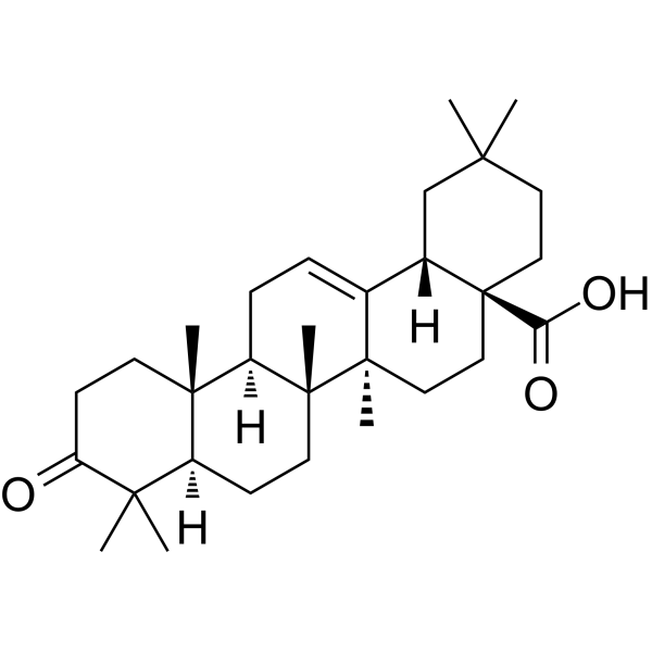
Oleanonic acid (3-Oxooleanolic acid) is an orally available triterpene that has anti-inflammatory and insecticidal properties. In vitro, oleanonic acid can improve oxidative stress, autophagy defects, ferroptosis, mitochondrial damage, and endoplasmic reticulum stress induced by Amyloid-β, and in vivo, it can alleviate myocardial hypertrophy in rats.
For research use only. We do not sell to patients.
- Purity : 99.91%
- CAS No.: 17990-42-0
- Formula: C30H46O3
- Molecular Weight:454.68
-
Storage:Powder -20°C, 3 years , 4°C, 2 years ; In solvent -80°C, 2 years , -20°C, 1 year

To place orders, for customer services and technical support, please contact: MedChemExpress USA
Tel: 609-228-6898 E-mail: [email protected] [email protected]


-
 Biological Activity
-
Chemical Information
-
Solvent & Solubility
- Protocols
- Purity & Documentation
- References
-
 Help & FAQs




Publications Citing Use of MedChemExpress (MCE) Oleanonic acid
More

Biological Activity
Description
IC50 & Target
|
HIV-1 |
Cellular Effect
|
Cell Line

|
Type | Value | Description | References |
|---|---|---|---|---|
| A549 | IC50 |
>20 μg/mL
Compound: Oleanonic acid
|
Cytotoxicity against human A549 cells by MTT assay
Cytotoxicity against human A549 cells by MTT assay
|
[PMID: 21106454] |
| A549 | IC50 |
49.5 μM
Compound: 4a
|
Cytotoxicity against human A549 cells incubated for 72 hrs by MTS assay
Cytotoxicity against human A549 cells incubated for 72 hrs by MTS assay
|
[PMID: 31677446] |
| A549 | IC50 |
50 μM
Compound: 3a
|
Cytotoxicity against human A549 cells assessed as reduction in cell viability incubated for 72 hrs by MTS assay
Cytotoxicity against human A549 cells assessed as reduction in cell viability incubated for 72 hrs by MTS assay
|
[PMID: 36174412] |
| BJ | IC50 |
>50 μM
Compound: 4a
|
Cytotoxicity against human BJ cells incubated for 72 hrs by MTS assay
Cytotoxicity against human BJ cells incubated for 72 hrs by MTS assay
|
[PMID: 31677446] |
| BJ | IC50 |
>50 μM
Compound: 3a
|
Cytotoxicity against human BJ cells assessed as reduction in cell viability incubated for 72 hrs by MTS assay
Cytotoxicity against human BJ cells assessed as reduction in cell viability incubated for 72 hrs by MTS assay
|
[PMID: 36174412] |
| Ca9-22 | IC50 |
18.04 μg/mL
Compound: Oleanonic acid
|
Cytotoxicity against human Ca9-22 cells by MTT assay
Cytotoxicity against human Ca9-22 cells by MTT assay
|
[PMID: 21106454] |
| CCRF-CEM | IC50 |
15.1 μM
Compound: 4a
|
Cytotoxicity against human CCRF-CEM cells incubated for 72 hrs by MTS assay
Cytotoxicity against human CCRF-CEM cells incubated for 72 hrs by MTS assay
|
[PMID: 31677446] |
| CCRF-CEM | IC50 |
9.5 μM
Compound: 3a
|
Cytotoxicity against human CCRF-CEM cells assessed as reduction in cell viability incubated for 72 hrs by MTS assay
Cytotoxicity against human CCRF-CEM cells assessed as reduction in cell viability incubated for 72 hrs by MTS assay
|
[PMID: 36174412] |
| CEM-DNR | IC50 |
17 μM
Compound: 3a
|
Cytotoxicity against human CEM-DNR cells assessed as reduction in cell viability incubated for 72 hrs by MTS assay
Cytotoxicity against human CEM-DNR cells assessed as reduction in cell viability incubated for 72 hrs by MTS assay
|
[PMID: 36174412] |
| H9 | EC50 |
0.11 μg/mL
Compound: 27
|
Cytotoxicity against mock-infected human H9 cells after 4 days
Cytotoxicity against mock-infected human H9 cells after 4 days
|
[PMID: 9748372] |
| H9 | IC50 |
0.8 μg/mL
Compound: 27
|
Antiviral activity against HIV1 3B in human H9 cells after 4 days by p24 antigen ELISA
Antiviral activity against HIV1 3B in human H9 cells after 4 days by p24 antigen ELISA
|
[PMID: 9748372] |
| HCT-116 | IC50 |
>50 μM
Compound: 4a
|
Cytotoxicity against p53 knockout human HCT116 cells incubated for 72 hrs by MTS assay
Cytotoxicity against p53 knockout human HCT116 cells incubated for 72 hrs by MTS assay
|
[PMID: 31677446] |
| HCT-116 | IC50 |
45 μM
Compound: 3a
|
Cytotoxicity against human HCT-116 cells assessed as reduction in cell viability incubated for 72 hrs by MTS assay
Cytotoxicity against human HCT-116 cells assessed as reduction in cell viability incubated for 72 hrs by MTS assay
|
[PMID: 36174412] |
| HCT-116 | IC50 |
45.1 μM
Compound: 4a
|
Cytotoxicity against human HCT116 cells incubated for 72 hrs by MTS assay
Cytotoxicity against human HCT116 cells incubated for 72 hrs by MTS assay
|
[PMID: 31677446] |
| Hep 3B2 | IC50 |
>20 μg/mL
Compound: Oleanonic acid
|
Cytotoxicity against human Hep3B cells by MTT assay
Cytotoxicity against human Hep3B cells by MTT assay
|
[PMID: 21106454] |
| HepG2 | IC50 |
>20 μg/mL
Compound: Oleanonic acid
|
Cytotoxicity against human HepG2 cells by MTT assay
Cytotoxicity against human HepG2 cells by MTT assay
|
[PMID: 21106454] |
| J774.A1 | IC50 |
38.8 μM
Compound: 3
|
Inhibition of LPS-induced NO production in mouse J774A1 cells compound preincubated for 1 hr before LPS treatment by Griess reaction
Inhibition of LPS-induced NO production in mouse J774A1 cells compound preincubated for 1 hr before LPS treatment by Griess reaction
|
[PMID: 25113933] |
| J774.A1 | IC50 |
87.72 μM
Compound: 3
|
Cytotoxicity against mouse J774A1 cells assessed as cell viability by MTT assay relative to control
Cytotoxicity against mouse J774A1 cells assessed as cell viability by MTT assay relative to control
|
[PMID: 25113933] |
| K562 | IC50 |
>50 μM
Compound: 4a
|
Cytotoxicity against human K562 cells incubated for 72 hrs by MTS assay
Cytotoxicity against human K562 cells incubated for 72 hrs by MTS assay
|
[PMID: 31677446] |
| K562 | IC50 |
>50 μM
Compound: 3a
|
Cytotoxicity against human K562 cells assessed as reduction in cell viability incubated for 72 hrs by MTS assay
Cytotoxicity against human K562 cells assessed as reduction in cell viability incubated for 72 hrs by MTS assay
|
[PMID: 36174412] |
| MCF7 | IC50 |
>20 μg/mL
Compound: Oleanonic acid
|
Cytotoxicity against human MCF7 cells by MTT assay
Cytotoxicity against human MCF7 cells by MTT assay
|
[PMID: 21106454] |
| MDA-MB-231 | IC50 |
>20 μg/mL
Compound: Oleanonic acid
|
Cytotoxicity against human MDA-MB-231 cells by MTT assay
Cytotoxicity against human MDA-MB-231 cells by MTT assay
|
[PMID: 21106454] |
| MRC5 | IC50 |
>50 μM
Compound: 4a
|
Cytotoxicity against human MRC5 cells incubated for 72 hrs by MTS assay
Cytotoxicity against human MRC5 cells incubated for 72 hrs by MTS assay
|
[PMID: 31677446] |
| MRC5 | IC50 |
>50 μM
Compound: 3a
|
Cytotoxicity against human MRC5 cells assessed as reduction in cell viability incubated for 72 hrs by MTS assay
Cytotoxicity against human MRC5 cells assessed as reduction in cell viability incubated for 72 hrs by MTS assay
|
[PMID: 36174412] |
| RAW264.7 | IC50 |
38.5 μM
Compound: 3
|
Inhibition of LPS-induced NO production in mouse RAW264.7 cells compound preincubated for 1 hr before LPS treatment by Griess reaction
Inhibition of LPS-induced NO production in mouse RAW264.7 cells compound preincubated for 1 hr before LPS treatment by Griess reaction
|
[PMID: 25113933] |
| RAW264.7 | IC50 |
89.6 μM
Compound: 3
|
Cytotoxicity mouse RAW264.7 cells assessed as cell viability by MTT assay relative to control
Cytotoxicity mouse RAW264.7 cells assessed as cell viability by MTT assay relative to control
|
[PMID: 25113933] |
| SK-MEL | IC50 |
31 μM
Compound: 19
|
Compound was tested for its cytotoxicity against human malignant melanoma cell line SK-MEL
Compound was tested for its cytotoxicity against human malignant melanoma cell line SK-MEL
|
[PMID: 10450948] |
| U2OS | IC50 |
48.5 μM
Compound: 4a
|
Cytotoxicity against human U2OS cells incubated for 72 hrs by MTS assay
Cytotoxicity against human U2OS cells incubated for 72 hrs by MTS assay
|
[PMID: 31677446] |
| U2OS | IC50 |
49 μM
Compound: 3a
|
Cytotoxicity against human U2OS cells assessed as reduction in cell viability incubated for 72 hrs by MTS assay
Cytotoxicity against human U2OS cells assessed as reduction in cell viability incubated for 72 hrs by MTS assay
|
[PMID: 36174412] |
In Vitro
Oleanonic acid inhibits HIV-1 infection in PBMCs in vitro, with EC50 values for naturally infected PBMCs and monocytes/macrophages being 22.7 mM, 24.6 mM, and 57.4 mM, respectively. Furthermore, it can inhibit the production of leukotriene B4 by mouse peritoneal leukocytes, with an IC50 of 17 μM[2].
Oleanonic acid has strong anti-insect activity against Rhipicephalus (Kreyszig) and Amazon ticks, with IC50 values of 18.5 µM and 29.9 µM[3].
Oleanonic acid (0-45 μM, 24 h) inhibits the increase of NF-κB transcription activity in cardiomyocytes treated with phenylephrine (PE) and reduces the mRNA expression of hypertrophic genes like atrial natriuretic factor (ANF) and brain natriuretic peptide (BNP) in a dose-dependent manner[4].
Oleanonic acid (15 μM, 24 h) inhibits the phosphorylation of protein kinase Cζ (PKCζ) at the Thr410 site in cardiomyocytes, subsequently reducing NF-κB activation in PE-treated cardiomyocytes[4].
Oleanonic acid (1-5 μM, 48 h) reduces APP expression in SH-SY5Y cells, lessens damage induced by oxidative stress, repairs autophagy defects, and inhibits ferroptosis[5].
MedChemExpress (MCE) has not independently confirmed the accuracy of these methods. They are for reference only. Further protocols information, click here.
-
Cell Line:NRCMs
-
Concentration:0, 5, 15, 45 μM
-
Incubation Time:24 h
-
Result:Inhibited NF-κB transcriptional activity and reduced the expression of hypertrophic genes such as Atrial Natriuretic Factor (ANF) and Brain Natriuretic Peptide (BNP).
-
Cell Line:NRCMs
-
Concentration:15 μM
-
Incubation Time:24 h
-
Result:Inhibited the phosphorylation of the Thr410 site protein kinase PKCζ, and suppressed NF-κB.
-
Cell Line:SH-SY5Y, SH-SY5Y-APP
-
Concentration:1, 2.5, 5 μM
-
Incubation Time:48 h
-
Result:Reduced APP expression, inhibited mTOR phosphorylation, increased autophagy markers ATG5 and LC3-II, suppressed Nrf2 and HO-1, and restored the levels of GPX4, NCOA, and COX2.
In Vivo
MedChemExpress (MCE) has not independently confirmed the accuracy of these methods. They are for reference only.
-
Animal Model:AAC rats[4]
-
Dosage:15, 45 mg/kg; daily; 8 weeks
-
Administration:Oral
-
Result:Reduced heart size and visibly decreased the thickness of the left ventricular wall, and in a dose-dependent manner, there was also a reduction in myocardial cell diameter and the heart weight to body weight ratio (HW/BW) under 400x HE staining.


Chemical Information
-
CAS No. 17990-42-0
-
Appearance Solid
-
Molecular Weight 454.68
-
Formula C30H46O3
-
Color White to off-white
-
SMILES
CC1(C)C(CC[C@]2(C)[C@@]3([H])CC=C4[C@]5([H])CC(C)(C)CC[C@@](C(O)=O)5CC[C@](C)4[C@@](C)3CC[C@@]12[H])=O
-
Synonyms
3-Oxooleanolic acid
-
Structure Classification
-
Initial Source
-
Shipping
Room temperature in continental US; may vary elsewhere.
-
Storage
Powder -20°C 3 years 4°C 2 years In solvent -80°C 2 years -20°C 1 year


Publications (2)
-
Journal Impact Factor
-
Most Recent
-
Pharmacol Res
2024 Jun:204:107208. PMID: 38729587 -
Vet Microbiol
The Chinese medicine monomer Schisandrin C inhibits PRRSV infection by regulating the OGT-PI3K/AKT/mTOR signaling pathway. [Abstract]2026 May:316:110992. PMID: 41865607


Solvent & Solubility
In Vitro:
DMSO : 50 mg/mL (109.97 mM; Need ultrasonic; Hygroscopic DMSO has a significant impact on the solubility of product, please use newly opened DMSO)
H2O : ≥ mg/mL
* "≥" means soluble, but saturation unknown.
Please refer to the solubility information to select the appropriate solvent. Once prepared, please aliquot and store the solution to prevent product inactivation from repeated freeze-thaw cycles.
Storage method and period of stock solution: -80°C, 2 years; -20°C, 1 year. When stored at -80°C, please use it within 2 years. When stored at -20°C, please use it within 1 year.
* Note: If you choose water as the stock solution, please dilute it to the working solution, then filter and sterilize it with a 0.22 μm filter before use.
Please refer to the solubility information to select the appropriate solvent. Once prepared, please aliquot and store the solution to prevent product inactivation from repeated freeze-thaw cycles.
Storage method and period of stock solution: -80°C, 2 years; -20°C, 1 year. When stored at -80°C, please use it within 2 years. When stored at -20°C, please use it within 1 year.
* Note: If you choose water as the stock solution, please dilute it to the working solution, then filter and sterilize it with a 0.22 μm filter before use.
Concentration (start) × Volume (start) = Concentration (final) × Volume (final)
In Vivo:
Select the appropriate dissolution method based on your experimental animal and administration route.
- For the following dissolution methods, please ensure to first prepare a clear stock solution using an In Vitro approach and then sequentially add co-solvents:
- To ensure reliable experimental results, the clarified stock solution can be appropriately stored based on storage conditions. As for the working solution for In Vivo experiments, it is recommended to prepare freshly and use it on the same day.
- The percentages shown for the solvents indicate their volumetric ratio in the final prepared solution. If precipitation or phase separation occurs during preparation, heat and/or sonication can be used to aid dissolution.
Add each solvent one by one: 10% DMSO 40% PEG300 5% Tween-80 45% Saline
Solubility: ≥ 2.5 mg/mL (5.50 mM); Clear solution
This protocol yields a clear solution of ≥ 2.5 mg/mL (saturation unknown).
Taking 1 mL working solution as an example, add 100 μL DMSO stock solution (25.0 mg/mL) to 400 μL PEG300, and mix evenly; then add 50 μL Tween-80 and mix evenly; then add 450 μL Saline to adjust the volume to 1 mL.
Preparation of Saline: Dissolve 0.9 g sodium chloride in ddH₂O and dilute to 100 mL to obtain a clear Saline solution.
Add each solvent one by one: 10% DMSO 90% (20% SBE-β-CD in Saline)
Solubility: 2.5 mg/mL (5.50 mM); Suspended solution; Need ultrasonic
This protocol yields a suspended solution of 2.5 mg/mL. Suspended solution can be used for oral and intraperitoneal injection.
Taking 1 mL working solution as an example, add 100 μL DMSO stock solution (25.0 mg/mL) to 900 μL 20% SBE-β-CD in Saline, and mix evenly.
Preparation of 20% SBE-β-CD in Saline (4°C, storage for one week): 2 g SBE-β-CD powder is dissolved in 10 mL Saline, completely dissolve until clear.
In Vivo Dissolution Calculator
Please enter the basic information of animal experiments:
-
-
-
-
Recommended: Prepare an additional quantity of animals to account for potential losses during experiments.
Please enter your animal formula composition:
-
%DMSO +
Recommended: Keep the proportion of DMSO in working solution below 2% if your animal is weak.
-
%+
-
+%Tween-80 + +
-
%Saline +
The co-solvents required include: DMSO, . All of co-solvents are available by MedChemExpress (MCE). , Tween 80. All of co-solvents are available by MedChemExpress (MCE).
Working solution concentration: 0.22 mg/mL
Method for preparing stock solution: mg drug dissolved in μL DMSO. Stock solution concentration: mg/mL.
1. Take μL DMSO stock solution;
2. Add μL .
μL , mix evenly;
3. Then add μL Tween 80, mix evenly;
4. Then add μL
Please ensure that the stock solution in the first step is dissolved to a clear state, and add co-solvents in sequence. You can use ultrasonic heating (ultrasonic cleaner, recommended frequency 20-40 kHz), vortexing, etc. to assist dissolution.

Protocols
-
ROS/oxidative-stress fluorescent staining
ROS/oxidative-stress fluorescent staining uses cell-permeant fluorogenic probes that become fluorescent after oxidation inside cells or tissues; commonly used examples include DCFH-DA/DCFDA for broad cellular oxidant detection, DHE for superoxide-related signal detection, MitoSOX for mitochondrial superoxide-related signal detection, and CellROX probes for oxidative-stress-associated fluorescence readouts. The assay detects probe oxidation rather than a single ROS species unless the probe and analysis method have been chemically validated for that species. DCFH-DA enters cells, is deacetylated by intracellular esterases to DCFH, and produces fluorescent DCF after oxidation, so the readout is used as an operational measure of total cellular oxidative stress rather than a species-specific ROS measurement. DHE and MitoSOX can report superoxide-related oxidation, but red fluorescence alone can include non-specific ethidium-like oxidation products; HPLC or optimized spectral approaches are
-
Autophagy
Autophagy is a process in which eukaryotic cells use lysosomes to degrade their own cytoplasmic proteins and damaged organelles under the regulation of autophagy related gene (Atg). Microtubule-associated proteins light chain 3 (LC3) is recognized as autophagy marker, which transfers from cytoplasmic LC3 (LC3-I) to membrane type (LC3-II). LC3-II/I ratio could be detected by Western Blot and fluorescence microscopy.
-
Research Protocol for Inflammation-related Diseases
The NLRP3 inflammasome is a cytosolic innate immune signaling platform that integrates priming signals and danger-signal activation to promote caspase-1 activation, maturation of IL-1β and IL-18, and gasdermin D-mediated pyroptotic cell death. The core experimental logic is to determine whether inflammatory disease phenotypes are driven by increased NLRP3 expression, ASC-containing inflammasome assembly, caspase-1 cleavage, GSDMD cleavage, and extracellular release of IL-1β/IL-18 rather than by nonspecific cell injury alone. The pathway is strongly linked to inflammation-related disease phenotypes because monosodium urate crystals activate NALP3/NLRP3 inflammasome signaling in gout-like crystal inflammation, cholesterol crystals activate NLRP3 inflammasomes in atherogenesis models, and DSS-induced intestinal inflammation has been reported to involve NLRP3 inflammasome activity. However, experimental colitis studies also show context-dependent protective effects of NLRP3 inflammasome co
-
Lysosome and acidic-vesicle live-cell staining
Lysosome and acidic-vesicle live-cell staining detects acidic intracellular compartments by using membrane-permeant acidotropic probes that accumulate in low-pH vesicles, including lysosomes, late endosomes, autolysosomes, and acidic phagosomes. LysoTracker staining is commonly used as an intensity-based readout of acidic lysosomal compartment abundance or enlargement, while acridine orange produces green fluorescence in less concentrated compartments and red fluorescence after concentration-dependent accumulation in acidic vesicular organelles. Loss or reduction of acridine-orange red signal can be used as a readout of lysosomal membrane permeabilization or reduced acidic-vesicle integrity. This protocol is designed for live cultured cells and can be adapted for fluorescence microscopy, high-content imaging, plate-reader readout, or flow cytometry when the selected literature supports the readout. Because these dyes report acidotropic accumulation rather than lysosome identity alone,
-
Ferroptosis Solutions
Ferroptosis is an iron-dependent, non-apoptotic form of regulated cell death characterized by lethal lipid peroxidation and sensitivity to suppression by iron chelators or lipophilic radical-trapping antioxidants. The core pathway links cystine uptake through system Xc−, glutathione availability, GPX4-dependent detoxification of phospholipid hydroperoxides, iron-dependent oxidative reactions, and polyunsaturated-phospholipid metabolism into a cell-death program that is biochemically and morphologically distinct from apoptosis, necrosis, and autophagy. The ferroptosis pathway is experimentally linked to phenotype through chemical and genetic perturbation. Erastin induces ferroptosis by inhibiting cystine uptake through system Xc− and weakening antioxidant defenses, while GPX4 inhibition or depletion causes lipid peroxide accumulation and ferroptotic cancer-cell death. ACSL4 and oxidizable arachidonoyl- or adrenoyl-containing phosphatidylethanolamines shape ferroptosis sensitivity by con
-
Macroautophagy Solutions
Macroautophagy is a conserved lysosome-dependent degradation pathway in which cytoplasmic material is sequestered into double-membrane autophagosomes and delivered to lysosomes for degradation and recycling. The pathway supports cellular homeostasis during nutrient limitation, organelle stress, protein-aggregate accumulation, infection, differentiation, and tissue remodeling by coupling cargo sequestration, autophagosome maturation, lysosomal fusion, and degradation of cargo-derived macromolecules. The core molecular sequence includes initiation by nutrient- and stress-regulated autophagy machinery, autophagosome nucleation, LC3/ATG8-family conjugation to autophagosomal membranes, cargo selection through receptors such as SQSTM1/p62, autophagosome-lysosome fusion, and lysosomal degradation. LC3 was identified as a mammalian homolog of yeast Atg8 that localizes to autophagosomal membranes after processing, and p62/SQSTM1 was shown to connect ubiquitinated cargo with autophagic degradati
-
Amyloid: Congo Red Amyloid Staining
Congo red amyloid staining is a histochemical method used to detect extracellular amyloid deposits in tissue sections based on the affinity of Congo red dye for β-pleated sheet-rich protein aggregates. When bound to amyloid, Congo red produces characteristic apple-green birefringence under polarized light microscopy, which is widely regarded as a diagnostic feature of amyloid deposition in histopathology. The diagnostic principle relies on the combination of dye binding (congophilia) and optical anisotropy under polarized illumination, which distinguishes amyloid from most non-amyloid eosinophilic extracellular deposits in routine histological evaluation. Amyloid identification by Congo red staining remains a cornerstone in diagnostic pathology despite the availability of adjunct methods such as immunohistochemistry and mass spectrometry, particularly because of its ability to localize deposits directly within tissue architecture. The specificity of Congo red-positive deposits is incre
-
Alzheimer’s Disease Modeling
Alzheimer’s Disease (AD) is a neurodegenerative disorder characterized by a progressive decline in cognitive functions and loss of specific types of neurons and synapses. Alzheimer's symptoms can be simulated in mice by injecting drugs (such as Aβ) or genetically modified.


Purity & Documentation
-
Data Sheet (284 KB)
-
SDS (396 KB)
- English - EN (396 KB)
- Français - FR (396 KB)
- Deutsch - DE (396 KB)
- Norwegian - NO (396 KB)
- Español - ES (396 KB)
- Swedish - SV (396 KB)
- Italian - IT (396 KB)
- Korean - KR (396 KB)
- Portuguese - PT (396 KB)
-
Handling Instructions (2659 KB)
References
[1]. Giner-Larza EM et al.Oleanonic acid, a 3-oxotriterpene from Pistacia, inhibits leukotriene synthesis and has anti-inflammatory activity.Eur J Pharmacol.Sep 28;428(1):137-43.doi:10.1016/S0014-2999(01)01290-0(2001) [Content Brief]
[2]. Fabio Mengoni.Anti-HIV Activity of Oleanolic Acid on Infected Human Mononuclear Cells.Planta Med 68(2): 111-114.DOI: 10.1055/s-2002-20256(2002) [Content Brief]
[4]. Hui Gao, et al. Oleanonic acid ameliorates pressure overload-induced cardiac hypertrophy in rats: The role of PKCζ-NF-κB pathway. Mol Cell Endocrinol. 2018 Jul 15:470:259-268. [Content Brief]
[5]. Liqing Tao, et al. Oleanonic acid ameliorates mutant Aβ precursor protein-induced oxidative stress, autophagy deficits, ferroptosis, mitochondrial damage, and ER stress in vitro. Biochim Biophys Acta Mol Basis Dis. 2024 Dec;1870(8):167459. [Content Brief]


Complete Stock Solution Preparation Table
Please refer to the solubility information to select the appropriate solvent. Once prepared, please aliquot and store the solution to prevent product inactivation from repeated freeze-thaw cycles.
Storage method and period of stock solution: -80°C, 2 years; -20°C, 1 year. When stored at -80°C, please use it within 2 years. When stored at -20°C, please use it within 1 year.
| Optional Solvent | Concentration Solvent Mass | 1 mg | 5 mg | 10 mg | 25 mg |
|---|---|---|---|---|---|
| DMSO | 1 mM | 2.1993 mL | 10.9967 mL | 21.9935 mL | 54.9837 mL |
| 5 mM | 0.4399 mL | 2.1993 mL | 4.3987 mL | 10.9967 mL | |
| 10 mM | 0.2199 mL | 1.0997 mL | 2.1993 mL | 5.4984 mL | |
| 15 mM | 0.1466 mL | 0.7331 mL | 1.4662 mL | 3.6656 mL | |
| 20 mM | 0.1100 mL | 0.5498 mL | 1.0997 mL | 2.7492 mL | |
| 25 mM | 0.0880 mL | 0.4399 mL | 0.8797 mL | 2.1993 mL | |
| 30 mM | 0.0733 mL | 0.3666 mL | 0.7331 mL | 1.8328 mL | |
| 40 mM | 0.0550 mL | 0.2749 mL | 0.5498 mL | 1.3746 mL | |
| 50 mM | 0.0440 mL | 0.2199 mL | 0.4399 mL | 1.0997 mL | |
| 60 mM | 0.0367 mL | 0.1833 mL | 0.3666 mL | 0.9164 mL | |
| 80 mM | 0.0275 mL | 0.1375 mL | 0.2749 mL | 0.6873 mL | |
| 100 mM | 0.0220 mL | 0.1100 mL | 0.2199 mL | 0.5498 mL |
* Note: If you choose water as the stock solution, please dilute it to the working solution, then filter and sterilize it with a 0.22 μm filter before use.


Oleanonic acid Related Classifications
HY-N1487 Related Classifications
 Powered by Bioz