



Simetryn

Based on 1 Customer Validation
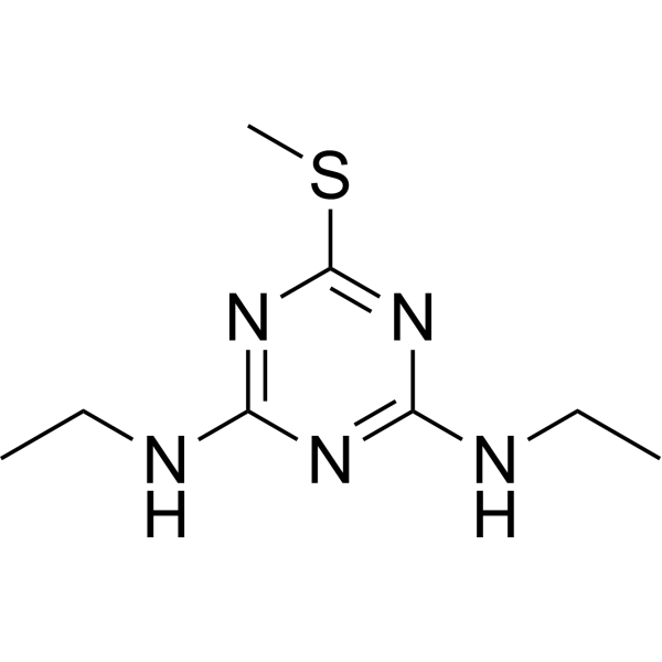
Simetryn is a triazine herbicide that exerts teratogenicity. Simetryn inhibits photosynthesis by blocking electron transfer in the chloroplast photosystem II and activates the MEK/Erk signaling pathway. Simetryn triggers vascular and developmental abnormalities in zebrafish and tadpoles, suppresses proliferation, enhances apoptosis, and induces malformations. Simetryn serves as a tool to establish a zebrafish model for studying arteriovenous malformations and related pathogenesis. Simetryn controls paddy weeds and can be used for research on developmental and vascular disorders.
For research use only. We do not sell to patients.
- Purity : 99.75%
- CAS No.: 1014-70-6
- Formula: C8H15N5S
- Molecular Weight:213.30
-
Storage:Powder -20°C, 3 years , 4°C, 2 years ; In solvent -80°C, 2 years , -20°C, 1 year

To place orders, for customer services and technical support, please contact: MedChemExpress USA
Tel: 609-228-6898 E-mail: [email protected] [email protected]


-
 Biological Activity
-
Chemical Information
-
Solvent & Solubility
- Protocols
- Purity & Documentation
- References
-
 Help & FAQs


All MEK Isoforms
More

Biological Activity
Description
In Vivo
MedChemExpress (MCE) has not independently confirmed the accuracy of these methods. They are for reference only.
-
Animal Model:AB, Tg(fli1a:GFP), Tg(gata1:Dsred), Tg(Flk1:GFP), Tg(kdrl:mcherry), Tg(lyve1b:YFP) (8-10 h post-fertilization embryos)[1]
-
Dosage:10 mg/L, 15 mg/L, 20 mg/L
-
Administration:waterborne exposure; continuous; 48 hours
-
Result:Induced blood stasis and abnormal artery-vein capillary fusion in the caudal vascular plexus (CVP).
Significantly increased dorsal aorta and largest venous diameters compared to controls at 48 hours post-exposure (10, 15, and 20 mg/L).
Caused erythrocyte accumulation at arteriovenous junctions in the CVP.
Reduced PH3-positive proliferating cells and increased TUNEL-positive apoptotic cells in the CVP in a concentration-dependent manner.
Upregulated p53 mRNA expression.
Impaired cerebral vascular development, with absence of the dorsal longitudinal vein and reduced central venous bud.
Induced abnormal morphology and reduced fluorescent signal intensity in caudal lymphatic vessels.
Increased phosphorylated Erk (p-Erk) and total Erk protein levels.
- Reproductive and Developmental Toxicity Study
- Detection of Apoptosis
- TUNEL staining for apoptotic DNA fragmentation
- Annexin V plus membrane-impermeant dye apoptosis staining
- How to Select a Suitable Non-Mouse Animal Model
- Apoptosis Solutions
- Cell Viability Determination by MTT Colorimetric Assay
- How to Choose the Right Model Animal


Chemical Information
-
CAS No. 1014-70-6
-
Appearance Solid
-
Molecular Weight 213.30
-
Formula C8H15N5S
-
Color White to off-white
-
SMILES
CSC1=NC(NCC)=NC(NCC)=N1
-
Shipping
Room temperature in continental US; may vary elsewhere.
-
Storage
Powder -20°C 3 years 4°C 2 years In solvent -80°C 2 years -20°C 1 year


Solvent & Solubility
In Vitro:
DMSO : ≥ 37 mg/mL (173.46 mM; Hygroscopic DMSO has a significant impact on the solubility of product, please use newly opened DMSO)
* "≥" means soluble, but saturation unknown.
Please refer to the solubility information to select the appropriate solvent. Once prepared, please aliquot and store the solution to prevent product inactivation from repeated freeze-thaw cycles.
Storage method and period of stock solution: -80°C, 2 years; -20°C, 1 year. When stored at -80°C, please use it within 2 years. When stored at -20°C, please use it within 1 year.
Please refer to the solubility information to select the appropriate solvent. Once prepared, please aliquot and store the solution to prevent product inactivation from repeated freeze-thaw cycles.
Storage method and period of stock solution: -80°C, 2 years; -20°C, 1 year. When stored at -80°C, please use it within 2 years. When stored at -20°C, please use it within 1 year.
Concentration (start) × Volume (start) = Concentration (final) × Volume (final)
In Vivo:
Select the appropriate dissolution method based on your experimental animal and administration route.
- For the following dissolution methods, please ensure to first prepare a clear stock solution using an In Vitro approach and then sequentially add co-solvents:
- To ensure reliable experimental results, the clarified stock solution can be appropriately stored based on storage conditions. As for the working solution for In Vivo experiments, it is recommended to prepare freshly and use it on the same day.
- The percentages shown for the solvents indicate their volumetric ratio in the final prepared solution. If precipitation or phase separation occurs during preparation, heat and/or sonication can be used to aid dissolution.
Add each solvent one by one: 10% DMSO 40% PEG300 5% Tween-80 45% Saline
Solubility: ≥ 2.5 mg/mL (11.72 mM); Clear solution
This protocol yields a clear solution of ≥ 2.5 mg/mL (saturation unknown).
Taking 1 mL working solution as an example, add 100 μL DMSO stock solution (25.0 mg/mL) to 400 μL PEG300, and mix evenly; then add 50 μL Tween-80 and mix evenly; then add 450 μL Saline to adjust the volume to 1 mL.
Preparation of Saline: Dissolve 0.9 g sodium chloride in ddH₂O and dilute to 100 mL to obtain a clear Saline solution.
In Vivo Dissolution Calculator
Please enter the basic information of animal experiments:
-
-
-
-
Recommended: Prepare an additional quantity of animals to account for potential losses during experiments.
Please enter your animal formula composition:
-
%DMSO +
Recommended: Keep the proportion of DMSO in working solution below 2% if your animal is weak.
-
%+
-
+%Tween-80 + +
-
%Saline +
The co-solvents required include: DMSO, . All of co-solvents are available by MedChemExpress (MCE). , Tween 80. All of co-solvents are available by MedChemExpress (MCE).
Working solution concentration: 0.22 mg/mL
Method for preparing stock solution: mg drug dissolved in μL DMSO. Stock solution concentration: mg/mL.
1. Take μL DMSO stock solution;
2. Add μL .
μL , mix evenly;
3. Then add μL Tween 80, mix evenly;
4. Then add μL
Please ensure that the stock solution in the first step is dissolved to a clear state, and add co-solvents in sequence. You can use ultrasonic heating (ultrasonic cleaner, recommended frequency 20-40 kHz), vortexing, etc. to assist dissolution.


Purity & Documentation
-
Data Sheet (272 KB)
-
SDS (597 KB)
- English - EN (597 KB)
- Français - FR (597 KB)
- Deutsch - DE (597 KB)
- Norwegian - NO (597 KB)
- Español - ES (597 KB)
- Swedish - SV (597 KB)
- Italian - IT (597 KB)
- Korean - KR (597 KB)
- Portuguese - PT (597 KB)
-
Handling Instructions (2659 KB)
References
[1]. Xiao Z, et al. Simetryn induces arteriovenous malformations in zebrafish embryos through activation of Erk signaling. Biochem Biophys Res Commun. 2026;804:153339. [Content Brief]
[2]. Saka M, et al. Application of an amphibian (Silurana tropicalis) metamorphosis assay to the testing of the chronic toxicity of three rice paddy herbicides: simetryn, mefenacet, and thiobencarb. Ecotoxicol Environ Saf. 2013;92:135-143. [Content Brief]


Complete Stock Solution Preparation Table
Please refer to the solubility information to select the appropriate solvent. Once prepared, please aliquot and store the solution to prevent product inactivation from repeated freeze-thaw cycles.
Storage method and period of stock solution: -80°C, 2 years; -20°C, 1 year. When stored at -80°C, please use it within 2 years. When stored at -20°C, please use it within 1 year.
| Optional Solvent | Concentration Solvent Mass | 1 mg | 5 mg | 10 mg | 25 mg |
|---|---|---|---|---|---|
| DMSO | 1 mM | 4.6882 mL | 23.4412 mL | 46.8823 mL | 117.2058 mL |
| 5 mM | 0.9376 mL | 4.6882 mL | 9.3765 mL | 23.4412 mL | |
| 10 mM | 0.4688 mL | 2.3441 mL | 4.6882 mL | 11.7206 mL | |
| 15 mM | 0.3125 mL | 1.5627 mL | 3.1255 mL | 7.8137 mL | |
| 20 mM | 0.2344 mL | 1.1721 mL | 2.3441 mL | 5.8603 mL | |
| 25 mM | 0.1875 mL | 0.9376 mL | 1.8753 mL | 4.6882 mL | |
| 30 mM | 0.1563 mL | 0.7814 mL | 1.5627 mL | 3.9069 mL | |
| 40 mM | 0.1172 mL | 0.5860 mL | 1.1721 mL | 2.9301 mL | |
| 50 mM | 0.0938 mL | 0.4688 mL | 0.9376 mL | 2.3441 mL | |
| 60 mM | 0.0781 mL | 0.3907 mL | 0.7814 mL | 1.9534 mL | |
| 80 mM | 0.0586 mL | 0.2930 mL | 0.5860 mL | 1.4651 mL | |
| 100 mM | 0.0469 mL | 0.2344 mL | 0.4688 mL | 1.1721 mL |


Simetryn Related Classifications
HY-B1853 Related Classifications
 Powered by Bioz
Keywords
- Simetryn
- 1014-70-6
- Environmental Pollutants
- Herbicide
- ERK
- Apoptosis
- MEK
- zebrafish embryos
- S1-RSA
- chloroplast photosystem II electron transport chain
- BALB/c mice
- freshwater plankton communities
- Silurana tropicalis tadpoles
- MEK/Erk signaling cascade
- arteriovenous malformations
- scoliosis
- Erk protein
- Inhibitor
- inhibitor
- inhibit