



WD6305 TFA
Based on 2 publication(s) in Google Scholar
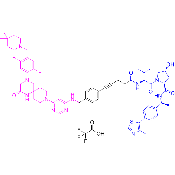
WD6305 TFA is a potent and selective METTL3-METTL14 PROTAC degrader. WD6305 TFA has DC50 values of 140 nM and 194 nM for METTL3 and METTL14, respectively. WD6305 TFA inhibits m6A modification and proliferation of AML cells, and induces apoptosis. WD6305 TFA has antitumor activity.
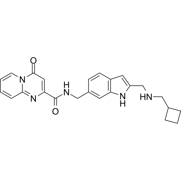
(Pink: METTL3-14 ligand (HY-115717); Blue: VHL ligand (HY-150803); Black: linker).
Nur für Forschungszwecke. Wir verkaufen nicht an Patienten.
- Reinheit : 99.81%
- Formel: C63H76F5N11O7S
- Molecular Weight:1226.40
-
Speicherung:
4°C, sealed storage, away from moisture
* In solvent : -80°C, 6 months; -20°C, 1 month (sealed storage, away from moisture)

To place orders, for customer services and technical support, please contact: MedChemExpress USA
Tel: 609-228-6898 E-mail: [email protected] [email protected]


-
 Biologische Aktivität
-
Chemical Information
-
Lösungsmittel & Löslichkeit
- Protokoll
- Reinheit & Dokumentation
- Verweise
-
 Help & FAQs



Publications Citing Use of MedChemExpress (MCE) WD6305 TFA
MoreAlle PROTACs Isoform-spezifische Produkte anzeigen
More

Biologische Aktivität
Beschreibung
IC50 & Target
DC50: 140 nM (METTL3); D max: 91.9% (METTL3) [1]
DC50: 194 nM (METTL14)[1]
In Vitro
Non-salt dose:
WD6305 (20-5000 nM; 24 h) reduces METTL3 protein levels in Mono-Mac-6 cells with a DC50 of 140 nM and a D max of 91.9%[1].
WD6305 (20-5000 nM; 24 h) reduces METTL14 protein levels in Mono-Mac-6 cells with a DC50 of 194 nM[1].
WD6305 (0.5-10 μM; 48 h) decreases the ratio of m6A to polyA mRNA, inhibits cell proliferation and induces cell apoptosis in Mono-Mac-6 cells[1].
MedChemExpress (MCE) has not independently confirmed the accuracy of these methods. They are for reference only.
-
Cell Line:Mono-Mac-6 cells
-
Concentration:20, 50, 100, 250, 500, 1000, 2000 and 5000 nM
-
Incubation Time:24 h
-
Result:Reduced METTL3 and METTL14 protein levels in a dose-dependent manner.
-
Cell Line:Mono-Mac-6 cells
-
Concentration:1 and 2.5 μM
-
Incubation Time:48 h
-
Result:Increased the levels of cleaved caspase-3 and cleaved PARP.


Chemical Information
-
Appearance Solid
-
Molecular Weight 1226.40
-
Formel C63H76F5N11O7S
-
Color White to light yellow
-
SMILES
O=C(N[C@@H](C(C)(C)C)C(N1[C@H](C(N[C@@H](C)C2=CC=C(C3=C(C)N=CS3)C=C2)=O)C[C@@H](O)C1)=O)CCC#CC(C=C4)=CC=C4CNC5=CC(N(CC6)CCC76NC(CN(C8=CC(F)=C(CN9CCC(C)(C)CC9)C=C8F)C7)=O)=NC=N5.O=C(O)C(F)(F)F
-
Versand
Room temperature in continental US; may vary elsewhere.
-
Speicherung
4°C, sealed storage, away from moisture
* In solvent : -80°C, 6 months; -20°C, 1 month (sealed storage, away from moisture)


Publications (2)
-
Journal Impact Factor
-
Most Recent
-
Cancer Res
2025 Aug 27. PMID: 40865050 -
RSC Med Chem
Structure-guided design of a methyltransferase-like 3 (METTL3) proteolysis targeting chimera (PROTAC) incorporating an indole-nicotinamide chemotype. [Abstract]2025 Jun 19. PMID: 40599585


Lösungsmittel & Löslichkeit
In Vitro:
DMSO : 95 mg/mL (77.46 mM; Need ultrasonic; Hygroscopic DMSO has a significant impact on the solubility of product, please use newly opened DMSO)
Please refer to the solubility information to select the appropriate solvent. Once prepared, please aliquot and store the solution to prevent product inactivation from repeated freeze-thaw cycles.
Storage method and period of stock solution: -80°C, 6 months; -20°C, 1 month (sealed storage, away from moisture). When stored at -80°C, please use it within 6 months. When stored at -20°C, please use it within 1 month.
Please refer to the solubility information to select the appropriate solvent. Once prepared, please aliquot and store the solution to prevent product inactivation from repeated freeze-thaw cycles.
Storage method and period of stock solution: -80°C, 6 months; -20°C, 1 month (sealed storage, away from moisture). When stored at -80°C, please use it within 6 months. When stored at -20°C, please use it within 1 month.
Konzentration (Stammlösung) × Volumen (Stammlösung) = Konzentration (Ziellösung) × Volumen (Ziellösung)


Reinheit & Dokumentation
-
Data Sheet (284 KB)
-
SDS (252 KB)
- English - EN (252 KB)
- Français - FR (252 KB)
- Deutsch - DE (252 KB)
- Norwegian - NO (252 KB)
- Español - ES (252 KB)
- Swedish - SV (252 KB)
- Italian - IT (252 KB)
- Korean - KR (252 KB)
- Portuguese - PT (252 KB)
-
Handling Instructions (2659 KB)
Verweise


Complete Stock Solution Preparation Table
Please refer to the solubility information to select the appropriate solvent. Once prepared, please aliquot and store the solution to prevent product inactivation from repeated freeze-thaw cycles.
Storage method and period of stock solution: -80°C, 6 months; -20°C, 1 month (sealed storage, away from moisture). When stored at -80°C, please use it within 6 months. When stored at -20°C, please use it within 1 month.
| Optional Solvent | Concentration Solvent Mass | 1 mg | 5 mg | 10 mg | 25 mg |
|---|---|---|---|---|---|
| DMSO | 1 mM | 0.8154 mL | 4.0770 mL | 8.1539 mL | 20.3849 mL |
| 5 mM | 0.1631 mL | 0.8154 mL | 1.6308 mL | 4.0770 mL | |
| 10 mM | 0.0815 mL | 0.4077 mL | 0.8154 mL | 2.0385 mL | |
| 15 mM | 0.0544 mL | 0.2718 mL | 0.5436 mL | 1.3590 mL | |
| 20 mM | 0.0408 mL | 0.2038 mL | 0.4077 mL | 1.0192 mL | |
| 25 mM | 0.0326 mL | 0.1631 mL | 0.3262 mL | 0.8154 mL | |
| 30 mM | 0.0272 mL | 0.1359 mL | 0.2718 mL | 0.6795 mL | |
| 40 mM | 0.0204 mL | 0.1019 mL | 0.2038 mL | 0.5096 mL | |
| 50 mM | 0.0163 mL | 0.0815 mL | 0.1631 mL | 0.4077 mL | |
| 60 mM | 0.0136 mL | 0.0679 mL | 0.1359 mL | 0.3397 mL |


WD6305 TFA Related Classifications
HY-160415A Related Classifications
 Powered by Bioz