



Aurantio-obtusin
Based on 1 publication(s) in Google Scholar
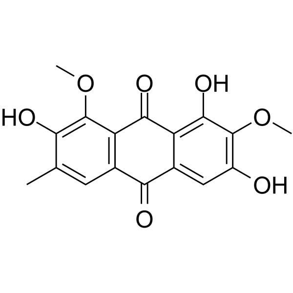
Aurantio-obtusin is a anthraquinone compound that can be extracted from cassia seed. Aurantio-obtusin has the effects of decreasing blood pressure, decreasing blood lipids and anti-inflammatory.Aurantio-obtusin is an orally active vasodilator. Aurantio-obtusin ameliorates hepatic steatosis through AMPK/ autophagy- and AMPK/TFEB mediated inhibition of lipid accumulation.
For research use only. We do not sell to patients.
- Purity : 99.39%
- CAS No.: 67979-25-3
- Formula: C17H14O7
- Molecular Weight:330.29
-
Storage:
4°C, protect from light
* In solvent : -80°C, 6 months; -20°C, 1 month (protect from light)

To place orders, for customer services and technical support, please contact: MedChemExpress USA
Tel: 609-228-6898 E-mail: [email protected] [email protected]


-
 Biological Activity
-
Chemical Information
-
Solvent & Solubility
- Protocols
- Purity & Documentation
- References
-
 Help & FAQs


-
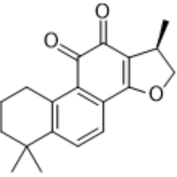
HY-N0174
CryptotanshinoneAutophagy Inducer -
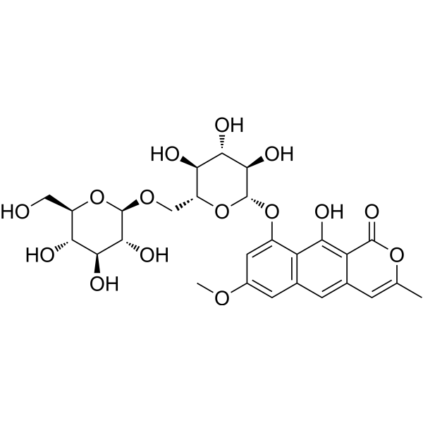
HY-N7628
Cassiaside CAGE Formation Inhibitor -
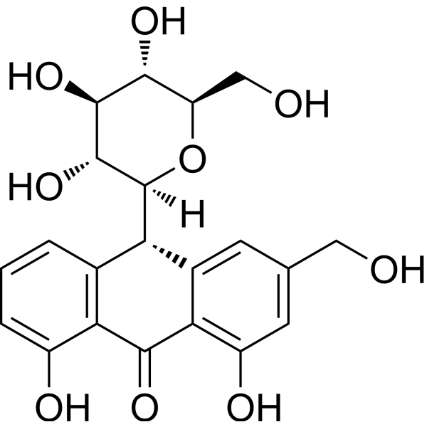
HY-N0123
Aloin-AAnticancer Agent -
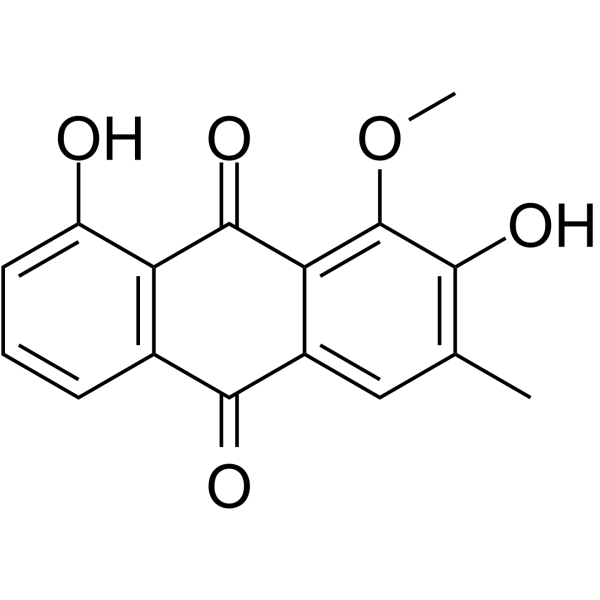
HY-N2098
ObtusifolinNF-kB Inhibitor -

HY-N0261R
Aurantio-obtusin (Standard)Vasodilator, Anti-inflammatory agent -
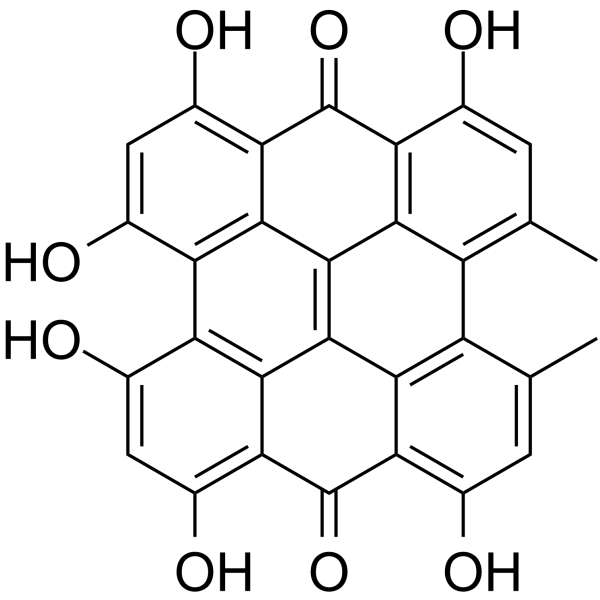
HY-N0453
HypericinPKC Inhibitor -
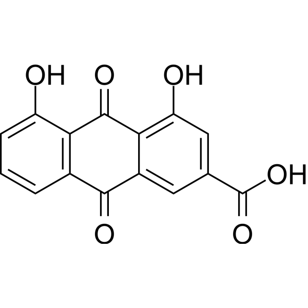
HY-N0105
RheinAntioxidant -
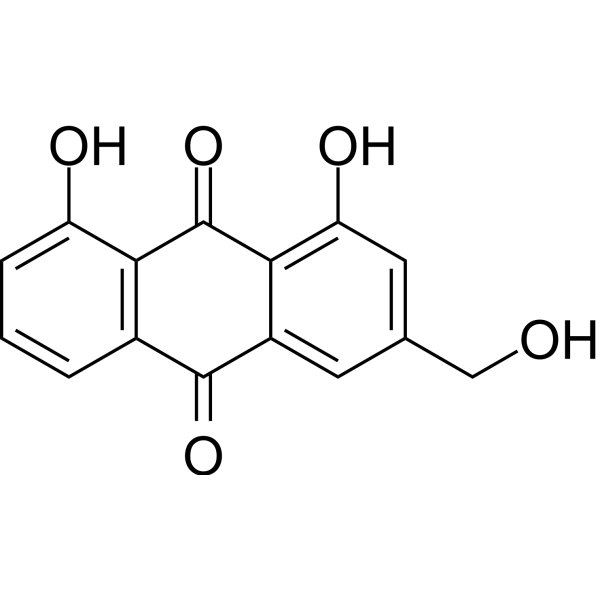
HY-N0189
Aloe emodinmTORC2 Inhibitor -
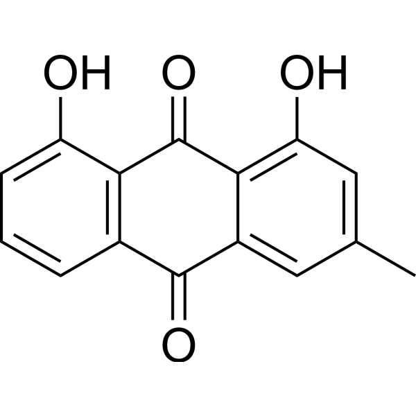
HY-13595
ChrysophanolEGFR Inhibitor -
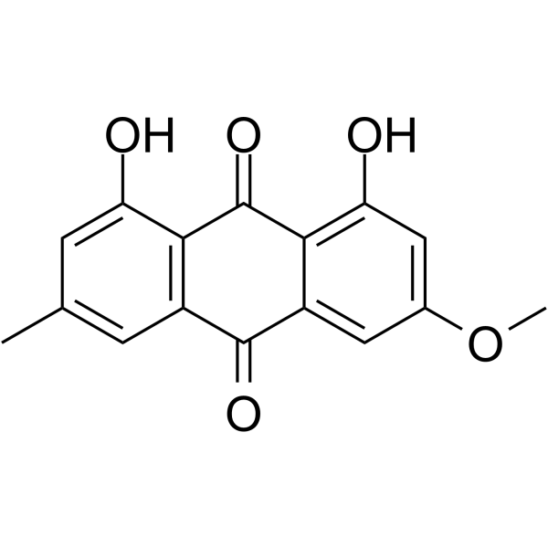
HY-N0108
PhyscionAnthraquinone -
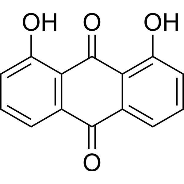
HY-B0923
DanthronAMPK Activator -
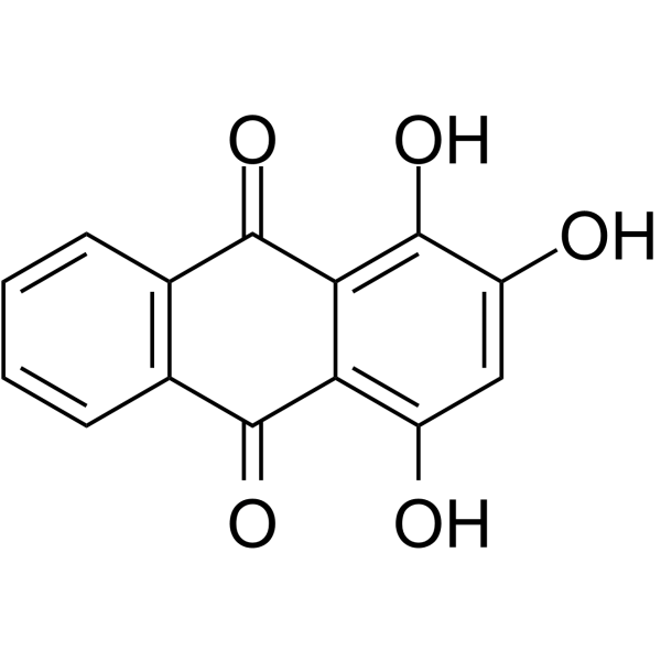
HY-N0571
PurpurinAntidepressant-like Drug
Publications Citing Use of MedChemExpress (MCE) Aurantio-obtusin
More

Biological Activity
Description
In Vitro
Aurantio-obtusin (6.25-100 μM; 24 h) significantly decreases the production of NO and PGE2, and inhibits the expression of IL-6, TNF-α, COX-2 protein in RAW264.7 cells treated with LPS (0.2µg/mL)[2].
Aurantio-obtusin (6.25-100 μM; 12 h) inhibits the activation of NF-κ B by inhibiting the phosphorylation of i-κB and IKK in RAW264.7 cells treated with LPS (0.2µg/mL)[1].
Aurantioobtusin (1-10000 nM) induces small resistance vasodilation in rat MA in a concentration-dependent manner[1].
MedChemExpress (MCE) has not independently confirmed the accuracy of these methods. They are for reference only. Further protocols information, click here.
In Vivo
Aurantio-obtusin (5-15 mg/kg; Oral administration; A single dose) inhibits fatty acid synthesis, promotes FAO and activates AMPK signaling and autophagy in HFSW-induced mice [3].
MedChemExpress (MCE) has not independently confirmed the accuracy of these methods. They are for reference only.
-
Animal Model:C57BL/6J mice model[3]
-
Dosage:5 mg/kg, 10 mg/kg, 15 mg/kg
-
Administration:Oral gavage (p.o.), feeding with HFSW diet for 4 weeks then giving different dose of Aurantio-obtusin for another 4 weeks.
-
Result:Decreased the levels of TG and TC in liver and TG, ALT and AST in serum induced by HSFW.
Reduced the number and size of fat droplets in liver cells.
Significantly increased AMPK phosphorylation in HFSW-induced mice.


Chemical Information
-
CAS No. 67979-25-3
-
Appearance Solid
-
Molecular Weight 330.29
-
Formula C17H14O7
-
Color Yellow to orange
-
SMILES
O=C1C2=C(C=C(C)C(O)=C2OC)C(C3=CC(O)=C(OC)C(O)=C13)=O
-
Structure Classification
-
Shipping
Room temperature in continental US; may vary elsewhere.
-
Storage
4°C, protect from light
* In solvent : -80°C, 6 months; -20°C, 1 month (protect from light)


Publications (1)
-
Journal Impact Factor
-
Most Recent
-
Biochem Genet
Aurantio-obtusin Alleviates Dry Eye Disease by Targeting NF-κB/NLRP3 Signaling in Rodent Models. [Abstract]2024 Apr;62(2):1-14. PMID: 37633872


Solvent & Solubility
In Vitro:
DMSO : 25 mg/mL (75.69 mM; ultrasonic and warming and heat to 60°C; Hygroscopic DMSO has a significant impact on the solubility of product, please use newly opened DMSO)
Please refer to the solubility information to select the appropriate solvent. Once prepared, please aliquot and store the solution to prevent product inactivation from repeated freeze-thaw cycles.
Storage method and period of stock solution: -80°C, 6 months; -20°C, 1 month (protect from light). When stored at -80°C, please use it within 6 months. When stored at -20°C, please use it within 1 month.
Please refer to the solubility information to select the appropriate solvent. Once prepared, please aliquot and store the solution to prevent product inactivation from repeated freeze-thaw cycles.
Storage method and period of stock solution: -80°C, 6 months; -20°C, 1 month (protect from light). When stored at -80°C, please use it within 6 months. When stored at -20°C, please use it within 1 month.
Concentration (start) × Volume (start) = Concentration (final) × Volume (final)
In Vivo:
Select the appropriate dissolution method based on your experimental animal and administration route.
- For the following dissolution methods, please ensure to first prepare a clear stock solution using an In Vitro approach and then sequentially add co-solvents:
- To ensure reliable experimental results, the clarified stock solution can be appropriately stored based on storage conditions. As for the working solution for In Vivo experiments, it is recommended to prepare freshly and use it on the same day.
- The percentages shown for the solvents indicate their volumetric ratio in the final prepared solution. If precipitation or phase separation occurs during preparation, heat and/or sonication can be used to aid dissolution.
Add each solvent one by one: 10% DMSO 40% PEG300 5% Tween-80 45% Saline
Solubility: ≥ 2.08 mg/mL (6.30 mM); Clear solution
This protocol yields a clear solution of ≥ 2.08 mg/mL (saturation unknown).
Taking 1 mL working solution as an example, add 100 μL DMSO stock solution (20.8 mg/mL) to 400 μL PEG300, and mix evenly; then add 50 μL Tween-80 and mix evenly; then add 450 μL Saline to adjust the volume to 1 mL.
Preparation of Saline: Dissolve 0.9 g sodium chloride in ddH₂O and dilute to 100 mL to obtain a clear Saline solution.
In Vivo Dissolution Calculator
Please enter the basic information of animal experiments:
-
-
-
-
Recommended: Prepare an additional quantity of animals to account for potential losses during experiments.
Please enter your animal formula composition:
-
%DMSO +
Recommended: Keep the proportion of DMSO in working solution below 2% if your animal is weak.
-
%+
-
+%Tween-80 + +
-
%Saline +
The co-solvents required include: DMSO, . All of co-solvents are available by MedChemExpress (MCE). , Tween 80. All of co-solvents are available by MedChemExpress (MCE).
Working solution concentration: 0.22 mg/mL
Method for preparing stock solution: mg drug dissolved in μL DMSO. Stock solution concentration: mg/mL. * In solvent : -80°C, 6 months; -20°C, 1 month (protect from light)
1. Take μL DMSO stock solution;
2. Add μL .
μL , mix evenly;
3. Then add μL Tween 80, mix evenly;
4. Then add μL
Please ensure that the stock solution in the first step is dissolved to a clear state, and add co-solvents in sequence. You can use ultrasonic heating (ultrasonic cleaner, recommended frequency 20-40 kHz), vortexing, etc. to assist dissolution.

Protocols
-
3T3-L1 preadipocyte-to-adipocyte differentiation
3T3-L1 preadipocytes are induced to differentiate after growth arrest using adipogenic media containing insulin, dexamethasone, and IBMX; differentiation is assessed by lipid-droplet accumulation, triglyceride increase, Oil Red O staining, and adipocyte-marker induction such as PPARγ and C/EBPα.
-
Autophagy
Autophagy is a process in which eukaryotic cells use lysosomes to degrade their own cytoplasmic proteins and damaged organelles under the regulation of autophagy related gene (Atg). Microtubule-associated proteins light chain 3 (LC3) is recognized as autophagy marker, which transfers from cytoplasmic LC3 (LC3-I) to membrane type (LC3-II). LC3-II/I ratio could be detected by Western Blot and fluorescence microscopy.
-
Research Protocol for Inflammation-related Diseases
The NLRP3 inflammasome is a cytosolic innate immune signaling platform that integrates priming signals and danger-signal activation to promote caspase-1 activation, maturation of IL-1β and IL-18, and gasdermin D-mediated pyroptotic cell death. The core experimental logic is to determine whether inflammatory disease phenotypes are driven by increased NLRP3 expression, ASC-containing inflammasome assembly, caspase-1 cleavage, GSDMD cleavage, and extracellular release of IL-1β/IL-18 rather than by nonspecific cell injury alone. The pathway is strongly linked to inflammation-related disease phenotypes because monosodium urate crystals activate NALP3/NLRP3 inflammasome signaling in gout-like crystal inflammation, cholesterol crystals activate NLRP3 inflammasomes in atherogenesis models, and DSS-induced intestinal inflammation has been reported to involve NLRP3 inflammasome activity. However, experimental colitis studies also show context-dependent protective effects of NLRP3 inflammasome co
-
Lysosome and acidic-vesicle live-cell staining
Lysosome and acidic-vesicle live-cell staining detects acidic intracellular compartments by using membrane-permeant acidotropic probes that accumulate in low-pH vesicles, including lysosomes, late endosomes, autolysosomes, and acidic phagosomes. LysoTracker staining is commonly used as an intensity-based readout of acidic lysosomal compartment abundance or enlargement, while acridine orange produces green fluorescence in less concentrated compartments and red fluorescence after concentration-dependent accumulation in acidic vesicular organelles. Loss or reduction of acridine-orange red signal can be used as a readout of lysosomal membrane permeabilization or reduced acidic-vesicle integrity. This protocol is designed for live cultured cells and can be adapted for fluorescence microscopy, high-content imaging, plate-reader readout, or flow cytometry when the selected literature supports the readout. Because these dyes report acidotropic accumulation rather than lysosome identity alone,
-
Macroautophagy Solutions
Macroautophagy is a conserved lysosome-dependent degradation pathway in which cytoplasmic material is sequestered into double-membrane autophagosomes and delivered to lysosomes for degradation and recycling. The pathway supports cellular homeostasis during nutrient limitation, organelle stress, protein-aggregate accumulation, infection, differentiation, and tissue remodeling by coupling cargo sequestration, autophagosome maturation, lysosomal fusion, and degradation of cargo-derived macromolecules. The core molecular sequence includes initiation by nutrient- and stress-regulated autophagy machinery, autophagosome nucleation, LC3/ATG8-family conjugation to autophagosomal membranes, cargo selection through receptors such as SQSTM1/p62, autophagosome-lysosome fusion, and lysosomal degradation. LC3 was identified as a mammalian homolog of yeast Atg8 that localizes to autophagosomal membranes after processing, and p62/SQSTM1 was shown to connect ubiquitinated cargo with autophagic degradati
-
Liver Histomorphometry
Liver histomorphometry is a quantitative histological approach used to measure structural alterations in hepatic tissue, including parenchymal loss, steatosis, fibrosis, and vascular remodeling, by combining stained tissue section analysis with stereological or computerized image-based measurements. Classical morphometric frameworks quantify volume fractions of liver compartments and fibrotic regions using systematic sampling and image analysis, enabling objective comparison of pathological changes across experimental groups. These approaches are widely applied in liver cirrhosis and fibrosis studies to reduce subjectivity in histological scoring and improve reproducibility of tissue evaluation. Recent methodological advances integrate automated image analysis and radiomics-based extraction of histological features from standard liver stains (e. g. , H&E and fibrotic stains), enabling quantitative correlation between morphometric features and fibrosis stages in non-alcoholic fatty live
-
Lipid Droplets: Oil Red O/Sudan Dye Lipid Staining
Lipid droplets are intracellular organelles with a neutral-lipid core that stores triacylglycerols and sterol esters, and Oil Red O or Sudan dyes detect these hydrophobic lipid deposits by partitioning into retained lipids in fresh or frozen specimens. Oil Red O stains neutral triglycerides and lipids in frozen tissue sections or air-dried cytologic preparations, while Sudan Black B has also been used as a histochemical fat stain for lipid-rich tissue structures.


Purity & Documentation
-
Data Sheet (278 KB)
-
SDS (393 KB)
- English - EN (393 KB)
- Français - FR (393 KB)
- Deutsch - DE (393 KB)
- Norwegian - NO (393 KB)
- Español - ES (393 KB)
- Swedish - SV (393 KB)
- Italian - IT (393 KB)
- Korean - KR (393 KB)
- Portuguese - PT (393 KB)
-
Handling Instructions (2659 KB)
References
[1]. Li, Shuzhen, et al. Aurantio-obtusin relaxes systemic arteries through endothelial PI3K/AKT/eNOS-dependent signaling pathway in rats. Journal of pharmacological sciences vol. 128,3 (2015): 108-15. [Content Brief]
[2]. Hou Jingyi, et al. Anti-Inflammatory Effects of Aurantio-Obtusin from Seed of Cassia obtusifolia L. through Modulation of the NF-κB Pathway. Molecules (Basel, Switzerland) vol. 23,12 3093. 27 Nov. 2018. [Content Brief]
[3]. Zhou Fei, et al. Aurantio-Obtusin Attenuates Non-Alcoholic Fatty Liver Disease Through AMPK-Mediated Autophagy and Fatty Acid Oxidation Pathways. Frontiers in pharmacology vol. 12 826628. 11 Jan. 2022. [Content Brief]


Complete Stock Solution Preparation Table
Please refer to the solubility information to select the appropriate solvent. Once prepared, please aliquot and store the solution to prevent product inactivation from repeated freeze-thaw cycles.
Storage method and period of stock solution: -80°C, 6 months; -20°C, 1 month (protect from light). When stored at -80°C, please use it within 6 months. When stored at -20°C, please use it within 1 month.
| Optional Solvent | Concentration Solvent Mass | 1 mg | 5 mg | 10 mg | 25 mg |
|---|---|---|---|---|---|
| DMSO | 1 mM | 3.0276 mL | 15.1382 mL | 30.2764 mL | 75.6911 mL |
| 5 mM | 0.6055 mL | 3.0276 mL | 6.0553 mL | 15.1382 mL | |
| 10 mM | 0.3028 mL | 1.5138 mL | 3.0276 mL | 7.5691 mL | |
| 15 mM | 0.2018 mL | 1.0092 mL | 2.0184 mL | 5.0461 mL | |
| 20 mM | 0.1514 mL | 0.7569 mL | 1.5138 mL | 3.7846 mL | |
| 25 mM | 0.1211 mL | 0.6055 mL | 1.2111 mL | 3.0276 mL | |
| 30 mM | 0.1009 mL | 0.5046 mL | 1.0092 mL | 2.5230 mL | |
| 40 mM | 0.0757 mL | 0.3785 mL | 0.7569 mL | 1.8923 mL | |
| 50 mM | 0.0606 mL | 0.3028 mL | 0.6055 mL | 1.5138 mL | |
| 60 mM | 0.0505 mL | 0.2523 mL | 0.5046 mL | 1.2615 mL |


Aurantio-obtusin Related Classifications
HY-N0261 Related Classifications
 Powered by Bioz