



Bexotegrast hydrochloride
Based on 4 publication(s) in Google Scholar
Bexotegrast hydrochloride (PLN-74809 hydrochloride) is a small molecule dual selective inhibitor with activity targeting αVβ1 and αVβ6. Bexotegrast hydrochloride is used for idiopathic pulmonary fibrosis (IPF) and primary sclerosing cholangitis (PSC). Bexotegrast hydrochloride inhibits the activation of TGF-β1 by blocking the function of these integrins, thereby preventing the growth of fibrous tissue in the lungs and bile ducts.
For research use only. We do not sell to patients.
- CAS No.: 2775365-40-5
- Formula: C27H38Cl2N6O3
- Molecular Weight:565.54
-
Storage:
Please store the product under the recommended conditions in the Certificate of Analysis.


To place orders, for customer services and technical support, please contact: MedChemExpress USA
Tel: 609-228-6898 E-mail: [email protected] [email protected]


-
 Biological Activity
-
Chemical Information
- Protocols
- Purity & Documentation
- References
-
 Help & FAQs


Publications Citing Use of MedChemExpress (MCE) Bexotegrast hydrochloride
More Customer Validation & Images

-
Cell Imaging/Staining
-
RT-PCR
-
IF


Biological Activity
Description


Chemical Information
-
CAS No. 2775365-40-5
-
Molecular Weight 565.54
-
Formula C27H38Cl2N6O3
-
SMILES
COCCN(CCCCC(N1)=CC=C2C1=NCCC2)CC[C@@H](C(O)=O)NC3=NC=NC4=C3C=CC=C4.Cl.Cl
-
Synonyms
PLN-74809 hydrochloride
-
Shipping
Room temperature in continental US; may vary elsewhere.
-
Storage
Please store the product under the recommended conditions in the Certificate of Analysis.


Publications (4)
-
Journal Impact Factor
-
Most Recent
-
Science
2026 Feb 5;391(6785):eadz4075. PMID: 41643022 -
Phytomedicine
Jiawei yanghe decoction alleviates osteoporotic osteoarthritis by promoting MSC osteogenic differentiation and homing via ITGB6/TGF-β/CXCR4 pathway. [Abstract]2025 Nov:147:157203. PMID: 40929885 -
-
bioRxiv
Bidirectional fibrogenic cross-talk revealed in a human iPSC-derived epithelial-mesenchymal co-culture model of pulmonary fibrosis. [Abstract]2026 Jan 31:2026.01.30.702837. PMID: 41659605
Bexotegrast hydrochloride purchased from MedChemExpress. Usage Cited in: bioRxiv. 2026 Jan 31:2026.01.30.702837. [Abstract]
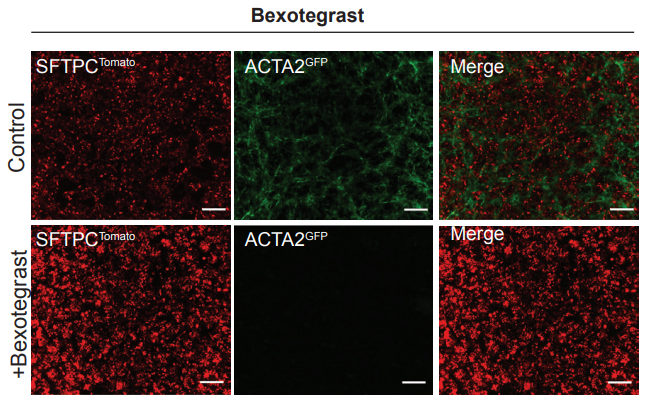
SFTPCtdTomato and ACTA2GFP fluorescence in live co-cultures of SFTPCI73T mutant iAT2s and iLM treated with 2 μM Bexotegrast for 1 week, as compared to vehicle control (treatment starting on the first day of co-culture). Scale bars = 400 μm.
Bexotegrast hydrochloride purchased from MedChemExpress. Usage Cited in: bioRxiv. 2026 Jan 31:2026.01.30.702837. [Abstract]
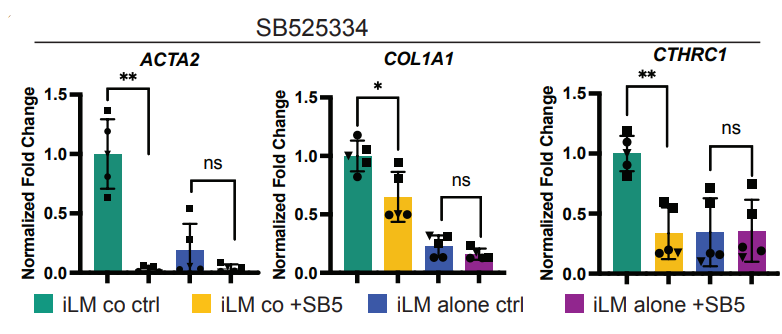
RT-qPCR analysis showing fold change expression (normalized to iLM co-cultured control) of fibrotic markers in EPCAM- sorted cells from co-cultures of iLM with SFTPCI73T mutant iAT2s or iLM cultured alone. N = 5 for SB525334 (1 μM; 1 week), N = 6 for Bexotegrast (2 μM; 1 week), symbol shapes indicate experimental replicates, i.e. multiple wells of the same experiment.
Bexotegrast hydrochloride purchased from MedChemExpress. Usage Cited in: bioRxiv. 2026 Jan 31:2026.01.30.702837. [Abstract]
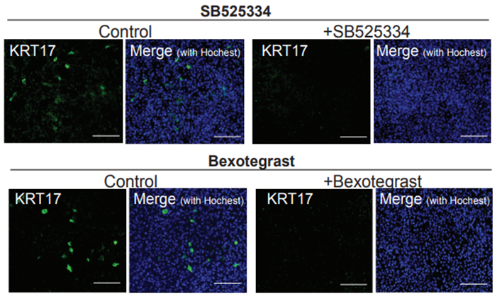
Immunofluorescence staining for KRT17 protein in co-cultures of SFTPCI73T mutant iAT2s and iLM. The results showed that treatment of co-cultures of SFTPCI73T mutant iAT2s and iLM for 7 days with either SB525334 (1 μM; 1 week) or Bexotegrast (2 μM; 1 week), compared to vehicle-treated controls, abolished KRT17 protein expression in co-cultured SFTPCI73T mutant iAT2s. Scale bars = 100 μm. Hoechst = nuclei.

Protocols
-
Fibrosis/Collagen Morphometry
Fibrosis and collagen morphometry is based on the quantitative visualization of fibrillar collagen deposition in tissue sections using histochemical stains such as Sirius Red (Picrosirius Red) or Masson's trichrome, followed by image-based or polarization-enhanced analysis to estimate collagen proportional area as a surrogate of extracellular matrix accumulation during fibrotic remodeling. Sirius Red combined with polarized light microscopy enhances detection of collagen fibers due to birefringence properties, enabling more specific visualization of collagen type I and III fibrils compared to conventional bright-field histology, while whole-section or region-restricted digital morphometry reduces field-selection bias in fibrosis assessment. Alternative quantitative approaches include second harmonic generation (SHG) and two-photon excited fluorescence microscopy, which enable label-free detection of fibrillar collagen and have been validated against histological staining and biochemica
-
Collagen: Sirius Red Staining
Sirius Red or picrosirius red staining is a histochemical method for visualizing collagen-rich extracellular matrix in tissue sections, and collagen fibers are detected as red-stained structures under bright-field microscopy with enhanced birefringence under polarized light. Picrosirius red is useful for assessing total collagen organization, distribution, and fibrosis burden, but polarized color should not be interpreted as a definitive collagen type I versus type III readout because color is affected by fiber orientation, thickness, and packing.
-
Connective Tissue: Masson's Trichrome/Collagen Trichrome Staining
Masson’s Trichrome (collagen/trichrome staining) is a histological technique that differentially stains tissue compartments using sequential acidic dyes to distinguish collagen from muscle and cytoplasmic components based on dye affinity and tissue permeability differences, enabling visualization of fibrosis and connective tissue architecture in histological sections. The classical formulation typically uses Weigert's iron hematoxylin for nuclear staining, Biebrich scarlet-acid fuchsin for cytoplasm and muscle, and aniline blue (or light green variants) for collagen, producing a characteristic blue/green collagen signal contrasted against red cytoplasm and dark nuclei. The staining principle relies on selective displacement of smaller dye molecules by larger anionic dyes in collagen-rich regions under controlled acidified conditions, which enhances collagen-specific dye retention. This property makes the method widely used for fibrosis assessment in organs such as heart, liver, lung, a


Purity & Documentation
References
[1]. Anindya Roy, et al. De novo design of highly selective miniprotein inhibitors of integrins αvβ6 and αvβ8. Nat Commun. 2023 Sep 13;14(1):5660. [Content Brief]
[2]. Bexotegrast in Patients with Idiopathic Pulmonary Fibrosis: The INTEGRIS-IPF Clinical Trial [Content Brief]
[3]. Martin L Decaris, et al. Dual inhibition of αvβ6 and αvβ1 reduces fibrogenesis in lung tissue explants from patients with IPF. Respir Res. 2021 Oct 19;22(1):265. [Content Brief]

Calculators
Concentration (start) × Volume (start) = Concentration (final) × Volume (final)


Bexotegrast hydrochloride Related Classifications
HY-137561A Related Classifications
 Powered by Bioz