
BMAP-28
BMAP-28 is an antibiotic peptide and an inducer of the mitochondrial permeability transition pore. BMAP-28 induces cell death through opening of the mitochondrial permeability transition pore. BMAP-28 can be used in study of microbial infections and cancer.
For research use only. We do not sell to patients.
- CAS No.: 184870-31-3
- Formula: C145H250N44O29
- Molecular Weight:3073.81
-
Storage:
Please store the product under the recommended conditions in the Certificate of Analysis.
All Antibiotic Isoforms
More
Biological Activity
Description
In Vitro
BMAP-28 (3 µM; 10 min) attenuates mitochondrial calcein fluorescence in U937 cells[1].
BMAP-28 causes depolarization of the inner mitochondrial membrane in single cells and in isolated mitochondria[1].
MedChemExpress (MCE) has not independently confirmed the accuracy of these methods. They are for reference only. Further protocols information, click here.
-
Cell Line:U937 cells
-
Concentration:3 µM
-
Incubation Time:10 min
-
Result:Caused a remarkable attenuation of the calcein fluorescence.
Chemical Information
-
CAS No. 184870-31-3
-
Molecular Weight 3073.81
-
Formula C145H250N44O29
-
Sequence
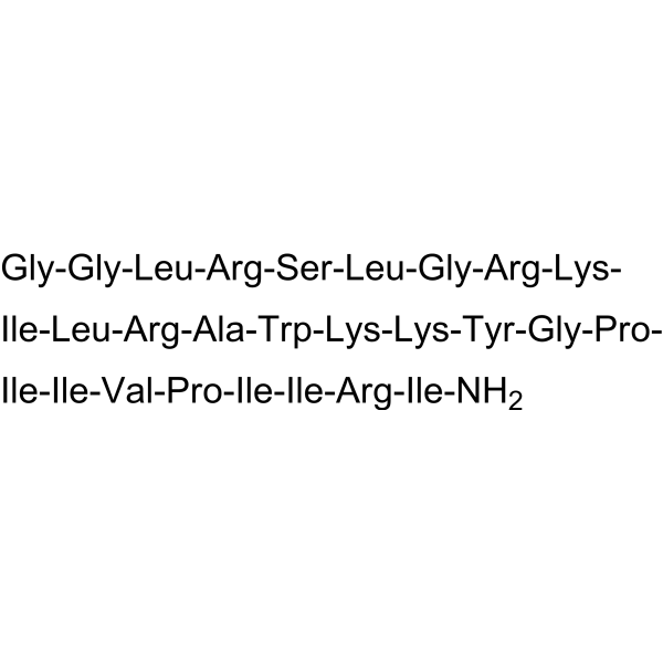
Gly-Gly-Leu-Arg-Ser-Leu-Gly-Arg-Lys-Ile-Leu-Arg-Ala-Trp-Lys-Lys-Tyr-Gly-Pro-Ile-Ile-Val-Pro-Ile-Ile-Arg-Ile-NH2
-
Sequence Shortening
GGLRSLGRKILRAWKKYGPIIVPIIRI-NH2
-
Shipping
Room temperature in continental US; may vary elsewhere.
-
Storage
Please store the product under the recommended conditions in the Certificate of Analysis.
Protocols
-
Cell Cytotoxicity Assay
Cytotoxicity assays are usually based on the assessment of cell membrane damage, which can also be indirectly detected by measuring cell viability. Detection methods include MTT assay, CKK-8 assay, LDH assay and ATP assay, etc.
-
Mammalian live/dead viability and cytotoxicity staining
Live/dead viability and cytotoxicity staining assays are based on the simultaneous detection of intracellular esterase activity in metabolically active (viable) cells and membrane integrity loss in non-viable cells. In commonly used dual-staining approaches, membrane-permeant fluorogenic substrates are converted by intracellular esterases into fluorescent products in live cells, while impermeant DNA-binding dyes selectively enter cells with compromised plasma membranes and label nucleic acids in dead or dying cells, enabling discrimination between viable and non-viable populations by fluorescence microscopy or flow cytometry.
-
Transepithelial/transendothelial electrical resistance assay
TEER measures electrical resistance across epithelial or endothelial monolayers cultured on permeable supports, and the readout reflects ionic conductance through the cell barrier, especially the paracellular pathway regulated by junctional integrity. TEER can be measured without destroying the monolayer and is commonly used before or during transport, permeability, barrier-disruption, and barrier-maturation experiments. TEER values are influenced by biological maturation and technical conditions; reported factors include temperature, medium formulation, passage number, electrode geometry, membrane properties, and junctional length during early monolayer maturation. Therefore, TEER should be interpreted with blank-insert subtraction, area normalization, repeated readings, and, when possible, orthogonal barrier readouts such as FITC-dextran flux or tight-junction staining.
-
Apoptosis Solutions
Apoptosis is a regulated, generally non-lytic cell-death pathway that removes unwanted, damaged, infected, or abnormal cells through coordinated morphological changes, caspase activation, DNA fragmentation, and membrane remodeling. The intrinsic apoptosis pathway is controlled mainly by mitochondrial outer membrane permeabilization, BCL-2 family proteins, cytochrome c release, apoptosome formation, caspase-9 activation, and downstream executioner caspase-3/7 activation. The extrinsic apoptosis pathway is initiated by death receptors such as Fas, TNFR, and TRAIL receptors, which recruit adaptor proteins and activate caspase-8 before engaging executioner caspases or mitochondrial amplification through BID cleavage. Apoptosis is linked to many phenotypes, including cancer cell killing, tissue homeostasis, immune regulation, neurodegeneration, infection response, and treatment-induced cytotoxicity; unresolved questions include how apoptosis interacts with necroptosis, pyroptosis, ferroptos
Purity & Documentation
References
Calculators
Concentration (start) × Volume (start) = Concentration (final) × Volume (final)

