



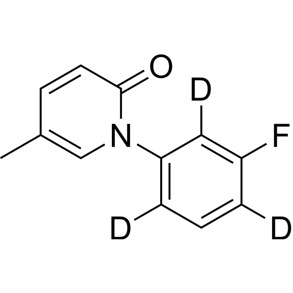
Fluorofenidone-d3
Fluorofenidone-d3 (AKF-PD-d3) is deuterium labeled Fluorofenidone (AKF-PD) (HY-121246). Fluorofenidone is an orally active compound with anti-fibrotic, antioxidant, and anti-inflammatory pharmacological effects. Fluorofenidone downregulates the expression of ACSL4, upregulates GPX4 expression and inhibits the NF-κB signaling pathway to alleviate inflammation and fibrosis. Fluorofenidone ameliorates cholestasis and fibrosis by inhibiting hepatic Erk/-Egr-1 signaling and Tgfβ1/Smad pathway in mice. Fluorofenidone demonstrates protective effects against chronic lung injury in mice. Fluorofenidone can be used for the study of chronic obstructive pulmonary disease (COPD), pulmonary interstitial fibrosis (PIF) and non-small cell lung cancer (NSCLC).
Nos produits utilisent uniquement pour la recherche. Nous ne vendons pas aux patients.
- Formule: C12H7D3FNO
- Masse moléculaire:206.23
-
Stockage:
Please store the product under the recommended conditions in the Certificate of Analysis.

To place orders, for customer services and technical support, please contact: MedChemExpress USA
Tel: 609-228-6898 E-mail: [email protected] [email protected]


-
 Activité biologique
-
Chemical Information
- Protocole
- Pureté et documentation
- Références
-
 Help & FAQs




Activité biologique
Description
IC50 & Target
|
ACSL4 |
In Vitro
Fluorofenidone (24 h) partially reverses the inhibition of cell viability and reduces the levels of inflammatory factors IL-1β, IL-6, and TNF-α in the culture supernatant induced by cigarette smoke extract (CSE) in BEAS-2B cells[1].
Fluorofenidone (24 h) partially reverses the increase in MDA level and decrease in GSH level in BEAS-2B cell supernatant caused by CSE[1].
Fluorofenidone (24 h) improves CSE-induced mitochondrial damage, downregulates the expression of ACSL4 and upregulates the expression of GPX4 in BEAS-2B cells[1].
Fluorofenidone (200-800 μg/mL, 24 h) inhibits proliferation of A549 and H1299 cells in a dose-dependent manner, with EC50 of 1030 μg/mL (A549) and 1118 μg/mL (H1299)[3].
Fluorofenidone (400-800 μg/mL, 3 h) significantly reduces the percentage of EdU-positive proliferating cells[3].
Fluorofenidone (200-800 μg/mL, 12-24 h) dose- and time-dependently inhibits cell migration and reduces the number of invasive cells in A549 and H1299 cells[3].
Fluorofenidone (200-800 μg/mL) upregulates the expression of epithelial marker E-cadherin and downregulates mesenchymal markers MMP9, vimentin, and SNAIL in A549 and H1299 cells[3].
Fluorofenidone (200-800 μg/mL) suppresses phosphorylation of JNK, ERK, P38 (MAPK pathway) and p-PI3K, p-AKT, p-mTOR (PI3K/AKT/mTOR pathway) in A549 and H1299 cells[3].
MedChemExpress (MCE) has not independently confirmed the accuracy of these methods. They are for reference only.
In Vivo
Fluorofenidone (0.45% in diet, p.o., 14 days) markedly alleviates hepatic inflammation in 3,5-diethoxycarbonyl-1,4-dihydroxychollidine (DDC)-induced cholestasis model of mice[2].
Fluorofenidone (400 mg/kg, p.o., daily, 3 weeks) combined with Cisplatin (HY-17394) significantly reduces the size, weight and volume of subcutaneous tumors in C57 mice bearing Lewis lung cancer cells[3].
MedChemExpress (MCE) has not independently confirmed the accuracy of these methods. They are for reference only.
-
Animal Model:C57BL/6 J mice (4-week-old, 18-20 g) were used to construct the chronic obstructive pulmonary disease (COPD) model[1]
-
Dosage:5 mg/kg
-
Administration:i.p., once daily, 7 weeks
-
Result:Alleviated CS/LPS-induced lung tissue morphological damage, including reducing interstitial inflammatory cell infiltration, alleviating lung overexpansion, thinning alveolar septa, and lowering lung injury score.
Reduced the levels of inflammatory factors IL-1β, IL-6, and TNF-α in bronchoalveolar lavage fluid (BALF) and serum.
Inhibited lung collagen deposition, decreased lung fibrosis score, downregulated mRNA and protein expressions of fibrosis markers α-SMA, FAP, and COL3A1 in lung tissue, and reduced lung hydroxyproline content.
Downregulated ACSL4 expression and upregulated GPX4 expression in lung tissue, reversed CS/LPS-induced increase in MDA level and decrease in GSH level in lung tissue and serum.
-
Animal Model:3,5-diethoxycarbonyl-1,4-dihydroxychollidine (DDC)-induced cholestasis model of 7-week-old male C57BL/6J mice[2]
-
Dosage:0.45% in diet
-
Administration:p.o., 14 days
-
Result:Reduced serum ALT, AST, ALP, and TBA levels, as well as hepatic BAs levels, and decreased the liver/body weight ratio.
Reduced mRNA levels of hepatic inflammatory cytokines and chemokines, lowered serum levels of these inflammatory factors, and decreased F4/80 expression to reduce macrophage infiltration.
Downregulated mRNA levels of profibrogenic markers, inhibited phosphorylation of Smad2/3, and reduced α-SMA protein level, thus mitigating hepatic fibrosis.
Downregulated expression of Cyp7a1 and Cyp27a1, upregulated expression of hepatic detoxification enzymes, and facilitated nuclear translocation.
Suppressed phosphorylation of Erk1/2, reduced Egr-1 protein level, and blocked the Tgfβ1/Smad pathway.
-
Animal Model:C57 mice (5-week-old, 16-18 g) were used to construct the Lewis lung cancer subcutaneous xenograft model by subcutaneously injecting 5 × 10⁵ Lewis lung cancer cells (C7205) into the axillary region[3]
-
Dosage:400 mg/kg combined with Cisplatin (5 mg/kg, i.p., weekly, 3 weeks)
-
Administration:p.o., daily, 3 weeks
-
Result:Reduced the size, weight and volume of subcutaneous tumors.
Downregulated the expression of mesenchymal marker N-cadherin.
Application
1. This compound can be used as a tracer
2. This compound can be used as an internal standard for quantitative analysis by NMR, GC-MS, or LC-MS.


Chemical Information
-
Unlabeled CAS 848353-85-5
-
Masse moléculaire 206.23
-
Formule C12H7D3FNO
-
SMILES
O=C1C=CC(C)=CN1C2=C([2H])C=C([2H])C(F)=C2[2H]
-
Synonyms
AKF-PD-d3
-
Livraison
Room temperature in continental US; may vary elsewhere.
-
Stockage
Please store the product under the recommended conditions in the Certificate of Analysis.


Pureté et documentation
Références
[1]. Wu Y, et al. Fluorofenidone alleviates cigarette smoke exposure-induced chronic lung injury by targeting ferroptosis. Sci Rep. 2024 Dec 30;14(1):32149. [Content Brief]
[2]. Wang H,et al. Fluorofenidone ameliorates cholestasis and fibrosis by inhibiting hepatic Erk/-Egr-1 signaling and Tgfβ1/Smad pathway in mice. Biochim Biophys Acta Mol Basis Dis. 2022 Dec 1;1868(12):166556. [Content Brief]
[3]. Wang S, et al. Fluorofenidone enhances cisplatin efficacy in non-small cell lung cancer: a novel approach to inhibiting cancer progression. Transl Lung Cancer Res. 2024 Nov 30;13(11):3175-3188. [Content Brief]

Calculators
Concentration (start) × Volume (start) = Concentration (final) × Volume (final)


Fluorofenidone-d3 Related Classifications
HY-121246S Related Classifications
 Powered by Bioz