



Gambogic amide
Based on 1 publication(s) in Google Scholar
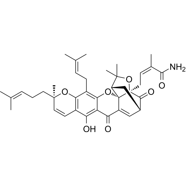
Gambogic amide is a potent and selective agonist of TrkA and also induces its tyrosine phosphorylation and activation of downstream signaling, including Akt and MAPK. Gambogic amide specifically interacts with the cytoplasmic juxtamembrane domain of the TrkA receptor and triggers its dimerization, leading to activation. Gambogic amide has neuroprotective activity preventing glutamate-induced neuronal cell death. Gambogic amide has improved efficacy in a transient middle cerebral artery occlusion model of stroke and could be used to study neurodegenerative diseases and stroke.
Nos produits utilisent uniquement pour la recherche. Nous ne vendons pas aux patients.
- Pureté : 95.76%
- CAS No.: 286935-60-2
- Formule: C38H45NO7
- Masse moléculaire:627.77
-
Stockage:Powder -20°C, 3 years , 4°C, 2 years ; In solvent -80°C, 6 months , -20°C, 1 month

To place orders, for customer services and technical support, please contact: MedChemExpress USA
Tel: 609-228-6898 E-mail: [email protected] [email protected]


-
 Activité biologique
-
Chemical Information
-
Solvant et solubilité
- Protocole
- Pureté et documentation
- Références
-
 Help & FAQs



Publications Citing Use of MedChemExpress (MCE) Gambogic amide
More

Activité biologique
Description
In Vitro
Gambogic amide (0.5 μM; 30 min) elicits TrkA tyrosine phosphorylation in hippocampal neurons[1].
MedChemExpress (MCE) has not independently confirmed the accuracy of these methods. They are for reference only. Further protocols information, click here.
-
Cell Line:Hippocampal neurons
-
Concentration:500 nM
-
Incubation Time:30 min
-
Result:Triggered TrkA Y490 phosphorylation.
In Vivo
MedChemExpress (MCE) has not independently confirmed the accuracy of these methods. They are for reference only.
-
Animal Model:MCAO mice model[1]
-
Dosage:2 mg/kg
-
Administration:s.c.; 5 min before the onset of reperfusion, after 2 h of MCAO followed by reperfusion; followed by 25 mg/kg Kainic acid (KA)
-
Result:Diminished Kainic acid-triggered hippocampal neuronal cell death.
Reduces infarct volume in MCAO rat brain.


Chemical Information
-
CAS No. 286935-60-2
-
Appearance Solid
-
Masse moléculaire 627.77
-
Formule C38H45NO7
-
Color Light yellow to yellow
-
SMILES
NC(/C(C)=C\C[C@@]12[C@]34C(C(C5=C(O)C(C=C[C@](CC/C=C(C)/C)(C)O6)=C6C(C/C=C(C)\C)=C5O3)=O)=C[C@H](C[C@H]4C(C)(C)O2)C1=O)=O
-
Livraison
Room temperature in continental US; may vary elsewhere.
-
Stockage
Powder -20°C 3 years 4°C 2 years In solvent -80°C 6 months -20°C 1 month


Publications (1)
-
Journal Impact Factor
-
Most Recent


Solvant et solubilité
In Vitro:
DMSO : 68 mg/mL (108.32 mM; Need ultrasonic; Hygroscopic DMSO has a significant impact on the solubility of product, please use newly opened DMSO)
Please refer to the solubility information to select the appropriate solvent. Once prepared, please aliquot and store the solution to prevent product inactivation from repeated freeze-thaw cycles.
Storage method and period of stock solution: -80°C, 6 months; -20°C, 1 month. When stored at -80°C, please use it within 6 months. When stored at -20°C, please use it within 1 month.
Please refer to the solubility information to select the appropriate solvent. Once prepared, please aliquot and store the solution to prevent product inactivation from repeated freeze-thaw cycles.
Storage method and period of stock solution: -80°C, 6 months; -20°C, 1 month. When stored at -80°C, please use it within 6 months. When stored at -20°C, please use it within 1 month.
Concentration (start) × Volume (start) = Concentration (final) × Volume (final)

Protocole
-
Kinase activity and phosphorylation assays
Kinase activity assays measure the ability of kinases to transfer phosphate groups from ATP to specific substrates, while phosphorylation assays detect the presence and levels of phosphorylated proteins. Common methods include radiolabeled ATP incorporation (e. g. ,), ADP release detection via bioluminescence (e. g. ,[3]), enzyme-linked immunosorbent assays (ELISA) for phospho-specific epitopes (e. g. ,[6]), and microtiter-based formats for high-throughput screening (e. g. ,[8]). The ADP-Glo assay quantifies kinase activity by measuring ADP produced during phosphorylation using a luciferase-based system. Radiometric assays involve autoradiography or scintillation counting after incorporation of 32P-labeled ATP into substrate proteins. ELISA-based approaches rely on phospho-specific antibodies to detect activated kinases in cell lysates or purified samples.
-
Western Blot
Western blotting (WB) is a commonly used experimental method in molecular biology, biochemistry, and immunogenetics for identifying and quantifying target proteins. It combines gel electrophoresis with immunoassay, enabling researchers to analyze protein expression, post-translational modifications, and molecular weight.
-
Cell Cytotoxicity Assay
Cytotoxicity assays are usually based on the assessment of cell membrane damage, which can also be indirectly detected by measuring cell viability. Detection methods include MTT assay, CKK-8 assay, LDH assay and ATP assay, etc.
-
Mammalian live/dead viability and cytotoxicity staining
Live/dead viability and cytotoxicity staining assays are based on the simultaneous detection of intracellular esterase activity in metabolically active (viable) cells and membrane integrity loss in non-viable cells. In commonly used dual-staining approaches, membrane-permeant fluorogenic substrates are converted by intracellular esterases into fluorescent products in live cells, while impermeant DNA-binding dyes selectively enter cells with compromised plasma membranes and label nucleic acids in dead or dying cells, enabling discrimination between viable and non-viable populations by fluorescence microscopy or flow cytometry.
-
Apoptosis Solutions
Apoptosis is a regulated, generally non-lytic cell-death pathway that removes unwanted, damaged, infected, or abnormal cells through coordinated morphological changes, caspase activation, DNA fragmentation, and membrane remodeling. The intrinsic apoptosis pathway is controlled mainly by mitochondrial outer membrane permeabilization, BCL-2 family proteins, cytochrome c release, apoptosome formation, caspase-9 activation, and downstream executioner caspase-3/7 activation. The extrinsic apoptosis pathway is initiated by death receptors such as Fas, TNFR, and TRAIL receptors, which recruit adaptor proteins and activate caspase-8 before engaging executioner caspases or mitochondrial amplification through BID cleavage. Apoptosis is linked to many phenotypes, including cancer cell killing, tissue homeostasis, immune regulation, neurodegeneration, infection response, and treatment-induced cytotoxicity; unresolved questions include how apoptosis interacts with necroptosis, pyroptosis, ferroptos
-
Protocol for Kinase activity and phosphorylation assays
Kinase activity assays measure transfer of phosphate from ATP to a protein or peptide substrate, generating phosphorylated substrate, ADP, or incorporated radiolabeled phosphate as the readout; phosphorylation assays measure site-specific phosphorylation in cells or tissues as a proxy for kinase-pathway activation, inhibition, or substrate regulation. Phosphorylation can be detected by phospho-specific Western blot, immunoprecipitation kinase assay, phospho-immunofluorescence, phospho-flow cytometry, luminescent ADP detection, radiolabeled ATP incorporation, or reporter-based pathway assays, and these readouts can be applied to cancer cells, primary neurons, mouse tumors, organoids, inflammatory macrophages, ferroptosis studies, and mitophagy studies when the kinase target is biologically relevant.


Pureté et documentation
-
Fiche technique (274 KB)
-
SDS (251 KB)
- English - EN (251 KB)
- Français - FR (251 KB)
- Deutsch - DE (251 KB)
- Norwegian - NO (251 KB)
- Español - ES (251 KB)
- Swedish - SV (251 KB)
- Italian - IT (251 KB)
- Korean - KR (251 KB)
- Portuguese - PT (251 KB)
-
Instruction de manipulation (2659 KB)
Références


Complete Stock Solution Preparation Table
Please refer to the solubility information to select the appropriate solvent. Once prepared, please aliquot and store the solution to prevent product inactivation from repeated freeze-thaw cycles.
Storage method and period of stock solution: -80°C, 6 months; -20°C, 1 month. When stored at -80°C, please use it within 6 months. When stored at -20°C, please use it within 1 month.
| Optional Solvent | Concentration Solvent Mass | 1 mg | 5 mg | 10 mg | 25 mg |
|---|---|---|---|---|---|
| DMSO | 1 mM | 1.5929 mL | 7.9647 mL | 15.9294 mL | 39.8235 mL |
| 5 mM | 0.3186 mL | 1.5929 mL | 3.1859 mL | 7.9647 mL | |
| 10 mM | 0.1593 mL | 0.7965 mL | 1.5929 mL | 3.9824 mL | |
| 15 mM | 0.1062 mL | 0.5310 mL | 1.0620 mL | 2.6549 mL | |
| 20 mM | 0.0796 mL | 0.3982 mL | 0.7965 mL | 1.9912 mL | |
| 25 mM | 0.0637 mL | 0.3186 mL | 0.6372 mL | 1.5929 mL | |
| 30 mM | 0.0531 mL | 0.2655 mL | 0.5310 mL | 1.3275 mL | |
| 40 mM | 0.0398 mL | 0.1991 mL | 0.3982 mL | 0.9956 mL | |
| 50 mM | 0.0319 mL | 0.1593 mL | 0.3186 mL | 0.7965 mL | |
| 60 mM | 0.0265 mL | 0.1327 mL | 0.2655 mL | 0.6637 mL | |
| 80 mM | 0.0199 mL | 0.0996 mL | 0.1991 mL | 0.4978 mL | |
| 100 mM | 0.0159 mL | 0.0796 mL | 0.1593 mL | 0.3982 mL |


Gambogic amide Related Classifications
HY-121833 Related Classifications
 Powered by Bioz