



IKK2-IN-4

Based on 1 Customer Validation
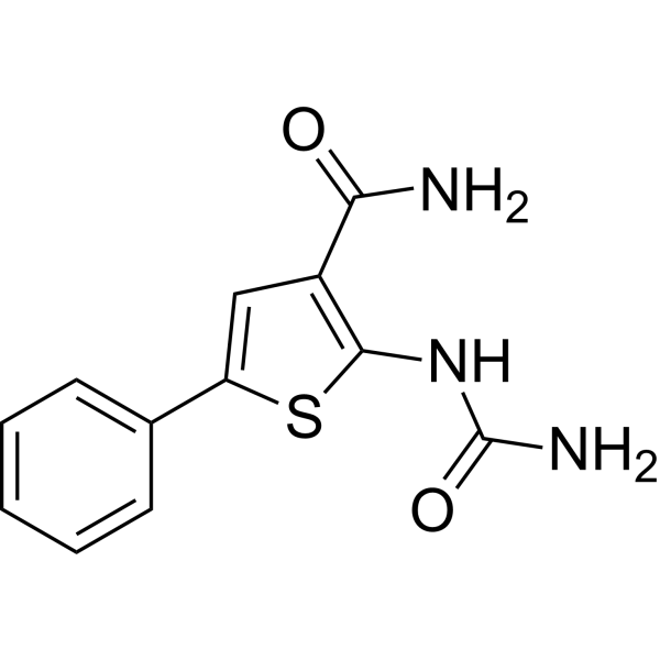
IKK2-IN-4 is an orally active and selective IKK-2 inhibitor (IC50 = 0.025 μM) that also inhibits JNK-1 (IC50 = 1.6 μM). IKK2-IN-4 blocks IκB phosphorylation and NF-κB activation, and inhibits JNK-1 kinase activity. IKK2-IN-4 inhibits TNF-α production in PBMCs. IKK2-IN-4 can be used for research on IKK-2-related diseases, such as autoimmune diseases.
Nos produits utilisent uniquement pour la recherche. Nous ne vendons pas aux patients.
- Pureté : 98.51%
- CAS No.: 354811-10-2
- Formule: C12H11N3O2S
- Masse moléculaire:261.30
-
Stockage:
4°C, protect from light
* In solvent : -80°C, 6 months; -20°C, 1 month (protect from light)

To place orders, for customer services and technical support, please contact: MedChemExpress USA
Tel: 609-228-6898 E-mail: [email protected] [email protected]


-
 Activité biologique
-
Chemical Information
-
Solvant et solubilité
- Protocole
- Pureté et documentation
- Références
-
 Help & FAQs


-
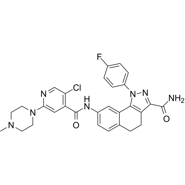
HY-14180
PHA 408IKK-2 Inhibitor -
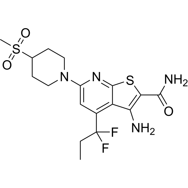
HY-13019
BI605906IKK Inhibitor -
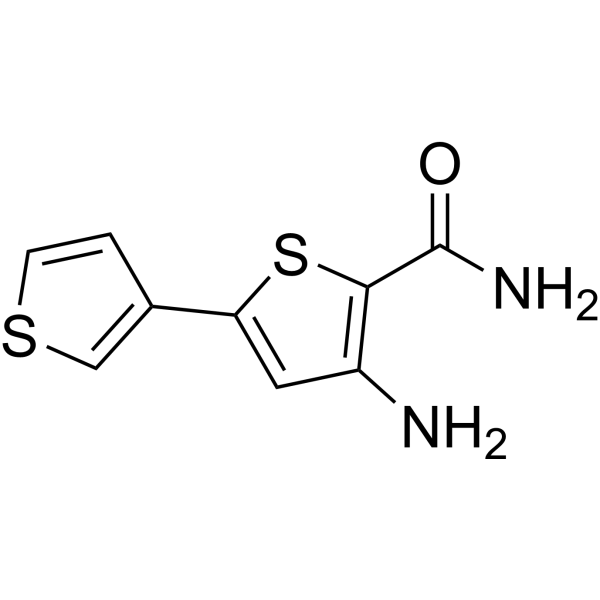
HY-13802
SC-514IKK Inhibitor -
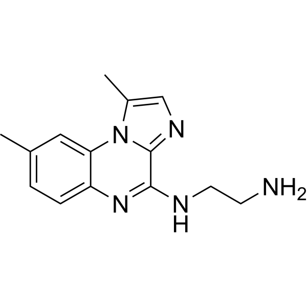
HY-10519
BMS-345541IKK-1/IKK-2 Inhibitor -
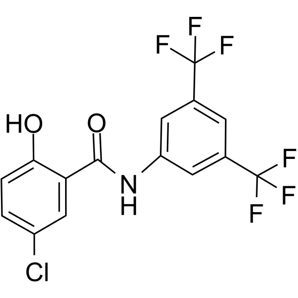
HY-10172
IMD-0354IKKβ Inhibitor -
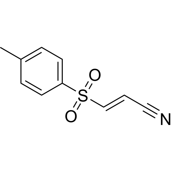
HY-13453
BAY 11-7082IκBα/NF-κB Inhibitor -
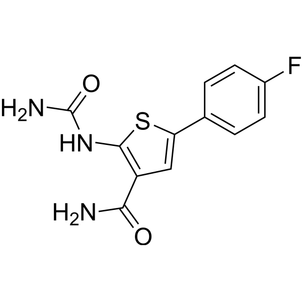
HY-10074
TPCA-1IKK-2 Inhibitor -
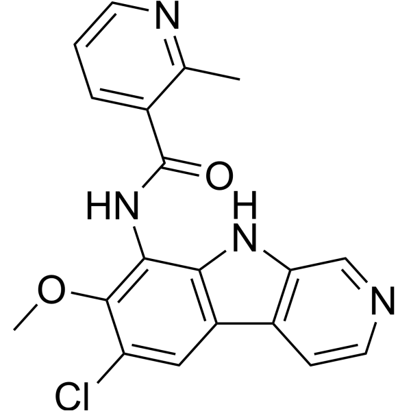
HY-15473
MLN120BIKKβ Inhibitor -
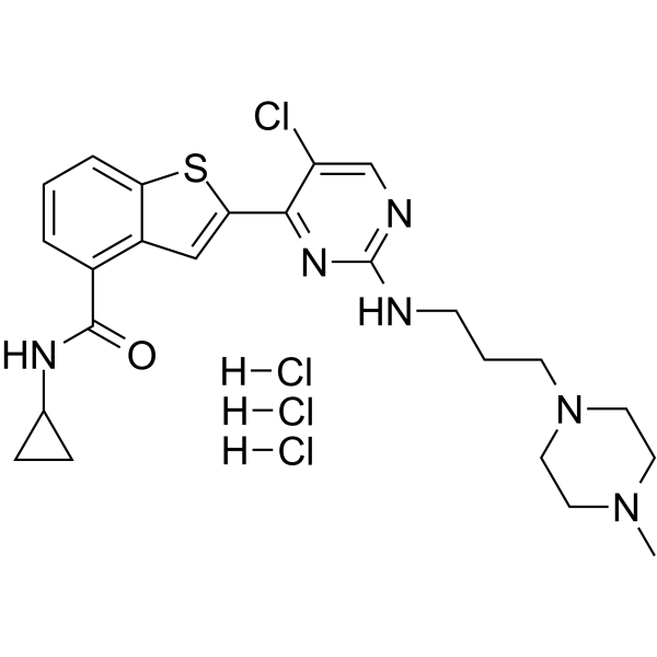
HY-B0788A
LY2409881 trihydrochlorideIKK2 Inhibitor -
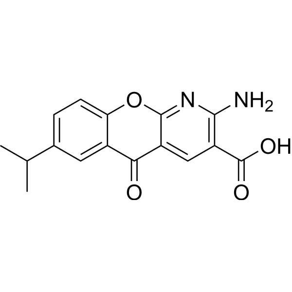
HY-B0713
AmlexanoxIKK Inhibitor -
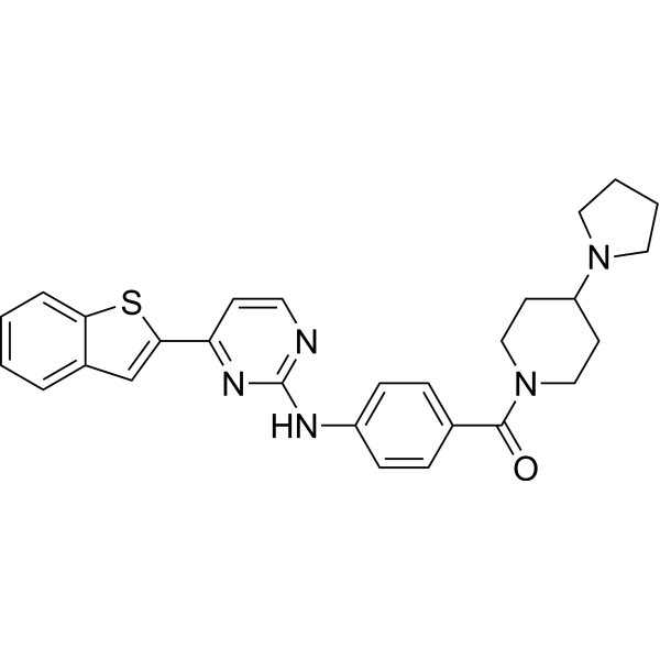
HY-13687
IKK 16IKK Inhibitor -
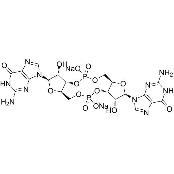
HY-110382
Cyclic-di-GMP disodiumSecond Messenger/STING Agonist
Voir tous les produits spécifiques à Isoform TNF Receptor
More

Activité biologique
Description
IC50 & Target
[1]|
IKK-2 0.025 μM (IC50) |
JNK1 1.6 μM (IC50) |
NF-κB |
TNF-α |
In Vitro
IKK2-IN-4 (compound 4) is a potent IKK-2 inhibitor with an IKK-2 IC50 of 0.025 μM; it inhibits IKK-1 with an IC50 of 1.0 μM, demonstrating 40-fold selectivity[1].
IKK2-IN-4 inhibits TNF-α production in PBMCs with a TNF cell IC50 of 0.25 μM; the WST-1 toxicity IC50 in PBMCs is 10 μM; Caco2 permeability is 17.7[1].
IKK2‑IN‑4 exhibits a clearance rate of 3 μL/min/106 cells in rat hepatocytes and a clearance rate of 27 μL/min/mg in human liver microsomes[1].
IKK2-IN-4 inhibits JNK-1 with an IC50 of 1.6 μM[1].
IKK2‑IN‑4 in complex with human JNK‑1 yields an X‑ray crystal structure at 2.1 Å resolution, which validates the modelled binding mode of IKK‑2, and all predicted hydrogen bonds are observed in the X‑ray structure[1].
MedChemExpress (MCE) has not independently confirmed the accuracy of these methods. They are for reference only. Further protocols information, click here.


Chemical Information
-
CAS No. 354811-10-2
-
Appearance Solid
-
Masse moléculaire 261.30
-
Formule C12H11N3O2S
-
Color Off-white to pink
-
SMILES
O=C(C1=C(NC(N)=O)SC(C2=CC=CC=C2)=C1)N
-
Livraison
Room temperature in continental US; may vary elsewhere.
-
Stockage
4°C, protect from light
* In solvent : -80°C, 6 months; -20°C, 1 month (protect from light)


Solvant et solubilité
In Vitro:
DMSO : 100 mg/mL (382.70 mM; Need ultrasonic; Hygroscopic DMSO has a significant impact on the solubility of product, please use newly opened DMSO)
Please refer to the solubility information to select the appropriate solvent. Once prepared, please aliquot and store the solution to prevent product inactivation from repeated freeze-thaw cycles.
Storage method and period of stock solution: -80°C, 6 months; -20°C, 1 month (protect from light). When stored at -80°C, please use it within 6 months. When stored at -20°C, please use it within 1 month.
Please refer to the solubility information to select the appropriate solvent. Once prepared, please aliquot and store the solution to prevent product inactivation from repeated freeze-thaw cycles.
Storage method and period of stock solution: -80°C, 6 months; -20°C, 1 month (protect from light). When stored at -80°C, please use it within 6 months. When stored at -20°C, please use it within 1 month.
Concentration (start) × Volume (start) = Concentration (final) × Volume (final)

Protocole
-
Kinase activity and phosphorylation assays
Kinase activity assays measure the ability of kinases to transfer phosphate groups from ATP to specific substrates, while phosphorylation assays detect the presence and levels of phosphorylated proteins. Common methods include radiolabeled ATP incorporation (e. g. ,), ADP release detection via bioluminescence (e. g. ,[3]), enzyme-linked immunosorbent assays (ELISA) for phospho-specific epitopes (e. g. ,[6]), and microtiter-based formats for high-throughput screening (e. g. ,[8]). The ADP-Glo assay quantifies kinase activity by measuring ADP produced during phosphorylation using a luciferase-based system. Radiometric assays involve autoradiography or scintillation counting after incorporation of 32P-labeled ATP into substrate proteins. ELISA-based approaches rely on phospho-specific antibodies to detect activated kinases in cell lysates or purified samples.
-
Western Blot
Western blotting (WB) is a commonly used experimental method in molecular biology, biochemistry, and immunogenetics for identifying and quantifying target proteins. It combines gel electrophoresis with immunoassay, enabling researchers to analyze protein expression, post-translational modifications, and molecular weight.
-
Cell Cytotoxicity Assay
Cytotoxicity assays are usually based on the assessment of cell membrane damage, which can also be indirectly detected by measuring cell viability. Detection methods include MTT assay, CKK-8 assay, LDH assay and ATP assay, etc.
-
Protocol for Kinase activity and phosphorylation assays
Kinase activity assays measure transfer of phosphate from ATP to a protein or peptide substrate, generating phosphorylated substrate, ADP, or incorporated radiolabeled phosphate as the readout; phosphorylation assays measure site-specific phosphorylation in cells or tissues as a proxy for kinase-pathway activation, inhibition, or substrate regulation. Phosphorylation can be detected by phospho-specific Western blot, immunoprecipitation kinase assay, phospho-immunofluorescence, phospho-flow cytometry, luminescent ADP detection, radiolabeled ATP incorporation, or reporter-based pathway assays, and these readouts can be applied to cancer cells, primary neurons, mouse tumors, organoids, inflammatory macrophages, ferroptosis studies, and mitophagy studies when the kinase target is biologically relevant.
-
Research Protocol for Inflammation-related Diseases
The NLRP3 inflammasome is a cytosolic innate immune signaling platform that integrates priming signals and danger-signal activation to promote caspase-1 activation, maturation of IL-1β and IL-18, and gasdermin D-mediated pyroptotic cell death. The core experimental logic is to determine whether inflammatory disease phenotypes are driven by increased NLRP3 expression, ASC-containing inflammasome assembly, caspase-1 cleavage, GSDMD cleavage, and extracellular release of IL-1β/IL-18 rather than by nonspecific cell injury alone. The pathway is strongly linked to inflammation-related disease phenotypes because monosodium urate crystals activate NALP3/NLRP3 inflammasome signaling in gout-like crystal inflammation, cholesterol crystals activate NLRP3 inflammasomes in atherogenesis models, and DSS-induced intestinal inflammation has been reported to involve NLRP3 inflammasome activity. However, experimental colitis studies also show context-dependent protective effects of NLRP3 inflammasome co


Pureté et documentation
-
Fiche technique (289 KB)
-
SDS (394 KB)
- English - EN (394 KB)
- Français - FR (394 KB)
- Deutsch - DE (394 KB)
- Norwegian - NO (394 KB)
- Español - ES (394 KB)
- Swedish - SV (394 KB)
- Italian - IT (394 KB)
- Korean - KR (394 KB)
- Portuguese - PT (394 KB)
-
Instruction de manipulation (2659 KB)
Références


Complete Stock Solution Preparation Table
Please refer to the solubility information to select the appropriate solvent. Once prepared, please aliquot and store the solution to prevent product inactivation from repeated freeze-thaw cycles.
Storage method and period of stock solution: -80°C, 6 months; -20°C, 1 month (protect from light). When stored at -80°C, please use it within 6 months. When stored at -20°C, please use it within 1 month.
| Optional Solvent | Concentration Solvent Mass | 1 mg | 5 mg | 10 mg | 25 mg |
|---|---|---|---|---|---|
| DMSO | 1 mM | 3.8270 mL | 19.1351 mL | 38.2702 mL | 95.6755 mL |
| 5 mM | 0.7654 mL | 3.8270 mL | 7.6540 mL | 19.1351 mL | |
| 10 mM | 0.3827 mL | 1.9135 mL | 3.8270 mL | 9.5675 mL | |
| 15 mM | 0.2551 mL | 1.2757 mL | 2.5513 mL | 6.3784 mL | |
| 20 mM | 0.1914 mL | 0.9568 mL | 1.9135 mL | 4.7838 mL | |
| 25 mM | 0.1531 mL | 0.7654 mL | 1.5308 mL | 3.8270 mL | |
| 30 mM | 0.1276 mL | 0.6378 mL | 1.2757 mL | 3.1892 mL | |
| 40 mM | 0.0957 mL | 0.4784 mL | 0.9568 mL | 2.3919 mL | |
| 50 mM | 0.0765 mL | 0.3827 mL | 0.7654 mL | 1.9135 mL | |
| 60 mM | 0.0638 mL | 0.3189 mL | 0.6378 mL | 1.5946 mL | |
| 80 mM | 0.0478 mL | 0.2392 mL | 0.4784 mL | 1.1959 mL | |
| 100 mM | 0.0383 mL | 0.1914 mL | 0.3827 mL | 0.9568 mL |


IKK2-IN-4 Related Classifications
HY-112456 Related Classifications
 Powered by Bioz