



Topics
Organoid Validation
Organoids are three-dimensional (3D) cell clusters derived entirely from primary tissues, embryonic stem cells (ESCs), or induced pluripotent stem cells (iPSCs). They can faithfully recapitulate the architecture and function of native organs in vitro. Organoids possess the capacity for self-renewal and self-organization, and exhibit organ-specific functions that closely resemble those of their tissues of origin.
The concept of organoids can be traced back to 1907, when Wilson and colleagues reported that dissociated sponge cells were capable of reaggregating and regenerating an entire organism. In 1987, researchers demonstrated that cultured mammary epithelial cells could differentiate into the characteristic ductal structures in vitro[1]. Thirty years later, organoid technology was recognized as the "Method of the Year 2017" by Nature Methods. Since then, organoid models representing a wide range of organs and diseases have been successfully established and widely adopted, significantly accelerating advances in basic research, drug discovery, precision medicine, and numerous other biomedical applications.
The establishment of organoid models primarily relies on two categories of tools: organoid culture tools and organoid validation tools.
Organoid culture tools include recombinant proteins, small-molecule inhibitors, extracellular matrix hydrogels (such as Matrigel), and specialized culture media. Directed differentiation is achieved by supplementing cultures with specific recombinant proteins or small-molecule inhibitors, while extracellular matrices and culture media provide the essential biochemical and structural support required for organoid growth, as well as microenvironments that mimic those of different native tissues.
Organoid validation tools primarily include antibodies, genomic analysis, transcriptomic/epigenomic sequencing, and other molecular characterization techniques. To verify that cultured organoids faithfully retain the genetic characteristics of their parental tissues, genomic and transcriptomic analyses are employed to assess their molecular profiles. In addition, antibody-based assays are used to detect the expression of key biomarkers within organoids, thereby confirming that they preserve the histopathological features and cellular identity of the original tissue.
Organoid validation can be broadly categorized into three complementary dimensions:
Histopathological validation – including hematoxylin and eosin (H&E) staining, immunohistochemistry (IHC), and related morphological analyses.
Transcriptional and epigenetic validation – including RNA sequencing, ATAC-seq, DNA methylation profiling, and other multi-omics approaches.
Functional biological validation – including both in vitro and in vivo functional assays to evaluate physiological and pathological responses.
Collectively, these validation strategies provide a comprehensive assessment of organoid fidelity. They confirm the morphological and phenotypic similarity between organoids and their corresponding native tissues, verify the expression of tissue-specific biomarkers, characterize cellular identity and composition, reveal the spatial organization of distinct cell populations, and evaluate molecular and functional homology with the original organ. Together, these approaches ensure that organoids faithfully recapitulate the structural, molecular, and functional characteristics of native tissues, supporting their reliability for disease modeling, drug discovery, and translational research.
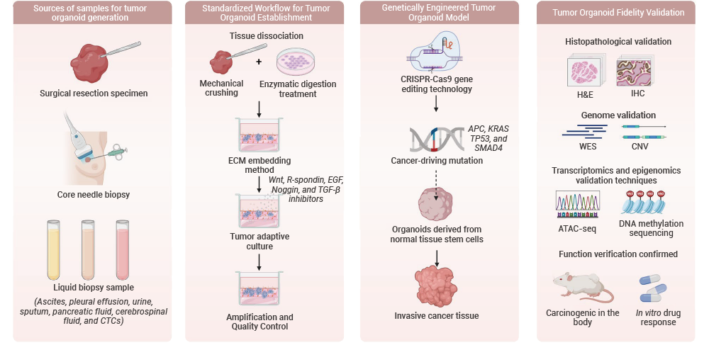
Figure 1. Schematic overview of the integrated workflow for tumor organoid establishment, genetic engineering, and validation.【2】
| Gastic organoid validation | Endometrial organoid validation |
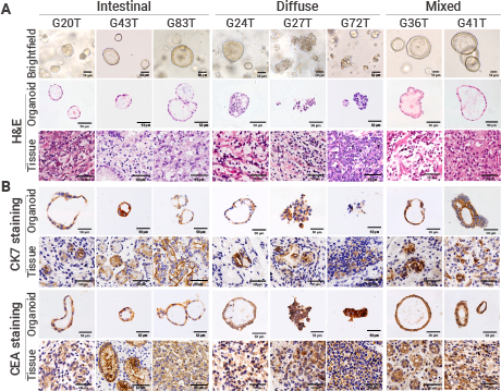 | 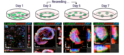 |
| Figure 2. Immunohistochemical staining of in vitro cultured gastric cancer organoids and their corresponding parental tumor tissues using anti-CK7 and anti-CEA antibodies. The results demonstrate that the gastric cancer organoids retained the histological architecture of the primary tumors, exhibiting characteristic glandular features, including glandular, non-cohesive, or solid growth patterns, as well as nuclear stratification.[3] | Figure 3. Immunofluorescence analysis of endometrial organoid development at different culture time points. Endometrial epithelial cells were labeled with an anti-E-cadherin antibody, whereas endometrial stromal cells were identified using an anti-vimentin antibody. Sequential imaging revealed the dynamic formation and maturation of three-dimensional (3D) organoid structures during culture.[4] |
| Liver organoid validation | Brain organoid validation |
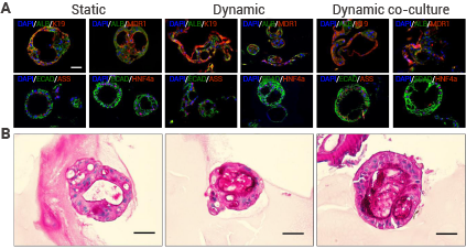 | 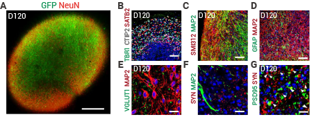 |
| Figure 4. Characterization of hepatic organoid differentiation by immunostaining and functional analysis. Anti-CK19 and anti-E-cadherin antibodies were used to label the cytoskeleton and cell membrane, respectively, confirming the formation of epithelial structures within the hepatic organoids. Expression of liver-specific markers, including ALB, MDR1, ASS, and HNF4, was detected using the corresponding antibodies, demonstrating differentiation of the organoids toward the hepatic lineage. In addition, periodic acid–Schiff (PAS) staining revealed glycogen storage capacity, indicating functional maturation of hepatocyte-like cells within the organoids.[5] | Figure 5. Immunofluorescence characterization of cerebral organoids cultured for 120 days. Layer-specific cortical neurons were identified using antibodies against TBR1, CTIP2, and SATB2, while mature neurons were labeled with antibodies against NeuN, MAP2, and SMI312. Astrocytes and excitatory glutamatergic neurons were detected using anti-GFAP and anti-VGLUT1 antibodies, respectively. The expression patterns of these lineage-specific markers demonstrated that the cerebral organoids closely recapitulated the development of the human fetal cerebral cortex.[6] |
| Human Cerebral Organoids Model Central Nervous System Oxygen Toxicity (IF = 11.6) | Gravity-Based Culture Combined with Microfluidics to Model the Tumor Microenvironment in Glioblastoma Organoids (IF = 5.1) |
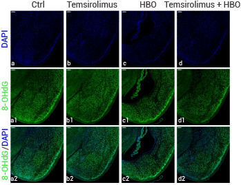 | 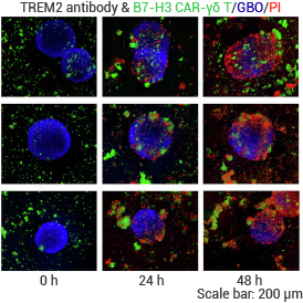 |
| Figure6. Exposure of human brain organoids to hyperbaric oxygen conditions recapitulated central nervous system oxygen toxicity, revealing pressure-dependent neurotoxicity mediated by the ROS–mTOR pathway. These findings were further confirmed in mouse brain tissue by detecting the DNA damage marker 8-OHdG (HY-P81140, 8-OHdG (DNA/RNA Damage) Antibody).[7] | Figure7. Under a gravity-based culture system integrated with a microfluidic platform, glioblastoma organoids exhibited histopathological features highly consistent with those of their matched patient tumors and demonstrated favorable responses to CSR-γδT cell-based combination immunotherapy (HY-P80920,TREM2 Antibody).[8] |
 |
| Product Name | Host | Reactivity | Application | |
|---|---|---|---|---|
| Gastric organoids | Rabbit | Human | WB, IHC-F, IHC-P, ICC/IF, IP, ELISA | |
| Rabbit | Human, Mouse, Rat | IHC-P, IHC-F, ICC/IF, FC, IF-Tissue, mIHC, ELISA | ||
| Colon organoids | Mouse | Human, Mouse, Rat, Rabbit | IHC-P, FC, ELISA | |
| Rabbit | Human, Mouse, Rat | WB, ICC/IF, IHC-P, FC, ELISA | ||
| Brain organoids | Rabbit | Human, Mouse, Rat | WB, IHC-P, IHC-F, ICC/IF, IP, IF-Tissue, mIHC, ELISA | |
| Rabbit | Human, Mouse, Rat | WB, IHC-P, ICC/IF, ELISA | ||
| Retinal organoids | Rabbit | Human, Rat | IHC-P, mIHC, ELISA | |
| Rabbit | Human, Rat | WB, IHC-P, ELISA | ||
| Heart organoids | Mouse | Human, Mouse, Rat, Rabbit, Monkey | WB, IHC-P, ICC/IF, FC, ELISA | |
| Rabbit | Human, Mouse, Rat | WB, IHC-P, ICC/IF, ELISA | ||
| Vescular organoids | Rabbit | Human, Mouse, Rat | WB, IHC-P, ICC/IF, IP, ELISA | |
| Mouse | Human | IHC-P, ICC/IF, ELISA | ||
| Liver organoids | Rabbit | Human, Mouse, Rat | WB, IHC-P, ICC/IF, IP, ELISA | |
| Rabbit | Human, Mouse, Rat | WB, IHC-P, IF-Tissue, mIHC, ELISA | ||
| Lung organoids | Rabbit | Human, Mouse, Rat | WB, IHC-P, ICC/IF, ELISA | |
| Rabbit | Human, Mouse, Rat | WB,IHC-P, ICC/IF, FC, ELISA | ||
| Thymus organoids | Rabbit | Human | WB, IHC-P, ICC/IF, FC, IF-Tissue, mIHC, ELISA | |
| Rabbit | Human | WB, IHC-F, IHC-P, ICC/IF, IP, ELISA | ||
| Cartilage organoids | Rabbit | Human, Mouse, Rat | WB, IHC-P, FC, ELISA | |
| Mouse | Human, Mouse, Rat | WB, IHC-P, ELISA | ||
| Kidney organoids | Rabbit | Human, Mouse | WB, IHC-P, FC, ELISA | |
| Mouse | Human, Mouse, Rat | WB, IHC-P, ICC/IF, ELISA |
 LinkedIn
 Researchgate
 Instagram
 X
X Facebook
 WhatsApp
 Reddit
 Mail
MailRéférences
[1]. Li ML, et al. Proc Natl Acad Sci USA, 1987, 84(1): 136-140. [Content Brief]
[2]. Zhang J, et al. Mol Biomed. 2026 Jun 30;7(1):103. [Content Brief]
[3]. Zhao Y, et al.Cell Rep Med. 2024 Jul 16;5(7):101627. [Content Brief]
[4]. Fujimura T, et al. Development. 2025 May 1;152(9):dev204461. [Content Brief]
[5]. Nguyen VVT, et al. J Extracell Vesicles. 2022 Nov;11(11):e12280. [Content Brief]
[6]. Hu N, et al. Nat Commun. 2024 Nov 6;15(1):9580. [Content Brief]
[7]. Xiaoying Ma, et al.Engineering,2025.
[8]. Zhu G, et al. Commun Biol. 2025 Dec 18;8(1):1791. [Content Brief]
