



Cephradine monohydrate
Based on 5 publication(s) in Google Scholar
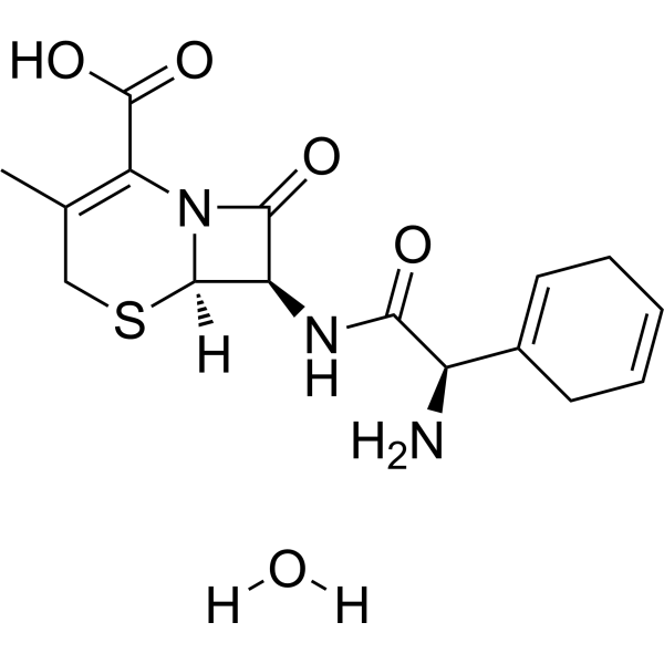
Cephradine (Cefradine) monohydrate is a broad-spectrum and orally active cephalosporin. Cephradine monohydrate is active against both grampositive and gram-negative pathogens and effective in eradicating most penicillinase-producing organisms known to be resistant to penicillin G, penicillin V, and ampicillin. Cephradine monohydrate has been used in the research of genitourinary, gastrointestinal and respiratory tract infections, and in infections of the skin and soft tissues. Cephradine monohydrate blocks solar-ultraviolet induced skin inflammation through direct inhibition of TOPK.
For research use only. We do not sell to patients.
- Purity : 91.02%
- CAS No.: 75975-70-1
- Formula: C16H21N3O5S
- Molecular Weight:367.42
-
Storage:
Store at room temperature, keep dry and cool.
In solvent -80°C, 1 year , -20°C, 6 months

To place orders, for customer services and technical support, please contact: MedChemExpress USA
Tel: 609-228-6898 E-mail: [email protected] [email protected]


-
 Biological Activity
-
Chemical Information
- Protocols
- Purity & Documentation
- References
-
 Help & FAQs


-
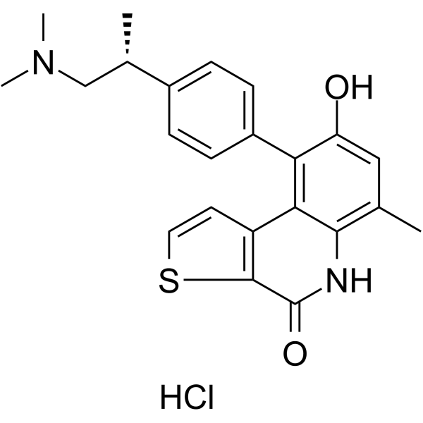
HY-12467
OTS964 hydrochlorideTOPK Inhibitor -
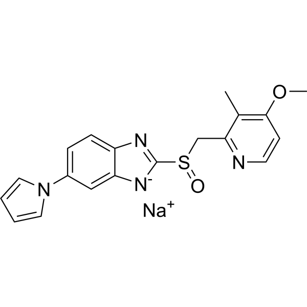
HY-B2145
Ilaprazole sodiumProton Pump Inhibitor -
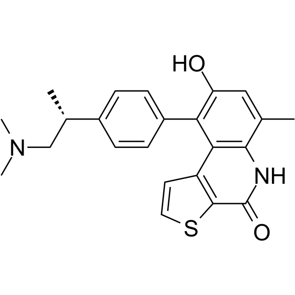
HY-19718
OTS964TOPK Inhibitor -
HY-B0441A
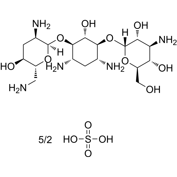
Tobramycin sulfateAminoglycoside Antibiotic -
HY-B0798
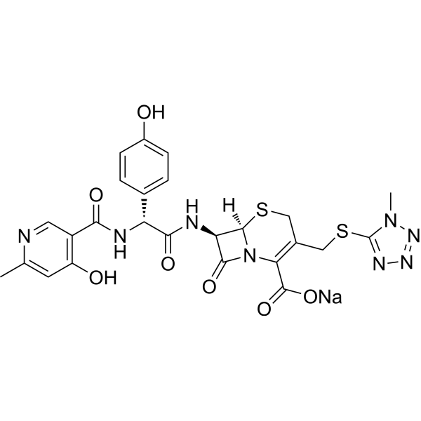
Cefpiramide sodiumAntibacterial Agent -

HY-B2145R
Ilaprazole sodium (Standard)Proton Pump Inhibitor -

HY-B0441AR
Tobramycin sulfate (Standard)Bacterial Inhibitor
Publications Citing Use of MedChemExpress (MCE) Cephradine monohydrate
MoreAll Antibiotic Isoforms
More

Biological Activity
Description
IC50 & Target
|
β-lactam |
In Vitro
Cephradine (0~8 μg/mL; 12 hours) makes bacterial viability rapidly increased at antibiotic concentrations below the MIC (minimum inhibitory concentration) of 0.70 µg/mL at both pH 7.4 and pH 5.5[4].
MedChemExpress (MCE) has not independently confirmed the accuracy of these methods. They are for reference only. Further protocols information, click here.
In Vivo
MedChemExpress (MCE) has not independently confirmed the accuracy of these methods. They are for reference only.
-
Animal Model:Nondiabetic mice
-
Dosage:25 mg/kg
-
Administration:S.c.; 11 days
-
Result:Reduced the bacterial density and counts in the abscesses.
Clinical Trial
| NCT Number | Sponsor | Condition | Start Date |
Phase

|
|---|---|---|---|---|
| NCT01329991 | Plexxikon| | 2011-05 | PHASE1 |


Chemical Information
-
CAS No. 75975-70-1
-
Appearance Solid
-
Molecular Weight 367.42
-
Formula C16H21N3O5S
-
Color Off-white to light yellow
-
SMILES
O=C(C(N12)=C(C)CS[C@]2([H])[C@H](NC([C@H](N)C3=CCC=CC3)=O)C1=O)O.[H]O[H]
-
Synonyms
Cefradine monohydrate
-
Shipping
Room temperature in continental US; may vary elsewhere.
-
Storage
Store at room temperature, keep dry and cool
In solvent -80°C 1 year -20°C 6 months


Publications (5)
-
Journal Impact Factor
-
Most Recent
-
ACS Environ Au
Machine Learning-Assisted Recognition of Environmental Sulfur-Containing Chemicals in Nontargeted Mass Spectrometry Analysis of Inadequate Mass Resolution. [Abstract]2025 Aug 5;5(6):573-582. PMID: 41277996 -
Anal Chem
Exposome-Scale Investigation of Cl-/Br-Containing Chemicals Using High-Resolution Mass Spectrometry, Multistage Machine Learning, and Cloud Computing. [Abstract]2025 Jun 3;97(21):11099-11109. PMID: 40401576 -
Molecules
Seeking Correlation Among Porin Permeabilities and Minimum Inhibitory Concentrations Through Machine Learning: A Promising Route to the Essential Molecular Descriptors. [Abstract]2025 Mar 9;30(6):1224. PMID: 40142001 -
Microbiol Spectr
2025 Aug 29:e0059025. PMID: 40879403 -
Chemosphere
Mass-balance-model-based evaluation of sewage treatment plant contribution to residual pharmaceuticals in environmental waters. [Abstract]2019 Jun:225:378-387. PMID: 30884299

Protocols
-
Imiquimod-Induced Psoriasiform Dermatitis
Imiquimod (IMQ)-induced psoriasiform dermatitis is a widely used murine model in which topical application of IMQ, a Toll-like receptor 7 (TLR7) agonist, triggers innate immune activation in the skin and induces a psoriasis-like inflammatory cascade characterized by epidermal hyperplasia, immune cell infiltration, and cytokine production dominated by the IL-23/IL-17 axis. This inflammatory response is mediated through activation of dendritic cells and downstream induction of IL-23, IL-17A, IL-22, and related pro-inflammatory mediators, recapitulating key features of human plaque psoriasis and enabling mechanistic and therapeutic studies. The model is commonly induced using Aldara (5% IMQ cream) applied topically to murine skin, resulting in rapid onset of erythema, scaling, and thickening that can be quantified as disease severity indices and validated histologically.
-
TPA/Croton Oil Ear Edema and Dermatitis
The TPA (12-O-tetradecanoylphorbol-13-acetate) and croton oil-induced mouse ear edema model is a well-established acute cutaneous inflammation system used to evaluate topical anti-inflammatory activity by measuring edema formation, neutrophil infiltration, vascular permeability, and cytokine-mediated skin responses in vivo. The inflammatory response is triggered by topical application of phorbol esters (TPA) or croton oil constituents, leading to rapid activation of protein kinase C signaling, leukocyte recruitment, and increased vascular permeability, which can be quantified by ear thickness, weight, dye extravasation, and biochemical markers such as myeloperoxidase (MPO) activity and pro-inflammatory mediators in ear tissue homogenates. This model is widely used for screening anti-inflammatory agents, where reductions in edema and inflammatory biomarkers reflect suppression of acute dermal inflammation and immune cell infiltration. Histological evaluation typically confirms epidermal
-
Contact Hypersensitivity Dermatitis
Contact hypersensitivity (CHS) dermatitis is a T cell-mediated delayed-type (Type IV) immune reaction in which low-molecular-weight haptens applied to the skin bind host proteins to form complete antigens, triggering sensitization followed by a secondary inflammatory response upon re-exposure (elicitation phase), which is commonly quantified by ear swelling as a readout of skin inflammation in murine models. This model is widely used to study allergic contact dermatitis because it is antigen-specific, reproducible, and reflects key immunological events including dendritic cell activation, T cell priming in draining lymph nodes, and effector T cell-driven tissue inflammation. DNFB- and oxazolone-induced CHS models are standard systems for evaluating both acute and chronic T cell-dependent skin inflammation and for testing immunomodulatory interventions.
-
Gram Staining of Tissue Sections
Gram staining of tissue sections is a histochemical technique used to differentiate Gram-positive and Gram-negative bacteria within histological specimens based on differences in bacterial cell wall structure and dye retention, adapted from classical bacteriological Gram staining into tissue-compatible “histological Gram stain” variants. In tissue applications, modifications of the Brown-Hopps and Brown-Brenn methods are commonly used to improve differentiation of microorganisms embedded within host connective tissue and to reduce overstaining or loss of Gram-negative signal, which are known limitations of earlier approaches. The principle relies on crystal violet-iodine complex retention in Gram-positive organisms and subsequent decolorization and counterstaining steps that allow contrast visualization of Gram-negative organisms against tissue background.
-
Research Protocol for Inflammation-related Diseases
The NLRP3 inflammasome is a cytosolic innate immune signaling platform that integrates priming signals and danger-signal activation to promote caspase-1 activation, maturation of IL-1β and IL-18, and gasdermin D-mediated pyroptotic cell death. The core experimental logic is to determine whether inflammatory disease phenotypes are driven by increased NLRP3 expression, ASC-containing inflammasome assembly, caspase-1 cleavage, GSDMD cleavage, and extracellular release of IL-1β/IL-18 rather than by nonspecific cell injury alone. The pathway is strongly linked to inflammation-related disease phenotypes because monosodium urate crystals activate NALP3/NLRP3 inflammasome signaling in gout-like crystal inflammation, cholesterol crystals activate NLRP3 inflammasomes in atherogenesis models, and DSS-induced intestinal inflammation has been reported to involve NLRP3 inflammasome activity. However, experimental colitis studies also show context-dependent protective effects of NLRP3 inflammasome co


Purity & Documentation
-
Data Sheet (277 KB)
-
SDS (252 KB)
- English - EN (252 KB)
- Français - FR (252 KB)
- Deutsch - DE (252 KB)
- Norwegian - NO (252 KB)
- Español - ES (252 KB)
- Swedish - SV (252 KB)
- Italian - IT (252 KB)
- Korean - KR (252 KB)
- Portuguese - PT (252 KB)
-
Handling Instructions (2659 KB)
References
[1]. Schwinghammer TL, et al. Pharmacokinetics of cephradine administered intravenously and orally to young and elderly subjects. J Clin Pharmacol. 1990;30(10):893-899 [Content Brief]
[2]. Caloza DL Jr, et al. Intravenous use of cephradine and cefazolin against serious infections. Antimicrob Agents Chemother. 1979;15(1):119-122. [Content Brief]
[3]. Fan X, et al. Cefradine blocks solar-ultraviolet induced skin inflammation through direct inhibition of T-LAK cell-originated protein kinase. Oncotarget. 2016;7(17):24633-24645. [Content Brief]
[4]. Kang S, et al. In Vitro and In Vivo Antimicrobial Activity of Antibiotic-Conjugated Carriers with Rapid pH-Responsive Release Kinetics. Adv Healthc Mater. 2019;8(14):e1900247. [Content Brief]

Calculators
Concentration (start) × Volume (start) = Concentration (final) × Volume (final)


Cephradine monohydrate Related Classifications
HY-128449 Related Classifications
 Powered by Bioz