



Cetirizine-d8

Based on 1 Customer Validation
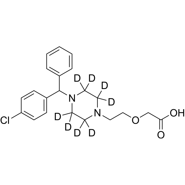
Cetirizine-d8 is a deuterium labeled Cetirizine. Cetirizine, a second-generation antihistamine and the carboxylated metabolite of hydroxyzine, is a specific, orally active and long-acting histamine H1-receptor antagonist. Cetirizine marks antiallergic properties and inhibits eosinophil chemotaxis during the allergic response.
For research use only. We do not sell to patients.
- Purity : 96.46%
- CAS No.: 774596-22-4
- Formula: C21H17D8ClN2O3
- Molecular Weight:396.94
-
Storage:Powder -20°C, 3 years ; In solvent -80°C, 6 months , -20°C, 1 month

To place orders, for customer services and technical support, please contact: MedChemExpress USA
Tel: 609-228-6898 E-mail: [email protected] [email protected]


-
 Biological Activity
-
Chemical Information
-
Solvent & Solubility
- Protocols
- Purity & Documentation
- References
-
 Help & FAQs


-
HY-10450S
Dapagliflozin-d5Stable Isotope -
HY-B0510S
Trimethoprim-d9Stable Isotope -
HY-17356S
Fenofibrate-d6Stable Isotope -
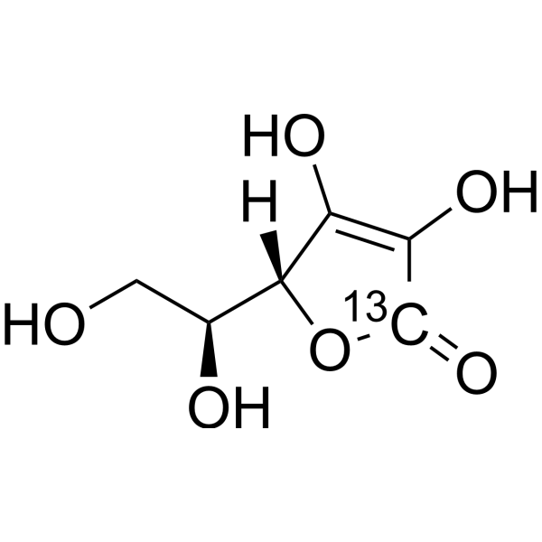
HY-B0166S1
L-Ascorbic acid-13CStable Isotope -
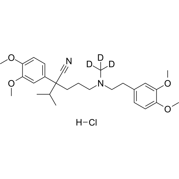
HY-A0064S
Verapamil-d3 hydrochlorideStable Isotope -
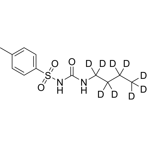
HY-B0401S
Tolbutamide-d9Stable Isotope -
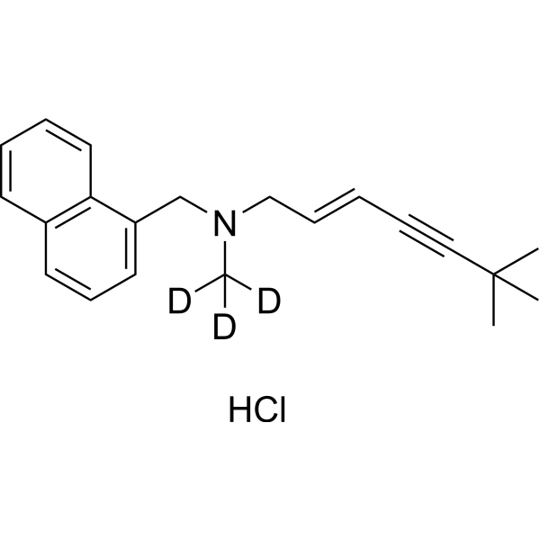
HY-17395S
Terbinafine-d3 hydrochlorideStable Isotope -
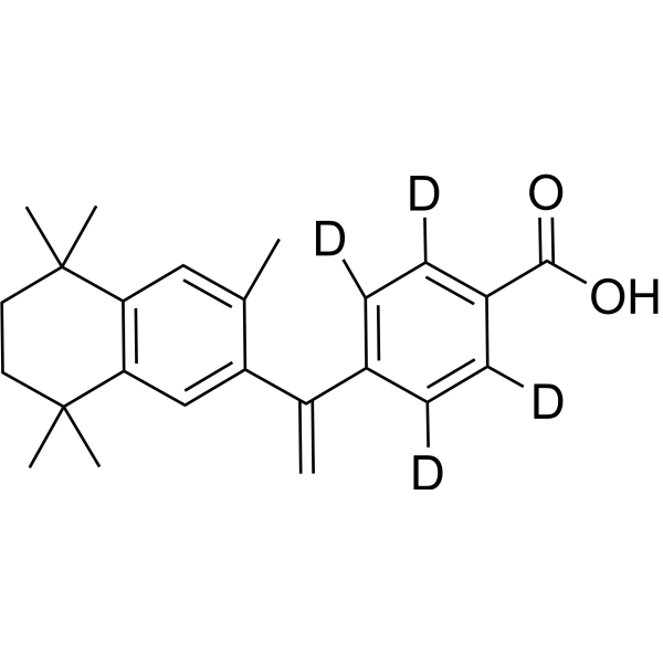
HY-14171S
Bexarotene-d4RXR Agonist -
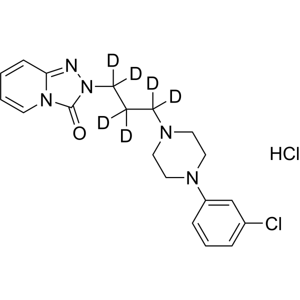
HY-B0478AS
Trazodone-d6 hydrochlorideStable Isotope -
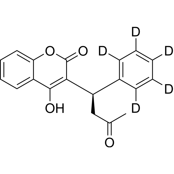
HY-W015998S
(R)-(+)-Warfarin-d5Stable Isotope -

HY-B0437AS
Sotalol-d6 hydrochlorideStable Isotope -
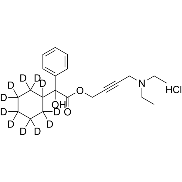
HY-B0267AS
Oxybutynin-d11 chlorideStable Isotope
All Histamine Receptor Isoforms
More

Biological Activity
Description
IC50 & Target
|
H1 Receptor |
Application
1. This compound can be used as a tracer
2. This compound can be used as an internal standard for quantitative analysis by NMR, GC-MS, or LC-MS.


Chemical Information
-
CAS No. 774596-22-4
-
Unlabeled CAS 83881-51-0
-
Appearance Solid
-
Molecular Weight 396.94
-
Formula C21H17D8ClN2O3
-
Color Off-white to light yellow
-
SMILES
O=C(O)COCCN1C([2H])([2H])C([2H])([2H])N(C(C2=CC=C(Cl)C=C2)C3=CC=CC=C3)C([2H])([2H])C1([2H])[2H]
-
Shipping
Room temperature in continental US; may vary elsewhere.
-
Storage
Powder -20°C 3 years In solvent -80°C 6 months -20°C 1 month


Solvent & Solubility
In Vitro:
DMSO : 100 mg/mL (251.93 mM; Need ultrasonic; Hygroscopic DMSO has a significant impact on the solubility of product, please use newly opened DMSO)
Please refer to the solubility information to select the appropriate solvent. Once prepared, please aliquot and store the solution to prevent product inactivation from repeated freeze-thaw cycles.
Storage method and period of stock solution: -80°C, 6 months; -20°C, 1 month. When stored at -80°C, please use it within 6 months. When stored at -20°C, please use it within 1 month.
Please refer to the solubility information to select the appropriate solvent. Once prepared, please aliquot and store the solution to prevent product inactivation from repeated freeze-thaw cycles.
Storage method and period of stock solution: -80°C, 6 months; -20°C, 1 month. When stored at -80°C, please use it within 6 months. When stored at -20°C, please use it within 1 month.
Concentration (start) × Volume (start) = Concentration (final) × Volume (final)
In Vivo:
Select the appropriate dissolution method based on your experimental animal and administration route.
- For the following dissolution methods, please ensure to first prepare a clear stock solution using an In Vitro approach and then sequentially add co-solvents:
- To ensure reliable experimental results, the clarified stock solution can be appropriately stored based on storage conditions. As for the working solution for In Vivo experiments, it is recommended to prepare freshly and use it on the same day.
- The percentages shown for the solvents indicate their volumetric ratio in the final prepared solution. If precipitation or phase separation occurs during preparation, heat and/or sonication can be used to aid dissolution.
Add each solvent one by one: 10% DMSO 40% PEG300 5% Tween-80 45% Saline
Solubility: ≥ 2.5 mg/mL (6.30 mM); Clear solution
This protocol yields a clear solution of ≥ 2.5 mg/mL (saturation unknown).
Taking 1 mL working solution as an example, add 100 μL DMSO stock solution (25.0 mg/mL) to 400 μL PEG300, and mix evenly; then add 50 μL Tween-80 and mix evenly; then add 450 μL Saline to adjust the volume to 1 mL.
Preparation of Saline: Dissolve 0.9 g sodium chloride in ddH₂O and dilute to 100 mL to obtain a clear Saline solution.
Add each solvent one by one: 10% DMSO 90% (20% SBE-β-CD in Saline)
Solubility: ≥ 2.5 mg/mL (6.30 mM); Clear solution
This protocol yields a clear solution of ≥ 2.5 mg/mL (saturation unknown).
Taking 1 mL working solution as an example, add 100 μL DMSO stock solution (25.0 mg/mL) to 900 μL 20% SBE-β-CD in Saline, and mix evenly.
Preparation of 20% SBE-β-CD in Saline (4°C, storage for one week): 2 g SBE-β-CD powder is dissolved in 10 mL Saline, completely dissolve until clear.
In Vivo Dissolution Calculator
Please enter the basic information of animal experiments:
-
-
-
-
Recommended: Prepare an additional quantity of animals to account for potential losses during experiments.
Please enter your animal formula composition:
-
%DMSO +
Recommended: Keep the proportion of DMSO in working solution below 2% if your animal is weak.
-
%+
-
+%Tween-80 + +
-
%Saline +
The co-solvents required include: DMSO, . All of co-solvents are available by MedChemExpress (MCE). , Tween 80. All of co-solvents are available by MedChemExpress (MCE).
Working solution concentration: 0.22 mg/mL
Method for preparing stock solution: mg drug dissolved in μL DMSO. Stock solution concentration: mg/mL.
1. Take μL DMSO stock solution;
2. Add μL .
μL , mix evenly;
3. Then add μL Tween 80, mix evenly;
4. Then add μL
Please ensure that the stock solution in the first step is dissolved to a clear state, and add co-solvents in sequence. You can use ultrasonic heating (ultrasonic cleaner, recommended frequency 20-40 kHz), vortexing, etc. to assist dissolution.

Protocols
-
Cell migration
Cell migration is a method that plays an important role in wound healing, cell differentiation, embryonic development, etc.
-
Chemotaxis Gradient Chamber Assay 1
The chemotaxis gradient chamber assay is based on the principle of observing directional cell migration in response to a stable, linear or quasi-linear concentration gradient of a chemoattractant formed between two fluid reservoirs separated by a narrow observation chamber. Cells placed within the chamber respond to the gradient by polarized movement toward higher chemoattractant concentrations, allowing quantification of chemotactic behavior in real time under microscopy. The classic Zigmond chamber design enables simultaneous visualization of gradient formation and individual cell trajectories, making it suitable for studying leukocyte chemotaxis and other motile cell types in vitro.
-
Under-Agarose Cell Migration Assay
The under-agarose cell migration assay is a classical in vitro chemotaxis method designed to evaluate directed cell movement through a semi-solid agarose matrix toward soluble chemoattractant gradients, enabling visualization and quantification of leukocyte or motile cell migration in a confined 2D-like environment. In this system, cells and chemoattractants are placed in separate wells cut into an agarose gel, allowing diffusion-driven gradient formation that guides directional migration, which is typically assessed by measuring migration distance, cell morphology changes, and accumulation toward the chemoattractant source. This assay has been widely used to study neutrophil and leukocyte chemotaxis as a simple alternative to filter-based migration systems and allows direct microscopic observation of migrating cells under near-physiological confinement conditions.
-
Transwell/Boyden Chamber Migration Assay
The Transwell/Boyden chamber migration assay measures cell movement through a porous membrane separating an upper and lower chamber, usually after a chemoattractant gradient is established by placing cells in the upper chamber and chemoattractant-containing medium in the lower chamber. The readout is generated by quantifying cells that traverse the membrane and appear on the lower membrane surface or in the lower chamber, depending on whether the cell type is adherent or non-adherent. This assay reflects chemotactic or haptotactic migration rather than matrix invasion unless an extracellular-matrix barrier is added to the membrane.
-
Chemotaxis Gradient Chamber Assay 2
Chemotaxis gradient chamber assays measure directional cell migration in response to a soluble chemical gradient by imaging cells as they move across a defined observation region; the readout is generated from time-lapse cell trajectories, displacement toward the gradient, forward migration index, trajectory plots, rose/polar plots, and statistical tests of non-random directionality. The Dunn chamber is a direct-viewing glass chamber in which cells migrate across a bridge between control and chemoattractant wells, allowing observation of cells in a linear concentration gradient; related direct-viewing formats include the Insall chamber, which supports defined unidirectional gradients and high numerical-aperture microscopy, and the μ-Slide Chemotaxis chamber, which supports long-term live-cell imaging and gradient characterization with fluorescent dye.


Purity & Documentation
-
Data Sheet (273 KB)
-
SDS (251 KB)
- English - EN (251 KB)
- Français - FR (251 KB)
- Deutsch - DE (251 KB)
- Norwegian - NO (251 KB)
- Español - ES (251 KB)
- Swedish - SV (251 KB)
- Italian - IT (251 KB)
- Korean - KR (251 KB)
- Portuguese - PT (251 KB)
-
Handling Instructions (2659 KB)
References
[1]. Caroline M. Spencer, et al. Cetirizine. Drugs 46 (6): 1055•1080, 1993.
[2]. Shih MY, et al. Influence of cetirizine and levocetirizine on two cytokines secretion in human airway epithelial cells. Allergy Asthma Proc. 2008 Sep-Oct;29(5):480-5. [Content Brief]
[3]. Shimizu T, et al. Cetirizine, an H1-receptor antagonist, suppresses the expression of macrophage migration inhibitory factor: its potential anti-inflammatory action. Clin Exp Allergy. 2004 Jan;34(1):103-9. [Content Brief]


Complete Stock Solution Preparation Table
Please refer to the solubility information to select the appropriate solvent. Once prepared, please aliquot and store the solution to prevent product inactivation from repeated freeze-thaw cycles.
Storage method and period of stock solution: -80°C, 6 months; -20°C, 1 month. When stored at -80°C, please use it within 6 months. When stored at -20°C, please use it within 1 month.
| Optional Solvent | Concentration Solvent Mass | 1 mg | 5 mg | 10 mg | 25 mg |
|---|---|---|---|---|---|
| DMSO | 1 mM | 2.5193 mL | 12.5964 mL | 25.1927 mL | 62.9818 mL |
| 5 mM | 0.5039 mL | 2.5193 mL | 5.0385 mL | 12.5964 mL | |
| 10 mM | 0.2519 mL | 1.2596 mL | 2.5193 mL | 6.2982 mL | |
| 15 mM | 0.1680 mL | 0.8398 mL | 1.6795 mL | 4.1988 mL | |
| 20 mM | 0.1260 mL | 0.6298 mL | 1.2596 mL | 3.1491 mL | |
| 25 mM | 0.1008 mL | 0.5039 mL | 1.0077 mL | 2.5193 mL | |
| 30 mM | 0.0840 mL | 0.4199 mL | 0.8398 mL | 2.0994 mL | |
| 40 mM | 0.0630 mL | 0.3149 mL | 0.6298 mL | 1.5745 mL | |
| 50 mM | 0.0504 mL | 0.2519 mL | 0.5039 mL | 1.2596 mL | |
| 60 mM | 0.0420 mL | 0.2099 mL | 0.4199 mL | 1.0497 mL | |
| 80 mM | 0.0315 mL | 0.1575 mL | 0.3149 mL | 0.7873 mL | |
| 100 mM | 0.0252 mL | 0.1260 mL | 0.2519 mL | 0.6298 mL |


Cetirizine-d8 Related Classifications
HY-17042S1 Related Classifications
 Powered by Bioz