



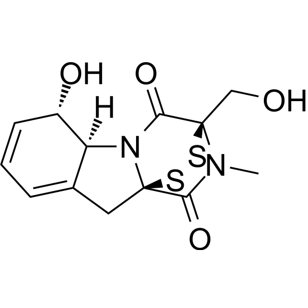
Gliotoxin
Based on 2 publication(s) in Google Scholar
Gliotoxin is a secondary metabolite, the most abundant mycotoxin secreted by A. fumigatus, inhibits the phagocytosis of macrophages and the immune functions of other immune cells . Gliotoxin inhibits inducible NF-κB activity by preventing IκB degradation, which consequently induces host-cell apoptosis. Gliotoxin activates PKA and increases intracellular cAMP concentration; modulates actin cytoskeleton rearrangement to facilitate A. fumigatus internalization into lung epithelial cells. Gliotoxin is a potent NOTCH2 transactivation inhibitor, can effectively induce apoptosis of chronic lymphocytic leukemia (CLL) cells.
Nur für Forschungszwecke. Wir verkaufen nicht an Patienten.
- Reinheit : 99.50%
- CAS. Nr.: 67-99-2
- Formel: C13H14N2O4S2
- Molecular Weight:326.39
-
Speicherung:Powder -20°C, 3 years ; In solvent -80°C, 6 months , -20°C, 1 month

To place orders, for customer services and technical support, please contact: MedChemExpress USA
Tel: 609-228-6898 E-mail: [email protected] [email protected]


-
 Biologische Aktivität
-
Chemical Information
-
Lösungsmittel & Löslichkeit
- Protokoll
- Reinheit & Dokumentation
- Verweise
-
 Help & FAQs


-

HY-15763
ErastinFerroptosis Inducer -
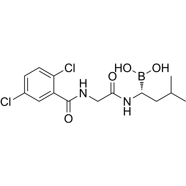
HY-10453
IxazomibProteasome Inhibitor -

HY-13259
MG-132Proteasome Inhibitor -
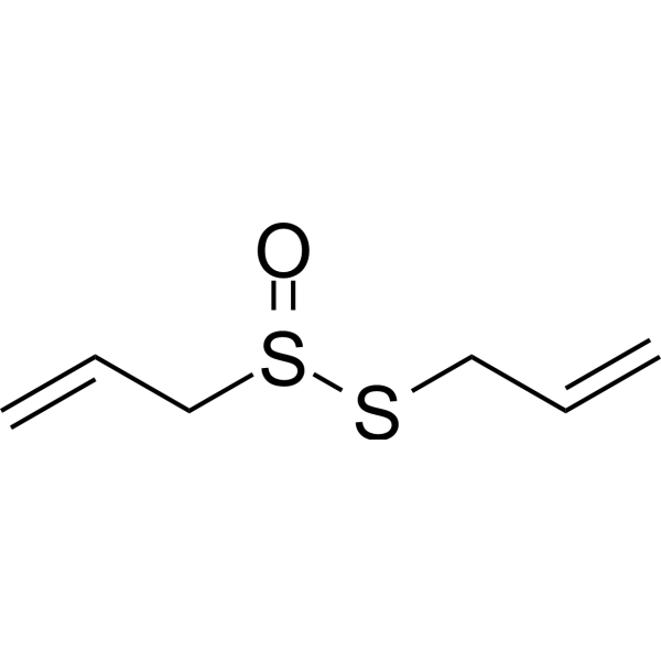
HY-N0315
AllicinBacterial Inhibitor -

HY-10227
BortezomibProteasome Inhibitor -
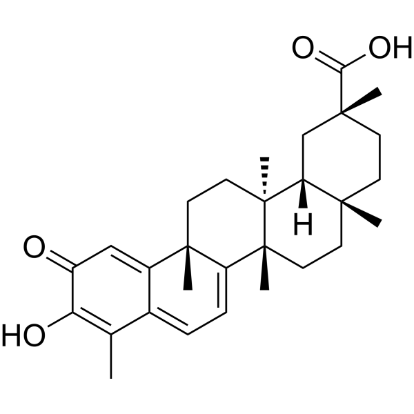
HY-13067
CelastrolAutophagy Inducer -
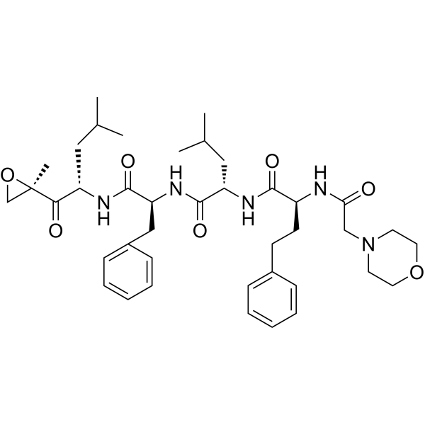
HY-10455
CarfilzomibProteasome Inhibitor -
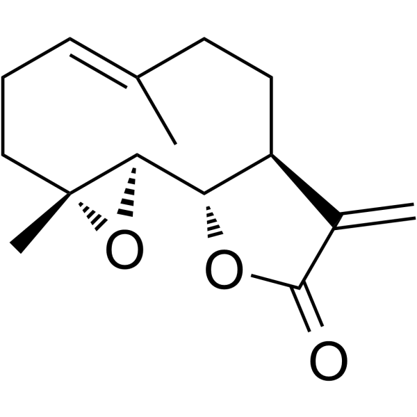
HY-N0141
ParthenolideNF-κB Inhibitor -
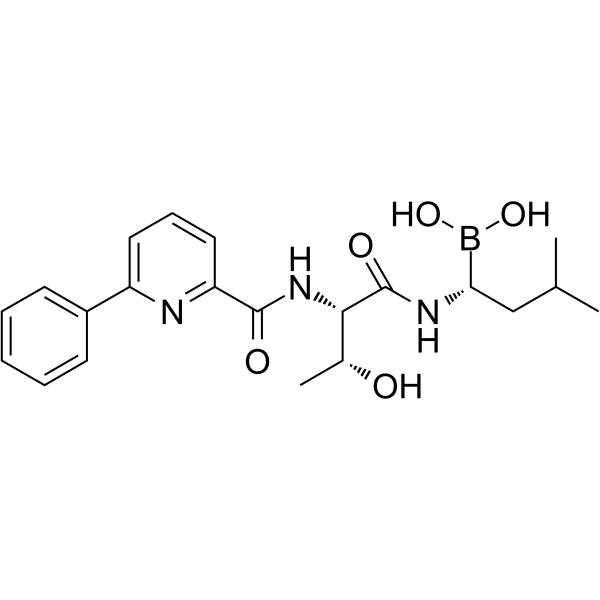
HY-10454
DelanzomibProteasome Inhibitor -
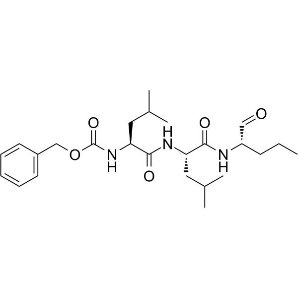
HY-108552
MG-115Proteasome Inhibitor -

HY-10227R
Bortezomib (Standard)Proteasome Inhibitor -
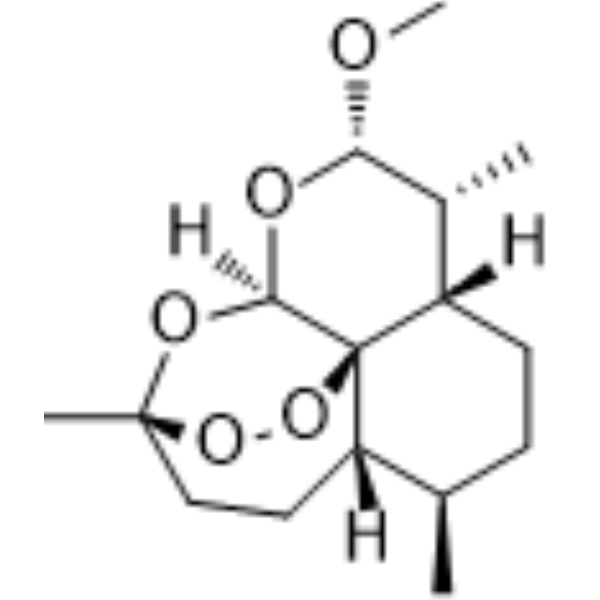
HY-N0402
ArtemetherParasite Inhibitor
Publications Citing Use of MedChemExpress (MCE) Gliotoxin
MoreAlle Antibiotic Isoform-spezifische Produkte anzeigen
More

Biologische Aktivität
Beschreibung
Cellular Effect
|
Cell Line

|
Type | Value | Description | References |
|---|---|---|---|---|
| A549 | IC50 |
0.003 μM
Compound: 10
|
Cytotoxicity against human A549 cells
Cytotoxicity against human A549 cells
|
[PMID: 20303767] |
| CCRF-CEM | IC50 |
0.15 μM
Compound: gliotoxin
|
Cytotoxicity against human CEM cells after 96 hrs
Cytotoxicity against human CEM cells after 96 hrs
|
[PMID: 16124785] |
| HCT-116 | IC50 |
0.41 μM
Compound: 311
|
Cytotoxicity against human HCT-116 cells assessed as reduction in cell viability incubated for 72 hrs by MTT assay
Cytotoxicity against human HCT-116 cells assessed as reduction in cell viability incubated for 72 hrs by MTT assay
|
[PMID: 38181652] |
| HCT-116 | IC50 |
1.24 μM
Compound: 311
|
Cytotoxicity against human HCT-116 cells assessed as reduction in cell viability incubated for 24 hrs by MTT assay
Cytotoxicity against human HCT-116 cells assessed as reduction in cell viability incubated for 24 hrs by MTT assay
|
[PMID: 38181652] |
| HEK293 | IC50 |
1.58 μM
Compound: 311
|
Cytotoxicity against HEK293 cells assessed as cell growth inhibition
Cytotoxicity against HEK293 cells assessed as cell growth inhibition
|
[PMID: 38181652] |
| HeLa | IC50 |
2.45 μM
Compound: 10
|
Cytotoxicity against human HeLa cells
Cytotoxicity against human HeLa cells
|
[PMID: 20303767] |
| HepG2 | IC50 |
0.21 μM
Compound: 311
|
Cytotoxicity against human HepG2 cells assessed as reduction in cell growth incubated for 48 hrs by SRB assay
Cytotoxicity against human HepG2 cells assessed as reduction in cell growth incubated for 48 hrs by SRB assay
|
[PMID: 38181652] |
| HepG2 | IC50 |
0.24 μM
Compound: 311
|
Cytotoxicity against human HepG2 cells assessed as reduction in cell growth incubated for 72 hrs by SRB assay
Cytotoxicity against human HepG2 cells assessed as reduction in cell growth incubated for 72 hrs by SRB assay
|
[PMID: 38181652] |
| HUVEC | IC50 |
0.123 μM
Compound: 10
|
Cytotoxicity against HUVEC
Cytotoxicity against HUVEC
|
[PMID: 20303767] |
| Jurkat | IC50 |
<1 μM
Compound: 10
|
Cytotoxicity against human Jurkat cells after 40 hrs bioluminescence assay
Cytotoxicity against human Jurkat cells after 40 hrs bioluminescence assay
|
[PMID: 20303767] |
| Macrophage | ED50 |
0.104 μM
Compound: 10
|
Immunomodulatory activity in macrophage assessed as inhibition of phagocytosis
Immunomodulatory activity in macrophage assessed as inhibition of phagocytosis
|
[PMID: 20303767] |
| MCF7 | IC50 |
0.08 μM
Compound: 311
|
Cytotoxicity against human MCF7 cells assessed as reduction in cell growth incubated for 48 hrs by SRB assay
Cytotoxicity against human MCF7 cells assessed as reduction in cell growth incubated for 48 hrs by SRB assay
|
[PMID: 38181652] |
| MCF7 | IC50 |
0.08 μM
Compound: 311
|
Cytotoxicity against human MCF7 cells assessed as reduction in cell growth incubated for 72 hrs by SRB assay
Cytotoxicity against human MCF7 cells assessed as reduction in cell growth incubated for 72 hrs by SRB assay
|
[PMID: 38181652] |
| NCI-H460 | IC50 |
0.21 μM
Compound: 311
|
Cytotoxicity against human NCI-H460 cells assessed as reduction in cell growth incubated for 72 hrs by SRB assay
Cytotoxicity against human NCI-H460 cells assessed as reduction in cell growth incubated for 72 hrs by SRB assay
|
[PMID: 38181652] |
| NCI-H460 | IC50 |
0.24 μM
Compound: 311
|
Cytotoxicity against human NCI-H460 cells assessed as reduction in cell growth incubated for 48 hrs by SRB assay
Cytotoxicity against human NCI-H460 cells assessed as reduction in cell growth incubated for 48 hrs by SRB assay
|
[PMID: 38181652] |
| P388 | IC50 |
0.024 μM
Compound: 6
|
Cytotoxicity against mouse P388 cells after 96 hrs by MTT assay
Cytotoxicity against mouse P388 cells after 96 hrs by MTT assay
|
[PMID: 22148349] |
| P815 | ED50 |
2 μM
Compound: Gliotoxin
|
Compound was evaluated for Thymidine incorporation in P815 cell lines
Compound was evaluated for Thymidine incorporation in P815 cell lines
|
10.1016/S0960-894X(97)10052-X |
| PC-3 | IC50 |
0.39 μM
Compound: 311
|
Cytotoxicity against human PC-3 cells assessed as reduction in cell viability
Cytotoxicity against human PC-3 cells assessed as reduction in cell viability
|
[PMID: 38181652] |
| PC-3 | IC50 |
15.87 μM
Compound: 311
|
Cytotoxicity against human PC-3 cells assessed as reduction in cell viability measured after 72 hrs by MTT assay
Cytotoxicity against human PC-3 cells assessed as reduction in cell viability measured after 72 hrs by MTT assay
|
[PMID: 38181652] |
| RKO | IC50 |
0.8 μM
Compound: 311
|
Cytotoxicity against human RKO cells assessed as inhibition of cell growth measured after 48 hrs by MTT assay
Cytotoxicity against human RKO cells assessed as inhibition of cell growth measured after 48 hrs by MTT assay
|
[PMID: 38181652] |
| SF-268 | IC50 |
0.24 μM
Compound: 311
|
Cytotoxicity against human SF-268 cells assessed as reduction in cell growth incubated for 48 hrs by SRB assay
Cytotoxicity against human SF-268 cells assessed as reduction in cell growth incubated for 48 hrs by SRB assay
|
[PMID: 38181652] |
| SF-268 | IC50 |
0.24 μM
Compound: 311
|
Cytotoxicity against human SF-268 cells assessed as reduction in cell growth incubated for 72 hrs by SRB assay
Cytotoxicity against human SF-268 cells assessed as reduction in cell growth incubated for 72 hrs by SRB assay
|
[PMID: 38181652] |
| U-937 | IC50 |
0.2 μM
Compound: 311
|
Cytotoxicity against human U-937 cells assessed as reduction in cell viability
Cytotoxicity against human U-937 cells assessed as reduction in cell viability
|
[PMID: 38181652] |
| U-937 | IC50 |
0.52 μM
Compound: 311
|
Cytotoxicity against human U-937 cells assessed as reduction in cell viability incubated for 72 hrs by MTT assay
Cytotoxicity against human U-937 cells assessed as reduction in cell viability incubated for 72 hrs by MTT assay
|
[PMID: 38181652] |
In Vitro
Gliotoxin (50 ng/ml, 48 hours) promotes the internalization of Aspergillus fumigatus by A549 type II human pneumocytes through inducing cofilin phosphorylation and actin cytoskeleton rearrangement[3]. Gliotoxin (2.5%, 5%, 10%, 20% culture filtrates of Aspergillus fumigatus containing Gliotoxin, 16 hours) induces apoptosis in EL4 thymoma cells through Bak-dependent mechanisms and activation of caspase 3, leading to phosphatidylserine translocation and cell death[5].
MedChemExpress (MCE) has not independently confirmed the accuracy of these methods. They are for reference only. Further protocols information, click here.
-
Cell Line:A549 type II human pneumocytes
-
Concentration:50 ng/ml
-
Incubation Time:48 hours
-
Result:Induced cofilin phosphorylation, promoting actin cytoskeleton rearrangement and facilitating internalization of Aspergillus fumigatus.
In Vivo
MedChemExpress (MCE) has not independently confirmed the accuracy of these methods. They are for reference only.
-
Animal Model:Hydrocortisone (HY-N0583)-induced immunosuppressed C57BL/6 mice[3]
-
Dosage:50 ng/ml
-
Administration:Intranasal administration, single dose
-
Result:Helped the gliP mutant strain (which cannot produce Gliotoxin) invade lung tissues, leading to increased fungal burden in the lungs of immunosuppressed mice.
-
Animal Model:Hydrocortisone (HY-N0583)-induced immunosuppressed BALB/c and 129/Sv mouse models[4]
-
Dosage:Gliotoxin concentration not explicitly mentioned, present in Aspergillus fumigatus culture filtrates
-
Administration:Intranasal administration of culture filtrates containing gliotoxin, single dose
-
Result:Increased lung tissue fungal burden and mortality in immunosuppressed mice. The gliP deletion strain, which cannot produce gliotoxin, showed reduced virulence.


Chemical Information
-
CAS. Nr. 67-99-2
-
Appearance Solid
-
Molecular Weight 326.39
-
Formel C13H14N2O4S2
-
Color White to off-white
-
SMILES
O=C1N([C@]2(C(N3[C@]1(CC4=CC=C[C@@H]([C@@]34[H])O)SS2)=O)CO)C
-
Synonyms
Aspergillin
-
Initial Source
Aspergillus fumigatus
-
Versand
Room temperature in continental US; may vary elsewhere.
-
Speicherung
Powder -20°C 3 years In solvent -80°C 6 months -20°C 1 month


Publications (2)
-
Journal Impact Factor
-
Most Recent
-
J Biol Chem
Mitotic spindle-organizing protein 2A (MZT2A) promotes cisplatin resistance through NEMO ubiquitination and NF-κB activation in lung adenocarcinoma. [Abstract]2026 Apr;302(4):111330. PMID: 41763311 -
J Nat Prod
Production of ETP-Type (Epipolythiodioxopiperazine) Mycotoxins in Clinical Fungal Isolates. [Abstract]2026 Jan 23;89(1):322-328. PMID: 41485188


Lösungsmittel & Löslichkeit
In Vitro:
DMSO : 5 mg/mL (15.32 mM; Need ultrasonic and warming; Hygroscopic DMSO has a significant impact on the solubility of product, please use newly opened DMSO)
DMF : 5 mg/mL (15.32 mM; Need ultrasonic and warming)
Please refer to the solubility information to select the appropriate solvent. Once prepared, please aliquot and store the solution to prevent product inactivation from repeated freeze-thaw cycles.
Storage method and period of stock solution: -80°C, 6 months; -20°C, 1 month. When stored at -80°C, please use it within 6 months. When stored at -20°C, please use it within 1 month.
Please refer to the solubility information to select the appropriate solvent. Once prepared, please aliquot and store the solution to prevent product inactivation from repeated freeze-thaw cycles.
Storage method and period of stock solution: -80°C, 6 months; -20°C, 1 month. When stored at -80°C, please use it within 6 months. When stored at -20°C, please use it within 1 month.
Konzentration (Stammlösung) × Volumen (Stammlösung) = Konzentration (Ziellösung) × Volumen (Ziellösung)

Protokoll
-
Phagocytosis Functional Assay
A phagocytosis functional assay measures the ability of phagocytic cells, such as neutrophils, macrophages, monocytes, or microglia/macrophages, to bind and internalize particulate targets including bacteria, yeast particles, beads, or myelin particles. Fluorescent flow-cytometry assays detect target uptake as fluorescence associated with gated phagocytes, while pH-sensitive dyes such as pHrodo increase signal in acidic phagosomal compartments and therefore preferentially report internalized particles rather than particles remaining outside the cell. Microscopy or high-content imaging can be used to confirm intracellular localization and, in some protocols, to follow uptake kinetics.
-
Apoptosis
Apoptosis, also called programmed cell death, is generally characterized by distinct morphological characteristics.
-
TUNEL staining for apoptotic DNA fragmentation
TUNEL staining detects DNA strand breaks by using terminal deoxynucleotidyl transferase to add labeled nucleotides to exposed 3′-OH DNA termini, generating either microscopic staining in fixed cells or tissue sections, or fluorescence/cytometric signal in cell suspensions. TUNEL positivity reflects DNA fragmentation but should not be interpreted alone as definitive apoptosis, because TUNEL can also label necrotic, autolytic, mechanically damaged, or DNA-repair-associated DNA breaks.
-
Annexin V plus membrane-impermeant dye apoptosis staining
Annexin V-based apoptosis assays rely on the detection of phosphatidylserine (PS) externalization from the inner leaflet of the plasma membrane to the outer leaflet, an early biochemical hallmark of apoptosis. Fluorescently labeled Annexin V binds PS in a calcium-dependent manner, enabling identification of early apoptotic cells by flow cytometry or fluorescence microscopy. When combined with a membrane-impermeant DNA-binding dye (e. g. , propidium iodide), this approach allows discrimination between viable (Annexin V−/dye−), early apoptotic (Annexin V+/dye−), and late apoptotic or necrotic (Annexin V+/dye+) cell populations by assessing membrane integrity and PS exposure.
-
Apoptosis Solutions
Apoptosis is a regulated, generally non-lytic cell-death pathway that removes unwanted, damaged, infected, or abnormal cells through coordinated morphological changes, caspase activation, DNA fragmentation, and membrane remodeling. The intrinsic apoptosis pathway is controlled mainly by mitochondrial outer membrane permeabilization, BCL-2 family proteins, cytochrome c release, apoptosome formation, caspase-9 activation, and downstream executioner caspase-3/7 activation. The extrinsic apoptosis pathway is initiated by death receptors such as Fas, TNFR, and TRAIL receptors, which recruit adaptor proteins and activate caspase-8 before engaging executioner caspases or mitochondrial amplification through BID cleavage. Apoptosis is linked to many phenotypes, including cancer cell killing, tissue homeostasis, immune regulation, neurodegeneration, infection response, and treatment-induced cytotoxicity; unresolved questions include how apoptosis interacts with necroptosis, pyroptosis, ferroptos
-
Notch Pathway Solutions
The Notch pathway is a contact-dependent signaling pathway that controls cell-fate decisions, differentiation, proliferation, and tissue patterning through interactions between membrane-bound Notch receptors and membrane-bound ligands on neighboring cells. Canonical Notch signaling is activated when ligand engagement triggers proteolytic release of the Notch intracellular domain, which enters the nucleus and regulates transcription together with DNA-binding transcriptional complexes. In the canonical mechanism, ligand-dependent Notch activation leads to release of the intracellular Notch domain, and presenilin-dependent γ-secretase activity is required for production of the active intracellular signaling fragment. The released intracellular domain functions as a nuclear signal that converts Notch receptor activation at the membrane into transcriptional regulation of target programs such as HES/HEY-family genes and other context-dependent downstream targets. The literature links Notch p
-
Research Protocol for Inflammation-related Diseases
The NLRP3 inflammasome is a cytosolic innate immune signaling platform that integrates priming signals and danger-signal activation to promote caspase-1 activation, maturation of IL-1β and IL-18, and gasdermin D-mediated pyroptotic cell death. The core experimental logic is to determine whether inflammatory disease phenotypes are driven by increased NLRP3 expression, ASC-containing inflammasome assembly, caspase-1 cleavage, GSDMD cleavage, and extracellular release of IL-1β/IL-18 rather than by nonspecific cell injury alone. The pathway is strongly linked to inflammation-related disease phenotypes because monosodium urate crystals activate NALP3/NLRP3 inflammasome signaling in gout-like crystal inflammation, cholesterol crystals activate NLRP3 inflammasomes in atherogenesis models, and DSS-induced intestinal inflammation has been reported to involve NLRP3 inflammasome activity. However, experimental colitis studies also show context-dependent protective effects of NLRP3 inflammasome co
-
Phalloidin F-actin cytoskeleton staining
Phalloidin F-actin staining detects polymerized filamentous actin in fixed and permeabilized specimens by using fluorescent phalloidin or phalloidin-derived phallotoxins that bind actin filaments and generate a fluorescence microscopy readout corresponding to F-actin organization, including stress fibers, cortical actin, filament bundles, and tissue-specific actin networks. Phalloidin stabilizes F-actin by reducing actin subunit dissociation from filament ends, and fluorescent phallotoxins were established as tools for visualizing actin-containing structures in eukaryotic cells.


Reinheit & Dokumentation
-
Data Sheet (285 KB)
-
SDS (419 KB)
- English - EN (419 KB)
- Français - FR (419 KB)
- Deutsch - DE (419 KB)
- Norwegian - NO (419 KB)
- Español - ES (419 KB)
- Swedish - SV (419 KB)
- Italian - IT (419 KB)
- Korean - KR (419 KB)
- Portuguese - PT (419 KB)
-
Handling Instructions (2659 KB)
Verweise
[1]. Schlam D, et al. Gliotoxin Suppresses Macrophage Immune Function by Subverting Phosphatidylinositol 3,4,5-Trisphosphate Homeostasis. MBio. 2016 Apr 5;7(2):e02242. [Content Brief]
[2]. Coméra C, et al. Gliotoxin from Aspergillus fumigatus affects phagocytosis and the organization of the actin cytoskeleton by distinct signalling pathways in human neutrophils. Microbes Infect. 2007 Jan;9(1):47-54. Epub 2006 Dec 12. [Content Brief]
[3]. Zhang C, et al. Gliotoxin Induces Cofilin Phosphorylation to Promote Actin Cytoskeleton Dynamics and Internalization of Aspergillus fumigatus Into Type II Human Pneumocyte Cells. Front Microbiol. 2019 Jun 18;10:1345. [Content Brief]
[4]. Hubmann R, et al. Gliotoxin is a potent NOTCH2 transactivation inhibitor and efficiently induces apoptosis in chronic lymphocytic leukaemia (CLL) cells. Br J Haematol. 2013 Mar;160(5):618-29. [Content Brief]
[5]. Sugui JA, et al. Gliotoxin is a virulence factor of Aspergillus fumigatus: gliP deletion attenuates virulence in mice immunosuppressed with hydrocortisone. Eukaryot Cell. 2007 Sep;6(9):1562-9. [Content Brief]


Complete Stock Solution Preparation Table
Please refer to the solubility information to select the appropriate solvent. Once prepared, please aliquot and store the solution to prevent product inactivation from repeated freeze-thaw cycles.
Storage method and period of stock solution: -80°C, 6 months; -20°C, 1 month. When stored at -80°C, please use it within 6 months. When stored at -20°C, please use it within 1 month.
| Optional Solvent | Concentration Solvent Mass | 1 mg | 5 mg | 10 mg | 25 mg |
|---|---|---|---|---|---|
| DMSO / DMF | 1 mM | 3.0638 mL | 15.3191 mL | 30.6382 mL | 76.5955 mL |
| 5 mM | 0.6128 mL | 3.0638 mL | 6.1276 mL | 15.3191 mL | |
| 10 mM | 0.3064 mL | 1.5319 mL | 3.0638 mL | 7.6595 mL | |
| 15 mM | 0.2043 mL | 1.0213 mL | 2.0425 mL | 5.1064 mL |


Gliotoxin Related Classifications
HY-N6727 Related Classifications
 Powered by Bioz