



Volociximab
Based on 3 publication(s) in Google Scholar
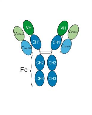
Volociximab (M200) is a chimeric human/murine IgG4 antibody IIA1 targeting integrin α5β1 (EC50=0.2 nM). Integrin α5β1 is a major fibronectin receptor involved in angiogenesis. Volociximab has antiangiogenic and antitumor activities and inhibits the proliferation of human umbilical vein vascular endothelial cells (HUVECs).
Nur für Forschungszwecke. Wir verkaufen nicht an Patienten.
- Reinheit : 98.597%
- CAS. Nr.: 558480-40-3
- Molecular Weight:145.5 kDa
-
Speicherung:
Please store the product under the recommended conditions in the Certificate of Analysis.


To place orders, for customer services and technical support, please contact: MedChemExpress USA
Tel: 609-228-6898 E-mail: [email protected] [email protected]


-
 Biologische Aktivität
-
Chemical Information
- Protokoll
- Reinheit & Dokumentation
- Verweise
-
 Help & FAQs


-

HY-P99291
Etaracizumabαvβ3 Integrin mAb -

HY-P99296
IntetumumabAnti-αv Integrin Monoclonal Antibody -

HY-P99003
Human IgG4 (S228P) kappa, Isotype ControlIgG4κ Isotype Control -
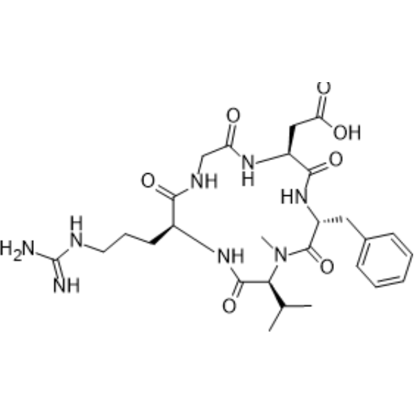
HY-16141
CilengitideIntegrin Inhibitor -
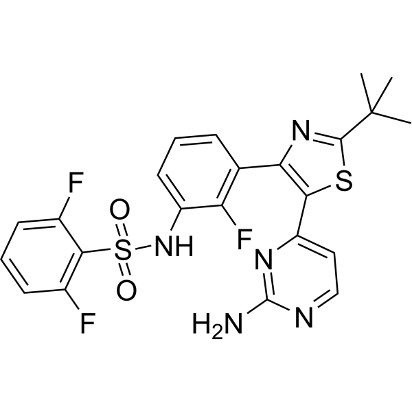
HY-14660
DabrafenibBRAF Inhibitor -

HY-P9911
Vedolizumab -

HY-P99320
TarextumabAnti-Notch2/3 Monoclonal Antibody -

HY-P9911A
Vedolizumab (anti-α4β7-integrin) -

HY-P99473
BersanlimabAnti-ICAM-1 Antibody -

HY-P99183
AbituzumabHumanised Anti-integrin αV mAb
Publications Citing Use of MedChemExpress (MCE) Volociximab
More

Biologische Aktivität
Beschreibung
Isotype
Human IgG4 kappa
Recommend Isotype Controls
Species Reactivity
Human
|
α5β1 0.2 nM (EC50) |
In Vitro
Volociximab (20, 40, 80, 160, 320 nM) specifically targets integrin α5β1 (Kd=0.367 nM), but not integrin α5α5 or β1β1[1].
Volociximab (0.01-100 nM; 4 d) inhibits the proliferation of human umbilical vein vascular endothelial cells (HUVEC) (IC50=0.2-0.5 nM) and (10 μg/mL; 16 h) induces apoptosis in schizogenic but not senescent Huvecs[1].
Volociximab (0.067-67 nM; 6 d) inhibits 0.1 μg/mL VEGF and/or bFGF induced HUVEC angiogenesis in vitro[1].
MedChemExpress (MCE) has not independently confirmed the accuracy of these methods. They are for reference only.
In Vivo
Volociximab (10 mg/kg; i.v.; twice weekly for 3 weeks) suppresses tumor progress in rabbits VX2 model[2].
MedChemExpress (MCE) has not independently confirmed the accuracy of these methods. They are for reference only.
Clinical Trial
| NCT Number | Sponsor | Condition | Start Date |
Phase

|
|---|---|---|---|---|
| NCT01329991 | Plexxikon| | 2011-05 | PHASE1 |
Gene ID
Accession
Conjugated
Unconjugated
Reconsititution
The product can be reconstituted/diluted with sterile PBS or saline.
Format
-
Human IgG4 kappa
Anwendung
ELISA, FACS, Functional assay
Verified Bioactivity
-
 Immobilized Integrin alpha 5 beta 1 Protein, Human (HEK293, Flag-His, HY-P73872) can bind Evinacumab. The EC50 for this effect is 13.19 ng/mL.


Chemical Information
-
CAS. Nr. 558480-40-3
-
Appearance Liquid
-
Molecular Weight 145.5 kDa
-
Color Colorless to light yellow
-
SMILES
[Volociximab]
-
Synonyms
M200; Eos 200-4
-
Versand
Shipping with dry ice.
-
Formulation
Please refer to the lot-specific COA for specific buffer information.
-
Speicherung
Please store the product under the recommended conditions in the Certificate of Analysis.


Publications (3)
-
Journal Impact Factor
-
Most Recent
-
Cancer Commun (Lond)
Blocking ITGA5 potentiates the efficacy of anti-PD-1 therapy on glioblastoma by remodeling tumor-associated macrophages. [Abstract]2025 Jun;45(6):677-701. PMID: 40084746 -
-
Oncogene
Fibroblast Activation Protein (FAP)+ cancer-associated fibroblasts induce macrophage M2-like polarization via the Fibronectin 1-Integrin α5β1 axis in breast cancer. [Abstract]2025 Jul;44(28):2396-2412. PMID: 40263422


Reinheit & Dokumentation
-
Data Sheet (261 KB)
-
SDS (251 KB)
- English - EN (251 KB)
- Français - FR (251 KB)
- Deutsch - DE (251 KB)
- Norwegian - NO (251 KB)
- Español - ES (251 KB)
- Swedish - SV (251 KB)
- Italian - IT (251 KB)
- Korean - KR (251 KB)
- Portuguese - PT (251 KB)
-
Inhibitory Antibodies User Guide (603 KB)
Verweise
[1]. Bhaskar V, et al. Volociximab, a chimeric integrin alpha5beta1 antibody, inhibits the growth of VX2 tumors in rabbits. Invest New Drugs. 2008 Feb;26(1):7-12. [Content Brief]
[2]. Ramakrishnan V, et al. Preclinical evaluation of an anti-alpha5beta1 integrin antibody as a novel anti-angiogenic agent. J Exp Ther Oncol. 2006;5(4):273-86. [Content Brief]

Calculators
Konzentration (Stammlösung) × Volumen (Stammlösung) = Konzentration (Ziellösung) × Volumen (Ziellösung)


Volociximab Related Classifications
HY-P99333 Related Classifications
 Powered by Bioz