



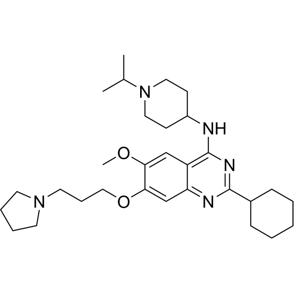
UNC0638
Based on 12 publication(s) in Google Scholar
UNC0638, a chemical probe, selectively inhibits G9a and GLP histone methyltransferases with IC50 of 15 nM and 19 nM, respectively. UNC0638 inhibits TNBC cell invasion and migration in vitro. UNC0638 is also an inhibitor of EHMT1/2 and induces fetal hemoglobin (HbF) expression in human erythroid progenitor cell culture. In addition, UNC0638 has anti-FMDV (foot-and-mouth disease virus) and anti-VSV (vesicular stomatitis virus) activities, with excellent potency and selectivity against multiple epigenetic and non-epigenetic targets.
Nos produits utilisent uniquement pour la recherche. Nous ne vendons pas aux patients.
- Pureté : 99.84%
- CAS No.: 1255580-76-7
- Formule: C30H47N5O2
- Masse moléculaire:509.73
-
Stockage:Powder -20°C, 3 years , 4°C, 2 years ; In solvent -80°C, 2 years , -20°C, 1 year

To place orders, for customer services and technical support, please contact: MedChemExpress USA
Tel: 609-228-6898 E-mail: [email protected] [email protected]


-
 Activité biologique
-
Chemical Information
-
Solvant et solubilité
- Protocole
- Pureté et documentation
- Références
-
 Help & FAQs


-
HY-10587
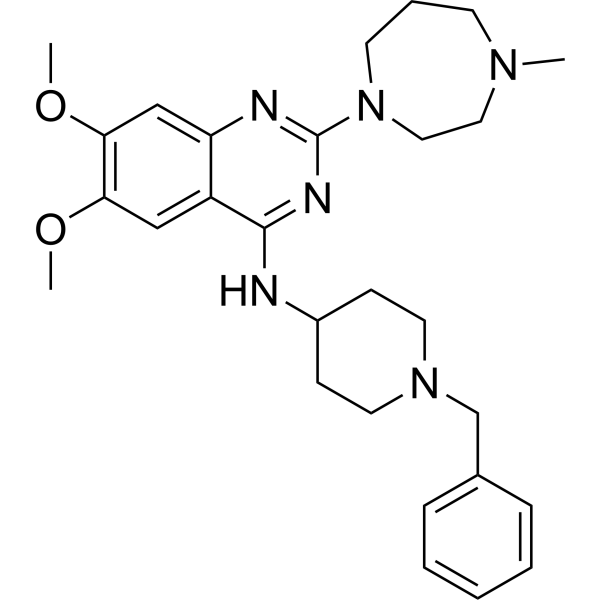
BIX-01294G9a/GLP Inhibitor -
HY-13980
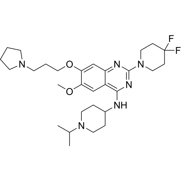
UNC0642G9a/GLP Inhibitor -
HY-12583
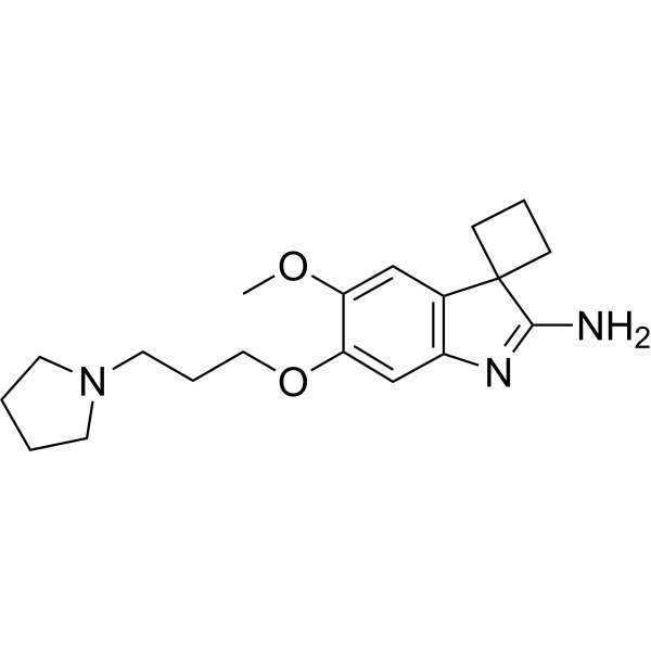
A-366G9a Inhibitor -
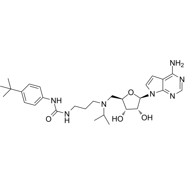
HY-15227
EPZ004777DOT1L Inhibitor -
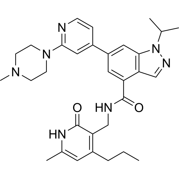
HY-13500
GSK343EZH2 Inhibitor -
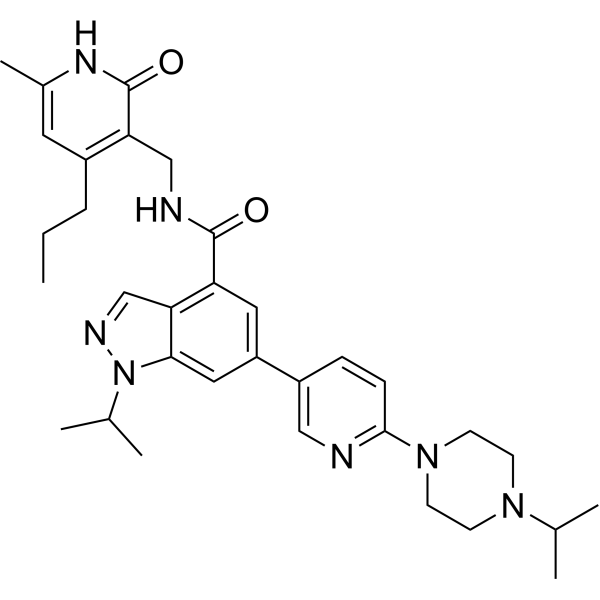
HY-15646
UNC1999EZH1/2 Inhibitor -
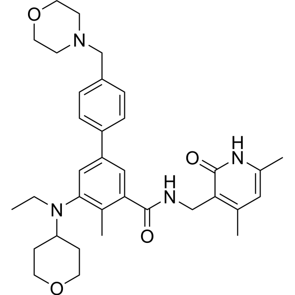
HY-13803
TazemetostatEZH2 Inhibitor -
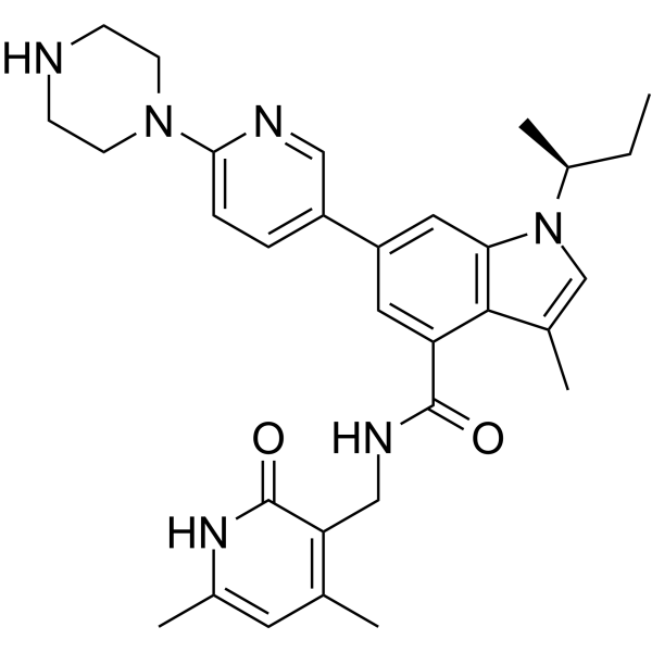
HY-13470
GSK126EZH2 Inhibitor -
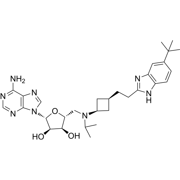
HY-15593
PinometostatDOT1L Inhibitor -
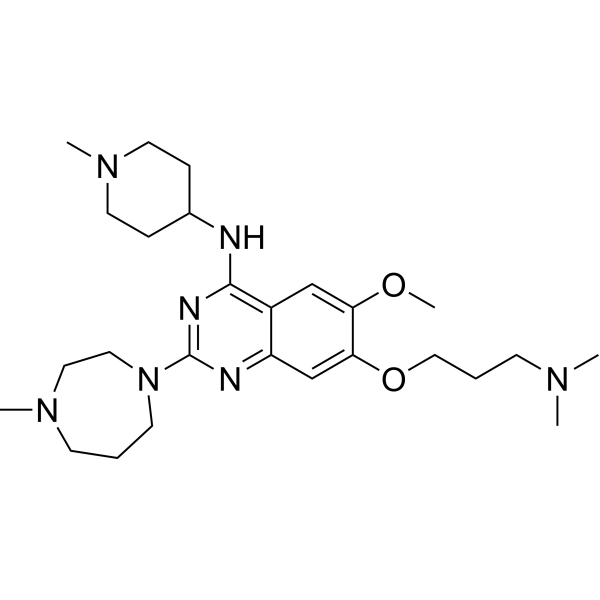
HY-10929
UNC0224G9a Inhibitor -
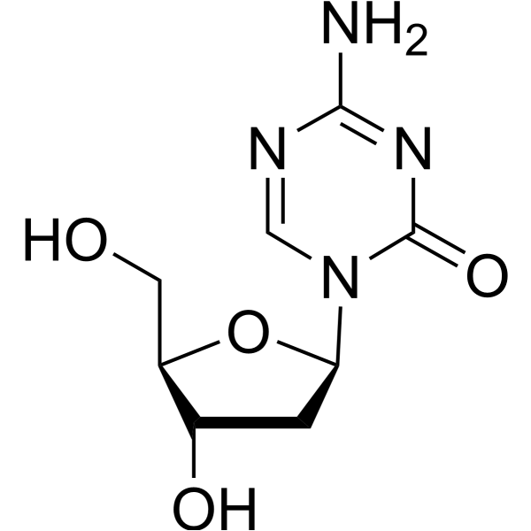
HY-A0004
DecitabineDNMT Inhibitor -
HY-15144
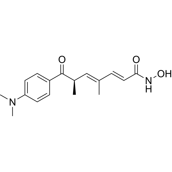
Trichostatin AHDAC Class I/II Inhibitor
Publications Citing Use of MedChemExpress (MCE) UNC0638
More- Cancer Res. 2026 Jun 10. [Abstract]
- Nat Commun. 2026 Feb 12;17(1):1214. [Abstract]
- Cell Death Dis. 2018 Jan 26;9(2):129. [Abstract]
- Acta Pharmacol Sin. 2025 Aug 19. [Abstract]
- Acta Pharmacol Sin. 2022 Feb;43(2):457-469. [Abstract]
- Proc Natl Acad Sci U S A. 2019 Feb 19;116(8):2961-2966. [Abstract]
- Cell Chem Biol. 2022 Jul 21;29(7):1153-1161.e5. [Abstract]
- Cell Biosci. 2023 Jan 12;13(1):7. [Abstract]
- Mol Med Rep. 2018 Feb;17(2):2239-2244. [Abstract]
- J Neurosci. 2025 Feb 5;45(6):e1790242024. [Abstract]
- Mol Carcinog. 2024 Nov;63(11):2119-2132. [Abstract]
- bioRxiv. 2023 Nov 17.
 Customer Validation & Images

-
WB
-
Cell Proliferation/Viability Assay
-
Cell Migration/Invasion Assay
-
Cell Proliferation/Viability Assay
-
RT-PCR
Voir tous les produits spécifiques à Isoform Histone Methyltransferase
More

Activité biologique
Description
IC50 & Target
|
EHMT2/G9a/KMT1C |
EHMT1/GLP/KMT1D |
Cellular Effect
|
Cell Line

|
Type | Value | Description | References |
|---|---|---|---|---|
| Calu-1 | EC50 |
1.3 μM
Compound: Chemical probe: UNC0638
|
Antiproliferative against human Calu-1 cells after 5 days by Cell-Titer Glo assay
Antiproliferative against human Calu-1 cells after 5 days by Cell-Titer Glo assay
|
[PMID: 26147105] |
| Calu-6 | EC50 |
1.5 μM
Compound: Chemical probe: UNC0638
|
Antiproliferative against human Calu-6 cells after 5 days by Cell-Titer Glo assay
Antiproliferative against human Calu-6 cells after 5 days by Cell-Titer Glo assay
|
[PMID: 26147105] |
| CWR22R | EC50 |
4.5 μM
Compound: 5, UNC0638
|
Cytotoxicity against human 22Rv1 cells assessed as cell viability after 48 hrs by MTT assay
Cytotoxicity against human 22Rv1 cells assessed as cell viability after 48 hrs by MTT assay
|
[PMID: 21780790] |
| CWR22R | IC50 |
0.048 μM
Compound: 5, UNC0638
|
Inhibition of G9a in human 22Rv1 cells assessed as reduction of H3K9me2 after 48 hrs by In-Cell Western assay
Inhibition of G9a in human 22Rv1 cells assessed as reduction of H3K9me2 after 48 hrs by In-Cell Western assay
|
[PMID: 21780790] |
| CWR22R | EC50 |
0.2 μM
Compound: Chemical probe: UNC0638
|
Antiproliferative against human 22RV1 cells after 5 days by Cell-Titer Glo assay
Antiproliferative against human 22RV1 cells after 5 days by Cell-Titer Glo assay
|
[PMID: 26147105] |
| Daoy | EC50 |
0.1 μM
Compound: Chemical probe: UNC0638
|
Antiproliferative against human Daoy cells after 7 days by Cell-Titer Glo assay
Antiproliferative against human Daoy cells after 7 days by Cell-Titer Glo assay
|
[PMID: 26147105] |
| DU-145 | EC50 |
0.7 μM
Compound: Chemical probe: UNC0638
|
Antiproliferative against human DU-145 cells after 5 days by Cell-Titer Glo assay
Antiproliferative against human DU-145 cells after 5 days by Cell-Titer Glo assay
|
[PMID: 26147105] |
| G-401 | EC50 |
0.4 μM
Compound: Chemical probe: UNC0638
|
Antiproliferative against human G-401 cells after 7 days by Cell-Titer Glo assay
Antiproliferative against human G-401 cells after 7 days by Cell-Titer Glo assay
|
[PMID: 26147105] |
| HCT-116 | EC50 |
11 μM
Compound: 5, UNC0638
|
Cytotoxicity against human HCT116 cells assessed as cell viability after 48 hrs by MTT assay
Cytotoxicity against human HCT116 cells assessed as cell viability after 48 hrs by MTT assay
|
[PMID: 21780790] |
| HCT-116 | EC50 |
11 μM
Compound: 5, UNC0638
|
Cytotoxicity against p53-deficient human HCT116 cells assessed as cell viability after 48 hrs by MTT assay
Cytotoxicity against p53-deficient human HCT116 cells assessed as cell viability after 48 hrs by MTT assay
|
[PMID: 21780790] |
| HCT-116 | IC50 |
0.21 μM
Compound: 5, UNC0638
|
Inhibition of G9a in human HCT116 cells assessed as reduction of H3K9me2 after 48 hrs by In-Cell Western assay
Inhibition of G9a in human HCT116 cells assessed as reduction of H3K9me2 after 48 hrs by In-Cell Western assay
|
[PMID: 21780790] |
| HCT-116 | IC50 |
0.24 μM
Compound: 5, UNC0638
|
Inhibition of G9a in p53-deficient human HCT116 cells assessed as reduction of H3K9me2 after 48 hrs by In-Cell Western assay
Inhibition of G9a in p53-deficient human HCT116 cells assessed as reduction of H3K9me2 after 48 hrs by In-Cell Western assay
|
[PMID: 21780790] |
| HCT-116 | IC50 |
3.35 μM
Compound: UNC0638
|
Antiproliferative activity against human HCT116 cells assessed as inhibition of cell viability after 72 hrs by MTT assay
Antiproliferative activity against human HCT116 cells assessed as inhibition of cell viability after 72 hrs by MTT assay
|
[PMID: 27720557] |
| HepG2 | IC50 |
3.7 μM
Compound: UNC0638
|
Antiproliferative activity against human HepG2 cells assessed as inhibition of cell viability after 72 hrs by MTT assay
Antiproliferative activity against human HepG2 cells assessed as inhibition of cell viability after 72 hrs by MTT assay
|
[PMID: 27720557] |
| HFF | IC50 |
2286 nM
Compound: 19; UNC0638
|
Cytotoxicity against HFF
Cytotoxicity against HFF
|
[PMID: 30366254] |
| HL-60 | IC50 |
23.75 μM
Compound: Chemical Probe: UNC0638
|
Cytotoxicity against human HL-60 cells assessed as decrease in cell viability measured after 48 hrs by MTT assay
Cytotoxicity against human HL-60 cells assessed as decrease in cell viability measured after 48 hrs by MTT assay
|
[PMID: 25978433] |
| HL-60 | IC50 |
27.38 μM
Compound: Chemical Probe: UNC0638
|
Cytotoxicity against human HL-60 cells assessed as decrease in cell viability measured after 24 hrs by MTT assay
Cytotoxicity against human HL-60 cells assessed as decrease in cell viability measured after 24 hrs by MTT assay
|
[PMID: 25978433] |
| HL-60 | EC50 |
6.5 μM
Compound: Chemical probe: UNC0638
|
Antiproliferative against human HL-60 cells after 5 days by Cell-Titer Glo assay
Antiproliferative against human HL-60 cells after 5 days by Cell-Titer Glo assay
|
[PMID: 26147105] |
| HT-1080 | EC50 |
0.5 nM
Compound: Chemical probe: UNC0638
|
Antiproliferative activity against human HT-1080 cells after 5 days by Cell-Titer-Glo assay
Antiproliferative activity against human HT-1080 cells after 5 days by Cell-Titer-Glo assay
|
[PMID: 26147105] |
| HT-1080 | EC50 |
0.5 μM
Compound: Chemical probe: UNC0638
|
Antiproliferative against human HT-1080 cells after 5 days by Cell-Titer Glo assay
Antiproliferative against human HT-1080 cells after 5 days by Cell-Titer Glo assay
|
[PMID: 26147105] |
| HT-1080 | EC50 |
1.2 nM
Compound: Chemical probe: UNC0638
|
Antiproliferative activity against human HT-1080 cells after 2 days by Cell-Titer-Glo assay
Antiproliferative activity against human HT-1080 cells after 2 days by Cell-Titer-Glo assay
|
[PMID: 26147105] |
| IMR-32 | EC50 |
>10 μM
Compound: Chemical probe: UNC0638
|
Antiproliferative against human IMR-32 cells after 7 days by Cell-Titer Glo assay
Antiproliferative against human IMR-32 cells after 7 days by Cell-Titer Glo assay
|
[PMID: 26147105] |
| IMR-90 | EC50 |
2.3 μM
Compound: 5, UNC0638
|
Cytotoxicity against human IMR90 cells assessed as cell viability after 48 hrs by MTT assay
Cytotoxicity against human IMR90 cells assessed as cell viability after 48 hrs by MTT assay
|
[PMID: 21780790] |
| IMR-90 | IC50 |
0.12 μM
Compound: 5, UNC0638
|
Inhibition of G9a in human IMR90 cells assessed as reduction of H3K9me2 after 48 hrs by In-Cell Western assay
Inhibition of G9a in human IMR90 cells assessed as reduction of H3K9me2 after 48 hrs by In-Cell Western assay
|
[PMID: 21780790] |
| K562 | EC50 |
5.3 μM
Compound: Chemical probe: UNC0638
|
Antiproliferative against human K562 cells after 5 days by Cell-Titer Glo assay
Antiproliferative against human K562 cells after 5 days by Cell-Titer Glo assay
|
[PMID: 26147105] |
| Kasumi 1 | EC50 |
1.1 μM
Compound: Chemical probe: UNC0638
|
Antiproliferative against human Kasumi-1 cells after 5 days by Cell-Titer Glo assay
Antiproliferative against human Kasumi-1 cells after 5 days by Cell-Titer Glo assay
|
[PMID: 26147105] |
| KG-1a | IC50 |
22.36 μM
Compound: Chemical Probe: UNC0638
|
Cytotoxicity against human KG-1a cells assessed as decrease in cell viability measured after 48 hrs by MTT assay
Cytotoxicity against human KG-1a cells assessed as decrease in cell viability measured after 48 hrs by MTT assay
|
[PMID: 25978433] |
| KG-1a | IC50 |
26.53 μM
Compound: Chemical Probe: UNC0638
|
Cytotoxicity against human KG-1a cells assessed as decrease in cell viability measured after 24 hrs by MTT assay
Cytotoxicity against human KG-1a cells assessed as decrease in cell viability measured after 24 hrs by MTT assay
|
[PMID: 25978433] |
| KMS-11 | EC50 |
4 μM
Compound: Chemical probe: UNC0638
|
Antiproliferative against human KMS-11 cells after 5 days by Cell-Titer Glo assay
Antiproliferative against human KMS-11 cells after 5 days by Cell-Titer Glo assay
|
[PMID: 26147105] |
| KMS-12-BM | EC50 |
4.4 μM
Compound: Chemical probe: UNC0638
|
Antiproliferative against human KMS-12-BM cells after 5 days by Cell-Titer Glo assay
Antiproliferative against human KMS-12-BM cells after 5 days by Cell-Titer Glo assay
|
[PMID: 26147105] |
| L-363 | EC50 |
3.9 μM
Compound: Chemical probe: UNC0638
|
Antiproliferative against human L-363 cells after 5 days by Cell-Titer Glo assay
Antiproliferative against human L-363 cells after 5 days by Cell-Titer Glo assay
|
[PMID: 26147105] |
| LN-229 | EC50 |
0.5 μM
Compound: Chemical probe: UNC0638
|
Antiproliferative against human LN-229 cells after 5 days by Cell-Titer Glo assay
Antiproliferative against human LN-229 cells after 5 days by Cell-Titer Glo assay
|
[PMID: 26147105] |
| LNCaP | EC50 |
0.3 μM
Compound: Chemical probe: UNC0638
|
Antiproliferative against human LNCaP cells after 5 days by Cell-Titer Glo assay
Antiproliferative against human LNCaP cells after 5 days by Cell-Titer Glo assay
|
[PMID: 26147105] |
| MCF7 | IC50 |
0.07 μM
Compound: 5, UNC0638
|
Inhibition of G9a in human MCF7 cells assessed as reduction of H3K9me2 after 48 hrs by In-Cell Western assay
Inhibition of G9a in human MCF7 cells assessed as reduction of H3K9me2 after 48 hrs by In-Cell Western assay
|
[PMID: 21780790] |
| MCF7 | IC50 |
81 nM
Compound: 11, UNC0638
|
Inhibition of G9a in human MCF7 cells after 48 hrs by clonogenic assay
Inhibition of G9a in human MCF7 cells after 48 hrs by clonogenic assay
|
[PMID: 22975593] |
| MCF7 | IC50 |
6.99 μM
Compound: UNC0638
|
Antiproliferative activity against human MCF7 cells assessed as inhibition of cell viability after 72 hrs by MTT assay
Antiproliferative activity against human MCF7 cells assessed as inhibition of cell viability after 72 hrs by MTT assay
|
[PMID: 27720557] |
| MDA-MB-231 | EC50 |
11 μM
Compound: 5, UNC0638
|
Cytotoxicity against human MDA-MB-231 cells assessed as cell viability after 48 hrs by MTT assay
Cytotoxicity against human MDA-MB-231 cells assessed as cell viability after 48 hrs by MTT assay
|
[PMID: 21780790] |
| MDA-MB-231 | IC50 |
0.081 μM
Compound: 5, UNC0638
|
Inhibition of G9a in human MDA-MB-231 cells assessed as reduction of H3K9me2 after 48 hrs by In-Cell Western assay
Inhibition of G9a in human MDA-MB-231 cells assessed as reduction of H3K9me2 after 48 hrs by In-Cell Western assay
|
[PMID: 21780790] |
| MDA-MB-231 | IC50 |
81 nM
Compound: 11, UNC0638
|
Inhibition of G9a in human MDA-MB-231 cells after 48 hrs by clonogenic assay
Inhibition of G9a in human MDA-MB-231 cells after 48 hrs by clonogenic assay
|
[PMID: 22975593] |
| MDA-MB-231 | IC50 |
81 nM
Compound: 5, UNC0638
|
Inhibition of lysine methyltransferase G9a in human MDA-MB-231 cells assessed as reduction of H3K9me2 cellular level by immunofluorescence in-cell Western assay
Inhibition of lysine methyltransferase G9a in human MDA-MB-231 cells assessed as reduction of H3K9me2 cellular level by immunofluorescence in-cell Western assay
|
[PMID: 24102134] |
| MDA-MB-231 | EC50 |
11 μM
Compound: 5, UNC0638
|
Cytotoxicity against human MDA-MB-231 cells after 48 hrs by Alamar Blue assay
Cytotoxicity against human MDA-MB-231 cells after 48 hrs by Alamar Blue assay
|
[PMID: 24102134] |
| MDA-MB-231 | IC50 |
3.56 μM
Compound: 47; UNC0638
|
Antiproliferative activity against human MDA-MB-231 cells assessed as inhibition of cell growth measured after 72 hrs by MTT assay
Antiproliferative activity against human MDA-MB-231 cells assessed as inhibition of cell growth measured after 72 hrs by MTT assay
|
[PMID: 35525212] |
| MDA-MB-231 | IC50 |
81 nM
Compound: 38; UNC0638
|
Reduction in H3K9me2 level in human MDA-MB-231 cells incubated for 48 hrs by ChIP assay
Reduction in H3K9me2 level in human MDA-MB-231 cells incubated for 48 hrs by ChIP assay
|
[PMID: 36528996] |
| MDA-MB-231 | EC50 |
11000 nM
Compound: 4
|
Antiproliferative activity against human MDA-MB-231 cells by MTT assay
Antiproliferative activity against human MDA-MB-231 cells by MTT assay
|
[PMID: 38799223] |
| MDA-MB-231 | IC50 |
81 nM
Compound: 4
|
Downregulation of H3K9me2 level in human MDA-MB-231 cells measured after 48 hrs by Western blot analysis
Downregulation of H3K9me2 level in human MDA-MB-231 cells measured after 48 hrs by Western blot analysis
|
[PMID: 38799223] |
| MDA-MB-231 | EC50 |
11 μM
Compound: 126, UNC0638
|
Cytotoxicity against human MDA-MB-231 cells by MTT assay
Cytotoxicity against human MDA-MB-231 cells by MTT assay
|
10.1039/C1MD00199J |
| MDA-MB-231 | IC50 |
81 nM
Compound: 126, UNC0638
|
Inhibition of G9a/GLP in human MDA-MB-231 cells assessed as reduction of H3K9me2 level after 48 hrs by flow cytometric analysis
Inhibition of G9a/GLP in human MDA-MB-231 cells assessed as reduction of H3K9me2 level after 48 hrs by flow cytometric analysis
|
10.1039/C1MD00199J |
| MOLT-16 | EC50 |
0.2 μM
Compound: Chemical probe: UNC0638
|
Antiproliferative against human MOLT-16 cells after 5 days by Cell-Titer Glo assay
Antiproliferative against human MOLT-16 cells after 5 days by Cell-Titer Glo assay
|
[PMID: 26147105] |
| MOLT-4 | GI50 |
188 nM
Compound: 5; UNC0638
|
Antiproliferative activity against human MOLT-4 cells assessed as inhibition of cell growth with replenishment of medium with compound on day 4 and 7 and measured after 12 days by ATPlite assay
Antiproliferative activity against human MOLT-4 cells assessed as inhibition of cell growth with replenishment of medium with compound on day 4 and 7 and measured after 12 days by ATPlite assay
|
[PMID: 36882960] |
| MOLT-4 | GI50 |
188 nM
Compound: 5; UNC0638
|
Antiproliferative activity against human MOLT-4 cells assessed as inhibition of cell growth with replenishment of medium with compound on day 4 and 7 by ATPlite assay
Antiproliferative activity against human MOLT-4 cells assessed as inhibition of cell growth with replenishment of medium with compound on day 4 and 7 by ATPlite assay
|
[PMID: 36882960] |
| MV4-11 | EC50 |
1.7 μM
Compound: Chemical probe: UNC0638
|
Antiproliferative against human MV4-11 cells after 5 days by Cell-Titer Glo assay
Antiproliferative against human MV4-11 cells after 5 days by Cell-Titer Glo assay
|
[PMID: 26147105] |
| NCI-H1975 | EC50 |
>10 μM
Compound: Chemical probe: UNC0638
|
Antiproliferative against human NCI-H1975 cells after 5 days by Cell-Titer Glo assay
Antiproliferative against human NCI-H1975 cells after 5 days by Cell-Titer Glo assay
|
[PMID: 26147105] |
| NCI-H2009 | EC50 |
3.9 μM
Compound: Chemical probe: UNC0638
|
Antiproliferative against human NCI-H2009 cells after 5 days by Cell-Titer Glo assay
Antiproliferative against human NCI-H2009 cells after 5 days by Cell-Titer Glo assay
|
[PMID: 26147105] |
| NCI-H358 | EC50 |
4.3 μM
Compound: Chemical probe: UNC0638
|
Antiproliferative against human NCI-H358 cells after 5 days by Cell-Titer Glo assay
Antiproliferative against human NCI-H358 cells after 5 days by Cell-Titer Glo assay
|
[PMID: 26147105] |
| NCI-H441 | EC50 |
8.7 μM
Compound: Chemical probe: UNC0638
|
Antiproliferative against human NCI-H441 cells after 5 days by Cell-Titer Glo assay
Antiproliferative against human NCI-H441 cells after 5 days by Cell-Titer Glo assay
|
[PMID: 26147105] |
| NCI-H522 | EC50 |
1.5 μM
Compound: Chemical probe: UNC0638
|
Antiproliferative against human NCI-H522 cells after 5 days by Cell-Titer Glo assay
Antiproliferative against human NCI-H522 cells after 5 days by Cell-Titer Glo assay
|
[PMID: 26147105] |
| NCI-H661 | EC50 |
4.9 μM
Compound: Chemical probe: UNC0638
|
Antiproliferative against human NCI-H661 cells after 5 days by Cell-Titer Glo assay
Antiproliferative against human NCI-H661 cells after 5 days by Cell-Titer Glo assay
|
[PMID: 26147105] |
| NCI-H929 | EC50 |
4.6 μM
Compound: Chemical probe: UNC0638
|
Antiproliferative against human NCI-H929 cells after 5 days by Cell-Titer Glo assay
Antiproliferative against human NCI-H929 cells after 5 days by Cell-Titer Glo assay
|
[PMID: 26147105] |
| OPM-2 | EC50 |
5.6 μM
Compound: Chemical probe: UNC0638
|
Antiproliferative against human OPM-2 cells after 5 days by Cell-Titer Glo assay
Antiproliferative against human OPM-2 cells after 5 days by Cell-Titer Glo assay
|
[PMID: 26147105] |
| PA-1 | EC50 |
0.2 μM
Compound: Chemical probe: UNC0638
|
Antiproliferative against human PA-1 cells after 7 days by Cell-Titer Glo assay
Antiproliferative against human PA-1 cells after 7 days by Cell-Titer Glo assay
|
[PMID: 26147105] |
| PC-3 | EC50 |
14 μM
Compound: 5, UNC0638
|
Cytotoxicity against human PC3 cells assessed as cell viability after 48 hrs by MTT assay
Cytotoxicity against human PC3 cells assessed as cell viability after 48 hrs by MTT assay
|
[PMID: 21780790] |
| PC-3 | IC50 |
0.059 μM
Compound: 5, UNC0638
|
Inhibition of G9a in human PC3 cells assessed as reduction of H3K9me2 after 48 hrs by In-Cell Western assay
Inhibition of G9a in human PC3 cells assessed as reduction of H3K9me2 after 48 hrs by In-Cell Western assay
|
[PMID: 21780790] |
| RD | IC50 |
11.17 μM
Compound: 47; UNC0638
|
Antiproliferative activity against human RD cells assessed as inhibition of cell growth incubated for 24 hrs
Antiproliferative activity against human RD cells assessed as inhibition of cell growth incubated for 24 hrs
|
[PMID: 35525212] |
| RD | IC50 |
2.91 μM
Compound: 47; UNC0638
|
Antiproliferative activity against human RD cells assessed as inhibition of cell growth incubated for 144 hrs
Antiproliferative activity against human RD cells assessed as inhibition of cell growth incubated for 144 hrs
|
[PMID: 35525212] |
| RD | IC50 |
3.05 μM
Compound: 47; UNC0638
|
Antiproliferative activity against human RD cells assessed as inhibition of cell growth incubated for 72 hrs
Antiproliferative activity against human RD cells assessed as inhibition of cell growth incubated for 72 hrs
|
[PMID: 35525212] |
| RD | CC50 |
34.2 μM
Compound: UNC-0638
|
Cytotoxicity against human RD cells assessed as reduction in cell viability incubated for 72 hrs by MTT assay
Cytotoxicity against human RD cells assessed as reduction in cell viability incubated for 72 hrs by MTT assay
|
[PMID: 37021456] |
| RPMI-8226 | EC50 |
5.1 μM
Compound: Chemical probe: UNC0638
|
Antiproliferative against human RPMI-8226 cells after 5 days by Cell-Titer Glo assay
Antiproliferative against human RPMI-8226 cells after 5 days by Cell-Titer Glo assay
|
[PMID: 26147105] |
| RS4-11 | EC50 |
3.1 μM
Compound: Chemical probe: UNC0638
|
Antiproliferative against human RS4-11 cells after 5 days by Cell-Titer Glo assay
Antiproliferative against human RS4-11 cells after 5 days by Cell-Titer Glo assay
|
[PMID: 26147105] |
| Sf9 | IC50 |
13 nM
Compound: UNC0638
|
Inhibition of N-terminal GST tagged human recombinant EHMT1 (794 to 1294 residues) expressed in baculovirus infected Sf9 insect cells assessed as reduction in conversion of SAH to AMP using histone H3 as substrate and SAH as cosubstrate incubated for 30 m
Inhibition of N-terminal GST tagged human recombinant EHMT1 (794 to 1294 residues) expressed in baculovirus infected Sf9 insect cells assessed as reduction in conversion of SAH to AMP using histone H3 as substrate and SAH as cosubstrate incubated for 30 m
|
[PMID: 31350126] |
| Sf9 | IC50 |
26 nM
Compound: UNC0638
|
Inhibition of N-terminal GST tagged human recombinant EHMT2 (785 to 1210 residues) expressed in baculovirus infected Sf9 insect cells assessed as reduction in conversion of SAH to AMP using histone H3 as substrate and SAH as cosubstrate incubated for 30 m
Inhibition of N-terminal GST tagged human recombinant EHMT2 (785 to 1210 residues) expressed in baculovirus infected Sf9 insect cells assessed as reduction in conversion of SAH to AMP using histone H3 as substrate and SAH as cosubstrate incubated for 30 m
|
[PMID: 31350126] |
| SJRH30 | IC50 |
2.48 μM
Compound: 47; UNC0638
|
Antiproliferative activity against human Rh30 cells assessed as inhibition of cell growth incubated for 144 hrs
Antiproliferative activity against human Rh30 cells assessed as inhibition of cell growth incubated for 144 hrs
|
[PMID: 35525212] |
| SJRH30 | IC50 |
2.64 μM
Compound: 47; UNC0638
|
Antiproliferative activity against human Rh30 cells assessed as inhibition of cell growth incubated for 72 hrs
Antiproliferative activity against human Rh30 cells assessed as inhibition of cell growth incubated for 72 hrs
|
[PMID: 35525212] |
| SJRH30 | IC50 |
5.01 μM
Compound: 47; UNC0638
|
Antiproliferative activity against human Rh30 cells assessed as inhibition of cell growth incubated for 24 hrs
Antiproliferative activity against human Rh30 cells assessed as inhibition of cell growth incubated for 24 hrs
|
[PMID: 35525212] |
| SK-BR-3 | EC50 |
0.6 μM
Compound: Chemical probe: UNC0638
|
Antiproliferative against human SK-BR-3 cells after 5 days by Cell-Titer Glo assay
Antiproliferative against human SK-BR-3 cells after 5 days by Cell-Titer Glo assay
|
[PMID: 26147105] |
| SK-N-MC | EC50 |
0.2 μM
Compound: Chemical probe: UNC0638
|
Antiproliferative against human SK-N-MC cells after 7 days by Cell-Titer Glo assay
Antiproliferative against human SK-N-MC cells after 7 days by Cell-Titer Glo assay
|
[PMID: 26147105] |
| SW982 | CC50 |
29.46 μM
Compound: UNC-0638
|
Cytotoxicity against human SW982 cells assessed as reduction in cell viability incubated for 72 hrs by MTT assay
Cytotoxicity against human SW982 cells assessed as reduction in cell viability incubated for 72 hrs by MTT assay
|
[PMID: 37021456] |
| THP-1 | EC50 |
4.9 μM
Compound: Chemical probe: UNC0638
|
Antiproliferative against human THP-1 cells after 5 days by Cell-Titer Glo assay
Antiproliferative against human THP-1 cells after 5 days by Cell-Titer Glo assay
|
[PMID: 26147105] |
| THP-1 | IC50 |
0.24 μM
Compound: 47; UNC0638
|
Antiproliferative activity against human THP-1 cells assessed as inhibition of cell growth incubated for 144 hrs by CellTiter-Glo luminescent cell viability assay
Antiproliferative activity against human THP-1 cells assessed as inhibition of cell growth incubated for 144 hrs by CellTiter-Glo luminescent cell viability assay
|
[PMID: 35525212] |
| THP-1 | IC50 |
0.34 μM
Compound: 47; UNC0638
|
Antiproliferative activity against human THP-1 cells assessed as inhibition of cell growth incubated for 72 hrs by CellTiter-Glo luminescent cell viability assay
Antiproliferative activity against human THP-1 cells assessed as inhibition of cell growth incubated for 72 hrs by CellTiter-Glo luminescent cell viability assay
|
[PMID: 35525212] |
| THP-1 | IC50 |
0.84 μM
Compound: 47; UNC0638
|
Antiproliferative activity against human THP-1 cells assessed as inhibition of cell growth incubated for 24 hrs by CellTiter-Glo luminescent cell viability assay
Antiproliferative activity against human THP-1 cells assessed as inhibition of cell growth incubated for 24 hrs by CellTiter-Glo luminescent cell viability assay
|
[PMID: 35525212] |
| U2OS | EC50 |
0.5 μM
Compound: Chemical probe: UNC0638
|
Antiproliferative against human U2OS cells after 7 days by Cell-Titer Glo assay
Antiproliferative against human U2OS cells after 7 days by Cell-Titer Glo assay
|
[PMID: 26147105] |
| U-937 | IC50 |
19.72 μM
Compound: Chemical Probe: UNC0638
|
Cytotoxicity against human U-937 cells assessed as decrease in cell viability measured after 48 hrs by MTT assay
Cytotoxicity against human U-937 cells assessed as decrease in cell viability measured after 48 hrs by MTT assay
|
[PMID: 25978433] |
| U-937 | IC50 |
21.4 μM
Compound: Chemical Probe: UNC0638
|
Cytotoxicity against human U-937 cells assessed as decrease in cell viability measured after 24 hrs by MTT assay
Cytotoxicity against human U-937 cells assessed as decrease in cell viability measured after 24 hrs by MTT assay
|
[PMID: 25978433] |
| ZR-75-30 | EC50 |
1.2 μM
Compound: Chemical probe: UNC0638
|
Antiproliferative against human ZR-75-30 cells after 7 days by Cell-Titer Glo assay
Antiproliferative against human ZR-75-30 cells after 7 days by Cell-Titer Glo assay
|
[PMID: 26147105] |
In Vitro
UNC0638, an inhibitor of G9a and GLP with excellent potency and selectivity over a wide range of epigenetic and non-epigenetic targets.The Ki of UNC0638 is determined to be 3.0±0.05 nM (n=2). Consistent with this, the Morrison Ki for UNC0638 is 3.7±0.2 nM (n=3). The selectivity of UNC0638 over a wide range of epigenetic targets is evaluated. Notably, UNC0638 is inactive against other H3K9 (SUV39H1 and SUV39H2), H3K27 (EZH2), H3K4 (SETD7, MLL and SMYD3), H3K79 (DOT1L) and H4K20 (SETD8) methyltransferases, as well as PRDM1, PRDM10 and PRDM12. In addition, UNC0638 is inactive against protein arginine methyltransferases PRMT1 and PRMT3, and HTATIP, a histone acetyltransferase. Of note, UNC0638 has weak but measurable activity against JMJD2E (IC50=4,500±1,100 nM), a Jumonji protein demethylase and DNA methyltransferase DNMT1 (IC50=107,000±6,000 nM). Nevertheless, the selectivity of UNC0638 for G9a and GLP over JMJD2E is >200-fold, and selectivity for G9a and GLP over DNMT1 is >5,000-fold[1]. UNC0638 is a type of small molecule that can specifically inhibit the enzyme activity of histone methyltransferase EHMT and reduce the H3K9 dimethylation (H3K9me2) levels in cells[2].
MedChemExpress (MCE) has not independently confirmed the accuracy of these methods. They are for reference only.
- Spheroid/Tumor Organoid Invasion Assay
- Ex Vivo Tissue Slice/Explant Invasion Assay
- RNA extraction experimental
- Patient-Derived Organoid Invasion Assay
- Cell migration
- Breast Cancer Modeling
- 3D Tumor Spheroid Invasion Assay
- Matrigel Transwell/Boyden Chamber Invasion Assay
- Invadopodia/Fluorescent Gelatin Degradation Assay
- Organotypic 3D Invasion Assay
- Cell invasion
- Matrigel/ECM Transwell Invasion Assay
- 3D Collagen/Hydrogel Matrix Invasion Assay


Chemical Information
-
CAS No. 1255580-76-7
-
Appearance Solid
-
Masse moléculaire 509.73
-
Formule C30H47N5O2
-
Color White to yellow
-
SMILES
CC(N1CCC(NC2=C3C=C(OC)C(OCCCN4CCCC4)=CC3=NC(C5CCCCC5)=N2)CC1)C
-
Livraison
Room temperature in continental US; may vary elsewhere.
-
Stockage
Powder -20°C 3 years 4°C 2 years In solvent -80°C 2 years -20°C 1 year


Publications (12)
-
Journal Impact Factor
-
Most Recent
-
Cancer Res
TNG917 is a Potent and Selective Inhibitor of Histone Lysine Methyltransferases EHMT1/2 that Enhances Anti-Tumor Immunity and Immunotherapy Efficacy. [Abstract]2026 Jun 10. PMID: 42268285 -
Nat Commun
Human iPSC-based Modeling of Pulmonary Fibrosis Reveals p300/CBP Inhibition Suppresses Alveolar Transitional Cell State. [Abstract]2026 Feb 12;17(1):1214. PMID: 41680175 -
Cell Death Dis
Targeting EHMT2 reverses EGFR-TKI resistance in NSCLC by epigenetically regulating the PTEN/AKT signaling pathway. [Abstract]2018 Jan 26;9(2):129. PMID: 29374157
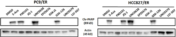
UNC0638 purchased from MedChemExpress. Usage Cited in: Cell Death Dis. 2018 Jan 26;9(2):129. [Abstract]
The effects of treatment with UNC0638 (10 μM) on cleaved PARP (Clv-PARP) expression in both PC9/ER and HCC827/ER cells. β-actin is used as a loading control.
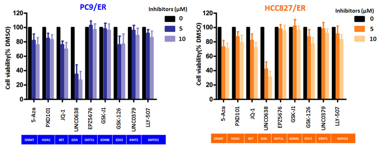
UNC0638 purchased from MedChemExpress. Usage Cited in: Cell Death Dis. 2018 Jan 26;9(2):129. [Abstract]
The EHMT2 inhibitor UNC0638 (5-10 μM; 48 h) effectively inhibited cell growth in both PC9/ER and HCC827/ER cells.
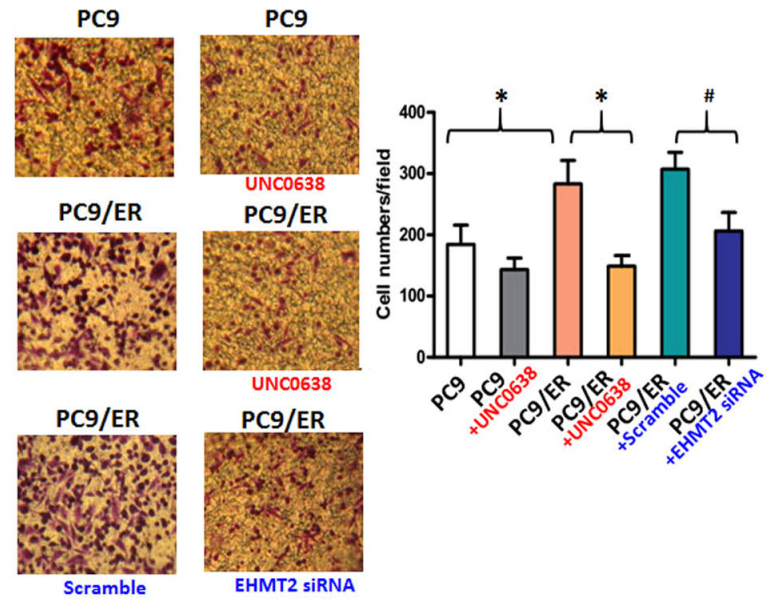
UNC0638 purchased from MedChemExpress. Usage Cited in: Cell Death Dis. 2018 Jan 26;9(2):129. [Abstract]
UNC0638 (100 nM; 48 h) suppressed cell migration in PC9 and PC9/ER cells.
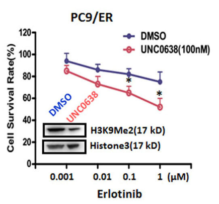
UNC0638 purchased from MedChemExpress. Usage Cited in: Cell Death Dis. 2018 Jan 26;9(2):129. [Abstract]
UNC0638 (100 nM; 48 h) pretreatment enhanced the sensitivity of PC9/ER cells to Erlotinib compared to non-pretreated cells.
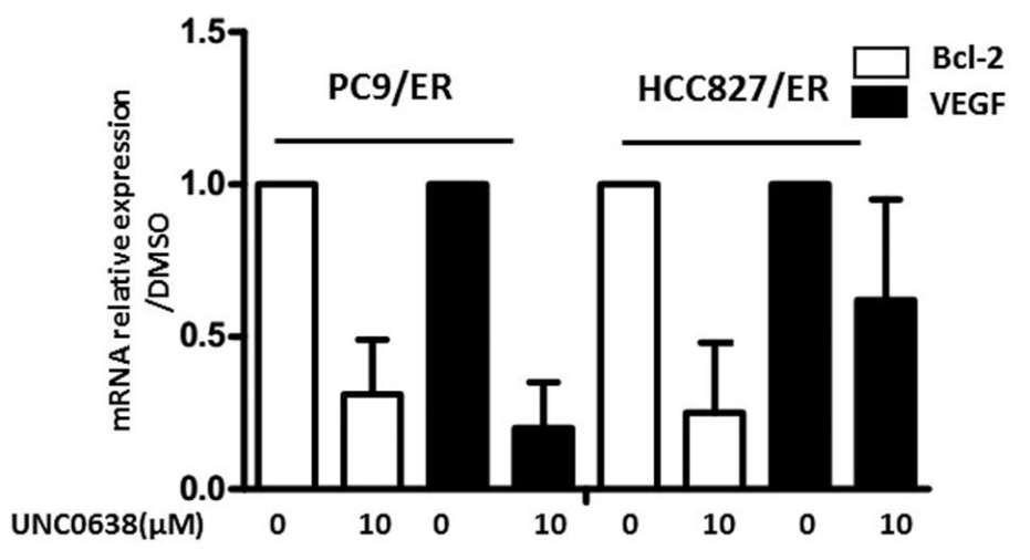
UNC0638 purchased from MedChemExpress. Usage Cited in: Cell Death Dis. 2018 Jan 26;9(2):129. [Abstract]
Treatment with UNC0638 (10 μM) resulted in the downregulation of Bcl-2 and VEGF in EGFR-TKI-resistant NSCLC cell lines.
-
Acta Pharmacol Sin
DNMT inhibition epigenetically restores the cGAS-STING pathway and activates RIG-I/MDA5-MAVS to enhance antitumor immunity. [Abstract]2025 Aug 19. PMID: 40830678 -
Acta Pharmacol Sin
2022 Feb;43(2):457-469. PMID: 33850273 -
Proc Natl Acad Sci U S A
2019 Feb 19;116(8):2961-2966. PMID: 30718431 -
Cell Chem Biol
Visualization of the dynamic interaction between nucleosomal histone H3K9 tri-methylation and HP1α chromodomain in living cells. [Abstract]2022 Jul 21;29(7):1153-1161.e5. PMID: 35728598 -
Cell Biosci
Activation of goblet cell Piezo1 alleviates mucus barrier damage in mice exposed to WAS by inhibiting H3K9me3 modification. [Abstract]2023 Jan 12;13(1):7. PMID: 36631841 -
Mol Med Rep
UNC0638, a G9a inhibitor, suppresses epithelial‑mesenchymal transition‑mediated cellular migration and invasion in triple negative breast cancer. [Abstract]2018 Feb;17(2):2239-2244. PMID: 29207160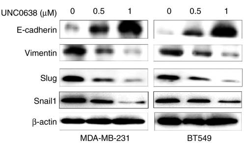
UNC0638 purchased from MedChemExpress. Usage Cited in: Mol Med Rep. 2018 Feb;17(2):2239-2244. [Abstract]
UNC0638 (0.5 and 1 µM; 24 h) suppressed triple negative breast cancer cell migration and invasion by regulating EMT-associated proteins. Western blots of EMT-associated proteins were performed.
-
J Neurosci
Histone Methyltransferase G9a in Primary Sensory Neurons Promotes Inflammatory Pain and Transcription of Trpa1 and Trpv1 via Bivalent Histone Modifications. [Abstract]2025 Feb 5;45(6):e1790242024. PMID: 39824634 -
Mol Carcinog
A novel high-risk model identified by epithelial-mesenchymal transition predicts prognosis and radioresistance in rectal cancer. [Abstract]2024 Nov;63(11):2119-2132. PMID: 39056517 -


Solvant et solubilité
In Vitro:
1M HCl : 100 mg/mL (196.18 mM; ultrasonic and adjust pH to 1 with HCl)
DMSO : 33.33 mg/mL (65.39 mM; Need ultrasonic; Hygroscopic DMSO has a significant impact on the solubility of product, please use newly opened DMSO)
Please refer to the solubility information to select the appropriate solvent. Once prepared, please aliquot and store the solution to prevent product inactivation from repeated freeze-thaw cycles.
Storage method and period of stock solution: -80°C, 2 years; -20°C, 1 year. When stored at -80°C, please use it within 2 years. When stored at -20°C, please use it within 1 year.
Please refer to the solubility information to select the appropriate solvent. Once prepared, please aliquot and store the solution to prevent product inactivation from repeated freeze-thaw cycles.
Storage method and period of stock solution: -80°C, 2 years; -20°C, 1 year. When stored at -80°C, please use it within 2 years. When stored at -20°C, please use it within 1 year.
Concentration (start) × Volume (start) = Concentration (final) × Volume (final)
In Vivo:
Select the appropriate dissolution method based on your experimental animal and administration route.
- For the following dissolution methods, please ensure to first prepare a clear stock solution using an In Vitro approach and then sequentially add co-solvents:
- To ensure reliable experimental results, the clarified stock solution can be appropriately stored based on storage conditions. As for the working solution for In Vivo experiments, it is recommended to prepare freshly and use it on the same day.
- The percentages shown for the solvents indicate their volumetric ratio in the final prepared solution. If precipitation or phase separation occurs during preparation, heat and/or sonication can be used to aid dissolution.
Add each solvent one by one: 10% DMSO 40% PEG300 5% Tween-80 45% Saline
Solubility: ≥ 2.5 mg/mL (4.90 mM); Clear solution
This protocol yields a clear solution of ≥ 2.5 mg/mL (saturation unknown).
Taking 1 mL working solution as an example, add 100 μL DMSO stock solution (25.0 mg/mL) to 400 μL PEG300, and mix evenly; then add 50 μL Tween-80 and mix evenly; then add 450 μL Saline to adjust the volume to 1 mL.
Preparation of Saline: Dissolve 0.9 g sodium chloride in ddH₂O and dilute to 100 mL to obtain a clear Saline solution.
Add each solvent one by one: 10% DMSO 90% (20% SBE-β-CD in Saline)
Solubility: ≥ 2.5 mg/mL (4.90 mM); Clear solution
This protocol yields a clear solution of ≥ 2.5 mg/mL (saturation unknown).
Taking 1 mL working solution as an example, add 100 μL DMSO stock solution (25.0 mg/mL) to 900 μL 20% SBE-β-CD in Saline, and mix evenly.
Preparation of 20% SBE-β-CD in Saline (4°C, storage for one week): 2 g SBE-β-CD powder is dissolved in 10 mL Saline, completely dissolve until clear.
In Vivo Dissolution Calculator
Please enter the basic information of animal experiments:
-
-
-
-
Recommended: Prepare an additional quantity of animals to account for potential losses during experiments.
Please enter your animal formula composition:
-
%DMSO +
Recommended: Keep the proportion of DMSO in working solution below 2% if your animal is weak.
-
%+
-
+%Tween-80 + +
-
%Saline +
The co-solvents required include: DMSO, . All of co-solvents are available by MedChemExpress (MCE). , Tween 80. All of co-solvents are available by MedChemExpress (MCE).
Working solution concentration: 0.22 mg/mL
Method for preparing stock solution: mg drug dissolved in μL DMSO. Stock solution concentration: mg/mL.
1. Take μL DMSO stock solution;
2. Add μL .
μL , mix evenly;
3. Then add μL Tween 80, mix evenly;
4. Then add μL
Please ensure that the stock solution in the first step is dissolved to a clear state, and add co-solvents in sequence. You can use ultrasonic heating (ultrasonic cleaner, recommended frequency 20-40 kHz), vortexing, etc. to assist dissolution.


Pureté et documentation
-
Fiche technique (277 KB)
-
SDS (392 KB)
- English - EN (392 KB)
- Français - FR (392 KB)
- Deutsch - DE (392 KB)
- Norwegian - NO (392 KB)
- Español - ES (392 KB)
- Swedish - SV (392 KB)
- Italian - IT (392 KB)
- Korean - KR (392 KB)
- Portuguese - PT (392 KB)
-
Instruction de manipulation (2659 KB)
Références
[1]. Vedadi M, et al. A chemical probe selectively inhibits G9a and GLP methyltransferase activity in cells. Nat Chem Biol. 2011 Jul 10;7(8):566-74. [Content Brief]
[2]. Fu L, et al. Effects of the Histone Methyltransferase Inhibitor UNC0638 on Histone H3K9 Dimethylation of Cultured Ovine Somatic Cells and Development of Resulting Early Cloned Embryos. Reprod Domest Anim. 2014 Apr;49(2):e21-5. [Content Brief]
[3]. Singh N, et al. Inhibition of EHMT2 Induces a Robust Antiviral Response Against Foot-and-Mouth Disease and Vesicular Stomatitis Virus Infections in Bovine Cells. J Interferon Cytokine Res. 2016 Jan;36(1):37-47. [Content Brief]
[4]. Nualkaew T, et al. UNC0638 induces high levels of fetal hemoglobin expression in β-thalassemia/HbE erythroid progenitor cells[J]. Annals of hematology, 2020, 99: 2027-2036. [Content Brief]


Complete Stock Solution Preparation Table
Please refer to the solubility information to select the appropriate solvent. Once prepared, please aliquot and store the solution to prevent product inactivation from repeated freeze-thaw cycles.
Storage method and period of stock solution: -80°C, 2 years; -20°C, 1 year. When stored at -80°C, please use it within 2 years. When stored at -20°C, please use it within 1 year.
| Optional Solvent | Concentration Solvent Mass | 1 mg | 5 mg | 10 mg | 25 mg |
|---|---|---|---|---|---|
| DMSO / 1M HCl | 1 mM | 1.9618 mL | 9.8091 mL | 19.6182 mL | 49.0456 mL |
| 5 mM | 0.3924 mL | 1.9618 mL | 3.9236 mL | 9.8091 mL | |
| 10 mM | 0.1962 mL | 0.9809 mL | 1.9618 mL | 4.9046 mL | |
| 15 mM | 0.1308 mL | 0.6539 mL | 1.3079 mL | 3.2697 mL | |
| 20 mM | 0.0981 mL | 0.4905 mL | 0.9809 mL | 2.4523 mL | |
| 25 mM | 0.0785 mL | 0.3924 mL | 0.7847 mL | 1.9618 mL | |
| 30 mM | 0.0654 mL | 0.3270 mL | 0.6539 mL | 1.6349 mL | |
| 40 mM | 0.0490 mL | 0.2452 mL | 0.4905 mL | 1.2261 mL | |
| 50 mM | 0.0392 mL | 0.1962 mL | 0.3924 mL | 0.9809 mL | |
| 60 mM | 0.0327 mL | 0.1635 mL | 0.3270 mL | 0.8174 mL | |
| 1M HCl | 80 mM | 0.0245 mL | 0.1226 mL | 0.2452 mL | 0.6131 mL |
| 100 mM | 0.0196 mL | 0.0981 mL | 0.1962 mL | 0.4905 mL |


UNC0638 Related Classifications
HY-15273 Related Classifications
 Powered by Bioz