



PC-766B
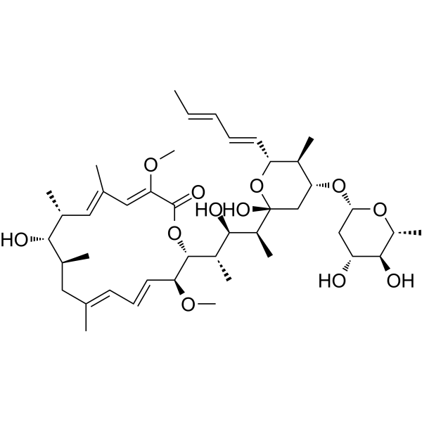
PC-766B is a macrolide antibiotic. PC-766B is active against Gram-positive bacteria, and some fungi and yeasts, but inactive against Gram-negative bacteria. PC-766B shows antitumor activity against murine tumor cells. PC-766B has weak inhibitory activity against Na+, K+-ATPase.
Nos produits utilisent uniquement pour la recherche. Nous ne vendons pas aux patients.
- CAS No.: 108375-77-5
- Formule: C43H68O12
- Masse moléculaire:776.99
-
Stockage:
Please store the product under the recommended conditions in the Certificate of Analysis.

To place orders, for customer services and technical support, please contact: MedChemExpress USA
Tel: 609-228-6898 E-mail: [email protected] [email protected]


-
 Activité biologique
-
Chemical Information
- Protocole
- Pureté et documentation
- Références
-
 Help & FAQs


Voir tous les produits spécifiques à Isoform Antibiotic
More

Activité biologique
Description
IC50 & Target
|
Macrolide |


Chemical Information
-
CAS No. 108375-77-5
-
Masse moléculaire 776.99
-
Formule C43H68O12
-
SMILES
O=C1O[C@@H]([C@H](/C=C/C=C(C[C@@H]([C@@H]([C@@H](/C=C(/C=C1\OC)C)C)O)C)\C)OC)[C@H]([C@H]([C@H](C)[C@]2(C[C@H]([C@@H]([C@H](O2)/C=C/C=C/C)C)O[C@H]3C[C@@H](O)[C@H](O)[C@H](O3)C)O)O)C
-
Synonyms
Antibiotic PC-766B
-
Structure Classification
-
Initial Source
Nocardia brasiliensis
-
Livraison
Room temperature in continental US; may vary elsewhere.
-
Stockage
Please store the product under the recommended conditions in the Certificate of Analysis.


Pureté et documentation
Références
[1]. Kumagai K, et, al. PC-766B, a new macrolide antibiotic produced by Nocardia brasiliensis. I. Taxonomy, fermentation and biological activity. J Antibiot (Tokyo). 1993 Jun;46(6):972-8. [Content Brief]
[2]. Kumagai K, et, al. PC-766B, a new macrolide antibiotic produced by Nocardia brasiliensis. II. Isolation, physico-chemical properties and structure elucidation. J Antibiot (Tokyo). 1993 Jul;46(7):1139-44. [Content Brief]

Calculators
Concentration (start) × Volume (start) = Concentration (final) × Volume (final)


PC-766B Related Classifications
HY-112661 Related Classifications
 Powered by Bioz