



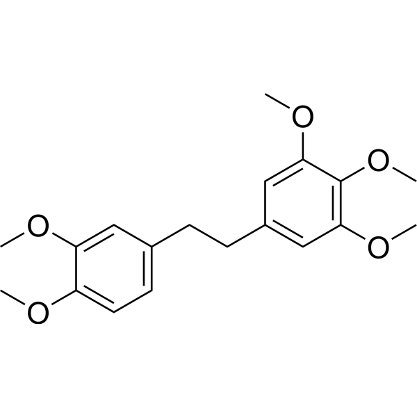
Chrysotobibenzyl
Chrysotobibenzyl can be isolated from stem of Dendrobium pulchellum. Chrysotobibenzyl inhibits lung cancer cell (H460 and H292) migration, invasion, filopodia formation via Cav-1, integrins β1, β3, and αν, and EMT suppressions. Chrysotobibenzyl also sensitizes lung cancer cell death mediated by Cisplatin (HY-17394).
연구목적의 판매만을 진행합니다. 환자를 대상으로 한 판매는 하지 않습니다.
- CAS No.: 108853-09-4
- 화학식: C19H24O5
- 분자량:332.39
-
보관:
Please store the product under the recommended conditions in the Certificate of Analysis.

To place orders, for customer services and technical support, please contact: MedChemExpress USA
Tel: 609-228-6898 E-mail: [email protected] [email protected]


-
 Biological Activity
-
Chemical Information
- Protocol
- 순도&문서
- References
-
 Help & FAQs




Biological Activity
제품 설명
Cellular Effect
|
Cell Line

|
Type | Value | Description | References |
|---|---|---|---|---|
| A549 | IC50 |
15 μM
Compound: 3a
|
Compound was tested for inhibition of cell growth of A-549 cells
Compound was tested for inhibition of cell growth of A-549 cells
|
10.1016/0960-894X(95)00008-H |
| HT-29 | IC50 |
15 μM
Compound: 3a
|
Compound was tested for inhibition of cell growth of HT-29 cells
Compound was tested for inhibition of cell growth of HT-29 cells
|
10.1016/0960-894X(95)00008-H |
| L1210 | IC50 |
20 μM
Compound: 5
|
Cytotoxicity against mouse L1210 cells after 24 hrs
Cytotoxicity against mouse L1210 cells after 24 hrs
|
[PMID: 3404149] |
| NCI-H23 | IC50 |
252.24 μM
Compound: 34
|
Anti-cancer activity against human NCI-H23 cells
Anti-cancer activity against human NCI-H23 cells
|
[PMID: 32711292] |
| P388 | IC50 |
15 μM
Compound: 3a
|
Compound was tested for inhibition of cell growth of P-388 cells
Compound was tested for inhibition of cell growth of P-388 cells
|
10.1016/0960-894X(95)00008-H |
| RAW | IC50 |
48.2 μM
Compound: 5
|
Inhibition of LPS and TNF-alpha-activated NO production in murine RAW 264.7 cells
Inhibition of LPS and TNF-alpha-activated NO production in murine RAW 264.7 cells
|
[PMID: 17253844] |


Chemical Information
-
CAS No. 108853-09-4
-
분자량 332.39
-
화학식 C19H24O5
-
SMILES
COC1=C(OC)C(OC)=CC(CCC2=CC=C(OC)C(OC)=C2)=C1
-
Structure Classification
-
선적
Room temperature in continental US; may vary elsewhere.
-
보관
Please store the product under the recommended conditions in the Certificate of Analysis.


순도&문서
References

Calculators
Concentration (start) × Volume (start) = Concentration (final) × Volume (final)


Chrysotobibenzyl Related Classifications
HY-122552 Related Classifications
 Powered by Bioz