



DI-1859
Based on 2 publication(s) in Google Scholar
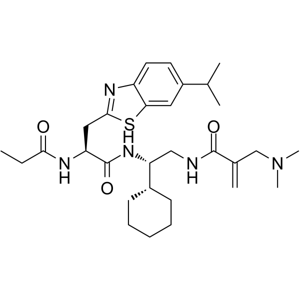
DI-1859 is a DCN1 and cullin 3 inhibitor with a Ki of <1 nM against human DCN1. DI-1859 forms a covalent bond with Cys115 of DCN1, cleaves its dimethylamino group, disrupts the DCN1-UBC12 interaction, and selectively inhibits the neddylation modification of cullin 3 relative to other cullins. DI-1859 inactivates CRL3, promotes the accumulation of NRF2 and SLC7A11, upregulates the expression of HO-1 and NQO1, reduces reactive oxygen species (ROS) levels, enhances insulin signaling, promotes insulin secretion, activates RhoA, regulates the phosphorylation of AKT, without completely blocking the neddylation modification of cullin 3 or restoring the expression of IRS-1. DI-1859 can be used in the research of Acetaminophen (HY-66005)-induced hepatotoxicity, hyperglycemia, metabolic dysfunction-associated steatotic liver disease, insulin resistance, breast cancer and lung cancer.
For research use only. We do not sell to patients.
- Purity : 99.62%
- CAS No.: 2247061-09-0
- Formula: C30H45N5O3S
- Molecular Weight:555.78
-
Storage:Powder -20°C, 3 years , 4°C, 2 years ; In solvent -80°C, 6 months , -20°C, 1 month

To place orders, for customer services and technical support, please contact: MedChemExpress USA
Tel: 609-228-6898 E-mail: [email protected] [email protected]


-
 Biological Activity
-
Chemical Information
-
Solvent & Solubility
- Protocols
- Purity & Documentation
- References
-
 Help & FAQs


-
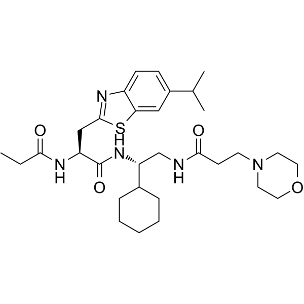
HY-124602
DI-591DCN1-UBC12 Inhibitor -
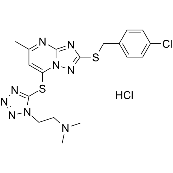
HY-126075A
WS-383DCN1-UBC12 Inhibitor -
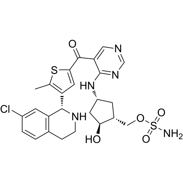
HY-111789
SubasumstatSUMOylation Inhibitor -
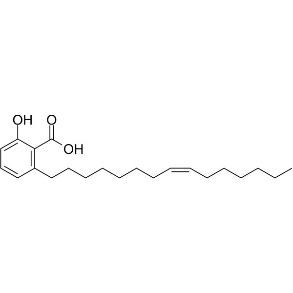
HY-N0077
Ginkgolic acidE1/E2/E3 Enzyme Inhibitor -
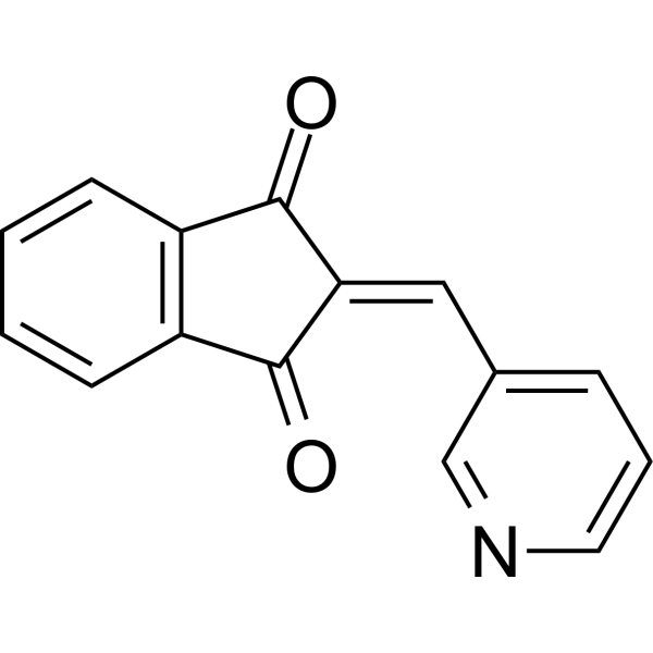
HY-19817
PRT4165E1/E2/E3 Enzyme Inhibitor -
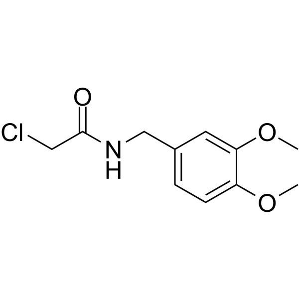
HY-101836
DKM 2-93UBA5 Inhibitor -
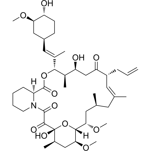
HY-13756
TacrolimusCalcineurin Inhibitor -
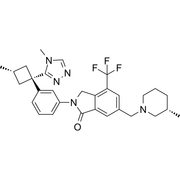
HY-141432
NX-1607Cbl-b Inhibitor -
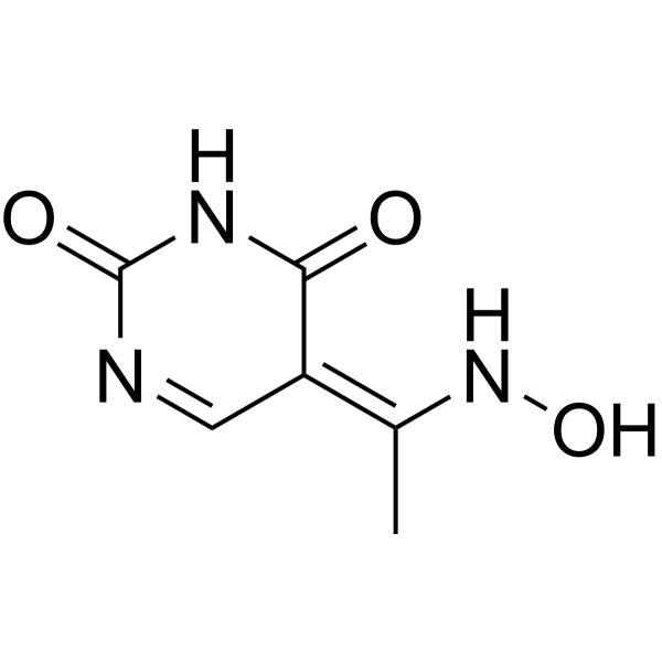
HY-103236
NSC232003UHRF1 Inhibitor -
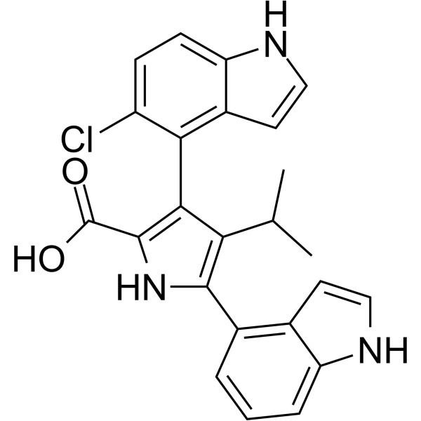
HY-139514
RB-3PRC1 Inhibitor -
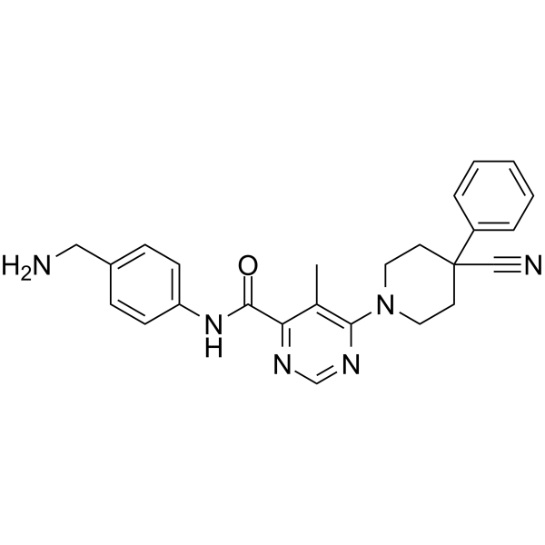
HY-120929
BI8622HUWE1 Inhibitor -
HY-141891
M435-1279UBE2T Inhibitor
Publications Citing Use of MedChemExpress (MCE) DI-1859
More

Biological Activity
Description
|
DCN1 <1 nM (Ki) |
cullin 3 |
Akt |
CRL3 |
Nrf2 |
SLC7A11 |
HO-1 |
NQO1 |
RhoA |
In Vitro
DI-1859 (1:1.2 protein-to-compound molar ratio; overnight) forms a covalent bond with Cys115 of recombinant human DCN1, with cleavage of its dimethylamino group, and maintains similar noncovalent interactions with DCN1 as the parent compound DI-591[1].
DI-1859 (1.2-fold molar excess relative to DCN3; 180 min) shows no detectable covalent reactivity with recombinant human DCN3 protein after 3 hours of incubation, demonstrating specificity for DCN1[1].
DI-1859 (250-500 nM; 1-2 h) selectively inhibits Cul3 neddylation, stabilizes IRS1 protein, and enhances insulin-stimulated AKT phosphorylation in AML12 mouse hepatocytes[2].
DI-1859 (250 nM; 1 h) inhibits Cul3 neddylation, stabilizes IRS1 protein, and enhances insulin-stimulated AKT phosphorylation in differentiated C2C12 mouse myotubes[2].
DI-1859 (100 nM; 1-3 h) potentiates glucose-stimulated insulin secretion in INS-1 832/13 rat pancreatic β cells via RhoA activation and cytoskeleton remodeling, as evidenced by the loss of effect when RhoA or F-actin dynamics are inhibited[2].
DI-1859 inhibits Cul3 neddylation and increases insulin secretion under both basal (3.5 mM) and glucose-stimulated (10 mM) conditions in cultured human pancreatic islets[2].
DI-1859 (100 nM; 1-2 h) does not promote glycolytic flux or overall glycolysis activity in INS-1 832/13 rat pancreatic β cells[2].
DI-1859 (All tested concentrations; 16 h) produces only modest restoration of insulin signaling via incomplete inhibition of CUL3 neddylation in H118Y SUN1-expressing Huh7 cells[3].
MedChemExpress (MCE) has not independently confirmed the accuracy of these methods. They are for reference only.
-
Cell Line:Osteosarcoma U2OS cells, immortalized THLE2 liver cells, breast cancer MDA-MB-231 cells, esophageal cancer KYSE70 cells, colon cancer HCT116 cells
-
Concentration:0.3-100 nM
-
Incubation Time:24 h
-
Result:Significantly reduced neddylated cullin 3 levels at concentrations as low as 0.3 nM, with profound inhibition achieved at 1-3 nM.
Was >300 times more potent than DI-591 across all tested cell lines.
Was ~30 times more potent than MLN4924 in THLE2 cells.
Had no effect on neddylation of cullin 1, 2, 4A, 4B, or 5 at concentrations up to 1000 nM.
Induced significant accumulation of NRF2 (a CRL3 substrate) at concentrations as low as 0.3-1 nM.
Dose-dependently upregulated HO-1 protein.
Had no effect on CRL1 substrates p21 and BIM, or CRL4A substrate CDT1 at concentrations up to 100 nM.
-
Cell Line:AML12 mouse hepatocytes
-
Concentration:250 nM (pre-incubation for insulin stimulation); 250-500 nM (Cul3 neddylation analysis)
-
Incubation Time:1 h (pre-incubation followed by 0-8 h insulin stimulation); 2 h (Cul3 neddylation analysis)
-
Result:Selectively inhibited Cul3 neddylation without affecting Cul1, Cul2, or Cul4B neddylation in cells treated for 2 hours.
Stabilized IRS1 protein in serum/ITS-starved cells pre-treated with 250 nM DI-1859.
Enhanced and prolonged insulin-stimulated AKT phosphorylation (S473) compared to vehicle-treated cells over 8 hours of insulin stimulation.
-
Cell Line:differentiated C2C12 mouse myotubes
-
Concentration:250 nM
-
Incubation Time:1 h (pre-incubation followed by 0-8 h insulin stimulation)
-
Result:Inhibited Cul3 neddylation in C2C12 myotubes over 8 hours of insulin stimulation.
Stabilized IRS1 protein in C2C12 myotubes over 8 hours of insulin stimulation.
Enhanced and prolonged insulin-stimulated AKT phosphorylation (S473) compared to vehicle-treated cells over 8 hours of insulin stimulation.
-
Cell Line:Huh7 cells stably expressing H118Y SUN1-mCherry, parental Huh7 cells, WT Huh7 cells
-
Concentration:All tested concentrations
-
Incubation Time:16 h
-
Result:Did not completely block CUL3 neddylation or restore IRS-1 expression in H118Y SUN1-expressing Huh7 cells.
Produced only a modest increase in insulin-stimulated AKT Ser473 phosphorylation in H118Y SUN1-expressing Huh7 cells.
Showed persistent CUL3 neddylation at all tested concentrations in WT, H118Y SUN1-expressing, and parental Huh7 cells.
-
Cell Line:BT549, MCF7, T47D, H358
-
Concentration:1-3 μM
-
Incubation Time:24 h
-
Result:Caused dose-dependent accumulation of SLC7A11 protein across all tested cell lines.
In Vivo
DI-1859 (10 mg/kg; i.p.; single dose) fails to lower blood glucose in streptozotocin-induced insulin-deficient diabetic male C57BL/6J mice, confirming its glucose-lowering effect is insulin-dependent[2].
MedChemExpress (MCE) has not independently confirmed the accuracy of these methods. They are for reference only.
-
Animal Model:C57BL/6 (male, ~8 weeks old)[1]
-
Dosage:25 mg/kg
-
Administration:i.p.; single dose
-
Result:Induced robust upregulation of NRF2 protein in mouse liver, with the effect persisting for >24 hours.
-
Animal Model:C57BL/6 (male, ~8 weeks old, acetaminophen-induced liver toxicity model)[1]
-
Dosage:25 mg/kg; 50 mg/kg
-
Administration:i.p.; once daily; 3 days
-
Result:Reduced acetaminophen-induced serum ALT elevation by 20% at 24 hours, largely abolished tissue distortion, and reduced liver damage at 25 mg/kg in prevention setting.
Reduced acetaminophen-induced serum ALT elevation by 90% at 24 hours and nearly completely prevented liver tissue damage at 50 mg/kg in prevention setting.
Reduced liver reactive oxygen species (ROS) levels at both doses in prevention setting.
Reduced acetaminophen-induced serum ALT elevation by ~50% at 24 hours, significantly reduced liver tissue damage, and lowered liver ROS levels at both 25 mg/kg and 50 mg/kg in treatment setting.
-
Animal Model:C57BL/6 (male, ~8 weeks old)[1]
-
Dosage:50 mg/kg
-
Administration:i.p.; daily; 14 days
-
Result:Was well tolerated, with no changes in body weight, organ weights, hematologic parameters, or observable tissue damage in liver, heart, kidney, lung, intestine, stomach, pancreas, colon, or spleen compared to vehicle-treated mice.
-
Animal Model:C57BL/6J (male, Western diet-induced obese)[2]
-
Dosage:10 mg/kg
-
Administration:i.p.; single dose
-
Result:Significantly decreased blood glucose (p<0.05).
Elevated blood insulin (p<0.05).
Elevated c-peptide (p<0.05).
Improved glucose tolerance relative to vehicle control.
-
Animal Model:C57BL/6J (male, streptozotocin-induced diabetic); C57BL/6J (male, non-diabetic control)[2]
-
Dosage:10 mg/kg
-
Administration:i.p.; single dose
-
Result:Did not significantly alter blood glucose levels in streptozotocin-induced diabetic mice.
Did not significantly alter insulin levels in streptozotocin-induced diabetic mice.
Significantly decreased blood glucose in non-diabetic control mice.
Significantly increased blood insulin in non-diabetic control mice.
- Kinase activity and phosphorylation assays
- RNA extraction experimental
- Western Blot
- ROS/oxidative-stress fluorescent staining
- Research Protocol for Endocrine Diseases
- Human Islet Cell Culture
- Ferroptosis Solutions
- Protocol for Kinase activity and phosphorylation assays
- Hepatotoxicity Study
- Research Protocol for Metabolic Diseases
- Breast Cancer Modeling


Chemical Information
-
CAS No. 2247061-09-0
-
Appearance Solid
-
Molecular Weight 555.78
-
Formula C30H45N5O3S
-
Color White to off-white
-
SMILES
CN(C)CC(C(NC[C@H](C1CCCCC1)NC([C@@H](NC(CC)=O)CC(SC2=C3)=NC2=CC=C3C(C)C)=O)=O)=C
-
Shipping
Room temperature in continental US; may vary elsewhere.
-
Storage
Powder -20°C 3 years 4°C 2 years In solvent -80°C 6 months -20°C 1 month


Publications (2)
-
Journal Impact Factor
-
Most Recent
-
Nat Commun
2026 Apr 29. PMID: 42056084 -
bioRxiv
The inner nuclear membrane protein SUN1 regulates cullin-3 neddylation to maintain insulin signaling. [Abstract]2026 Apr 20:2026.04.16.718478. PMID: 42079055


Solvent & Solubility
In Vitro:
DMSO : 100 mg/mL (179.93 mM; ultrasonic and warming and heat to 60°C; Hygroscopic DMSO has a significant impact on the solubility of product, please use newly opened DMSO)
Please refer to the solubility information to select the appropriate solvent. Once prepared, please aliquot and store the solution to prevent product inactivation from repeated freeze-thaw cycles.
Storage method and period of stock solution: -80°C, 6 months; -20°C, 1 month. When stored at -80°C, please use it within 6 months. When stored at -20°C, please use it within 1 month.
Please refer to the solubility information to select the appropriate solvent. Once prepared, please aliquot and store the solution to prevent product inactivation from repeated freeze-thaw cycles.
Storage method and period of stock solution: -80°C, 6 months; -20°C, 1 month. When stored at -80°C, please use it within 6 months. When stored at -20°C, please use it within 1 month.
Concentration (start) × Volume (start) = Concentration (final) × Volume (final)


Purity & Documentation
-
Data Sheet (301 KB)
-
SDS (254 KB)
- English - EN (254 KB)
- Français - FR (254 KB)
- Deutsch - DE (254 KB)
- Norwegian - NO (254 KB)
- Español - ES (254 KB)
- Swedish - SV (254 KB)
- Italian - IT (254 KB)
- Korean - KR (254 KB)
- Portuguese - PT (254 KB)
-
Handling Instructions (2659 KB)
References
[1]. Zhou H, et al. Selective inhibition of cullin 3 neddylation through covalent targeting DCN1 protects mice from acetaminophen-induced liver toxicity. Nature communications. 2021 May 11;12(1):2621. [Content Brief]
[2]. Gu L, et al. A selective Cullin 3 RING E3 ligase inhibitor attenuates hyperglycemia via dual insulin sensitizing and insulinotropic action. bioRxiv [Preprint]. 2026 Feb 1:2026.01.28.702366. [Content Brief]
[3]. Upadhyay KK, et al. The inner nuclear membrane protein SUN1 regulates cullin-3 neddylation to maintain insulin signaling. bioRxiv [Preprint]. 2026 Apr 20:2026.04.16.718478. [Content Brief]
[4]. Zhou Q, et al. The CRL3 ubiquitin ligase-USP18 axis coordinately regulates cystine uptake and ferroptosis by modulating SLC7A11. Proceedings of the National Academy of Sciences of the United States of America. 2024 Jul 09;121(28):e2320655121. [Content Brief]


Complete Stock Solution Preparation Table
Please refer to the solubility information to select the appropriate solvent. Once prepared, please aliquot and store the solution to prevent product inactivation from repeated freeze-thaw cycles.
Storage method and period of stock solution: -80°C, 6 months; -20°C, 1 month. When stored at -80°C, please use it within 6 months. When stored at -20°C, please use it within 1 month.
| Optional Solvent | Concentration Solvent Mass | 1 mg | 5 mg | 10 mg | 25 mg |
|---|---|---|---|---|---|
| DMSO | 1 mM | 1.7993 mL | 8.9964 mL | 17.9927 mL | 44.9818 mL |
| 5 mM | 0.3599 mL | 1.7993 mL | 3.5985 mL | 8.9964 mL | |
| 10 mM | 0.1799 mL | 0.8996 mL | 1.7993 mL | 4.4982 mL | |
| 15 mM | 0.1200 mL | 0.5998 mL | 1.1995 mL | 2.9988 mL | |
| 20 mM | 0.0900 mL | 0.4498 mL | 0.8996 mL | 2.2491 mL | |
| 25 mM | 0.0720 mL | 0.3599 mL | 0.7197 mL | 1.7993 mL | |
| 30 mM | 0.0600 mL | 0.2999 mL | 0.5998 mL | 1.4994 mL | |
| 40 mM | 0.0450 mL | 0.2249 mL | 0.4498 mL | 1.1245 mL | |
| 50 mM | 0.0360 mL | 0.1799 mL | 0.3599 mL | 0.8996 mL | |
| 60 mM | 0.0300 mL | 0.1499 mL | 0.2999 mL | 0.7497 mL | |
| 80 mM | 0.0225 mL | 0.1125 mL | 0.2249 mL | 0.5623 mL | |
| 100 mM | 0.0180 mL | 0.0900 mL | 0.1799 mL | 0.4498 mL |


DI-1859 Related Classifications
HY-145733 Related Classifications
 Powered by Bioz