



Bacoside A3
Based on 1 publication(s) in Google Scholar
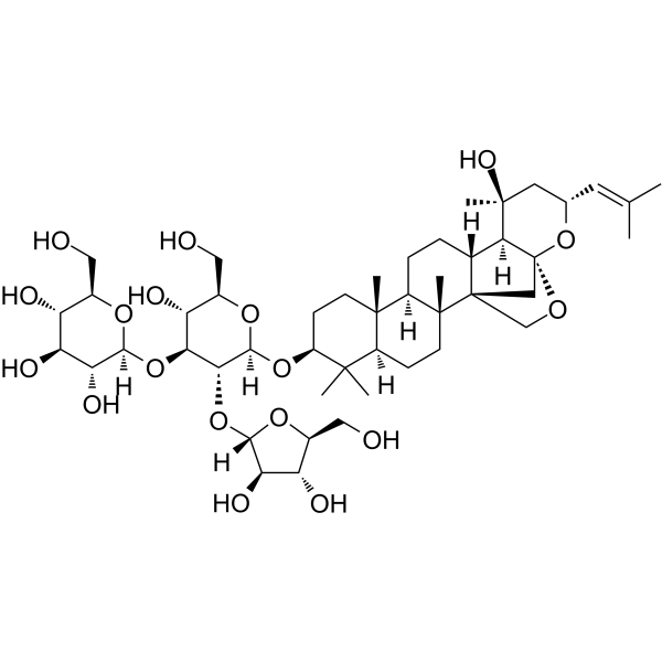
Bacoside A3 is a triterpenoid saponin isolated from Bacopa monnieri. Bacoside A3 has neuroprotective activity, downregulating β-amyloid-induced inflammatory responses and inhibiting neuronal apoptosis. Bacoside A3 can be used in the study of Alzheimer's disease.
For research use only. We do not sell to patients.
- Purity : 99.85%
- CAS No.: 157408-08-7
- Formula: C47H76O18
- Molecular Weight:929.10
-
Storage:
4°C, protect from light
* In solvent : -80°C, 6 months; -20°C, 1 month (protect from light)

To place orders, for customer services and technical support, please contact: MedChemExpress USA
Tel: 609-228-6898 E-mail: [email protected] [email protected]


-
 Biological Activity
-
Chemical Information
-
Solvent & Solubility
- Protocols
- Purity & Documentation
- References
-
 Help & FAQs


-
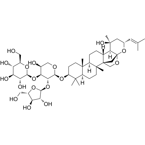
HY-N5140
Bacopaside XSaponin -

HY-10071
Y-27632ROCK Inhibitor -

HY-10431
SB-431542TGF-β Receptor Kinase Inhibitor -
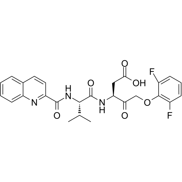
HY-12305
Q-VD-OPhPan-caspase Inhibitor -

HY-N5064R
Bacoside A3 (Standard)Neuroprotective Agent -
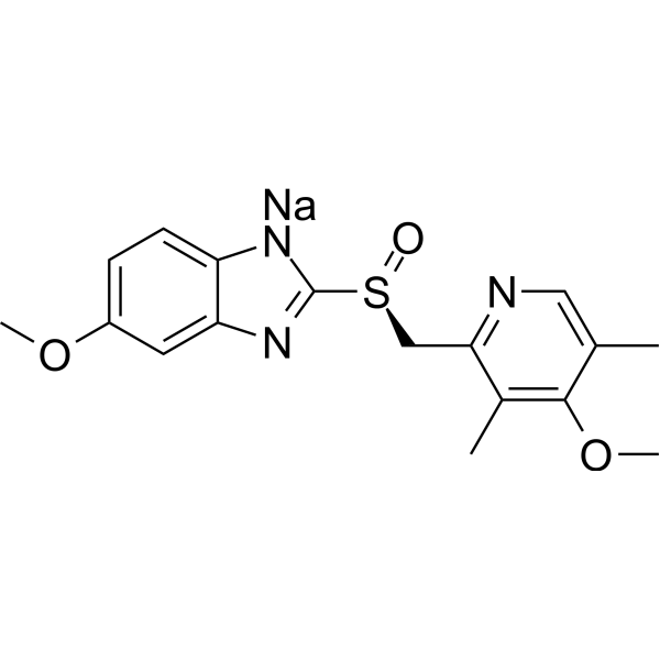
HY-17023
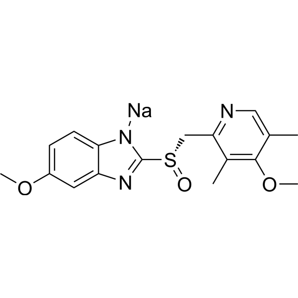
Esomeprazole sodiumProton Pump Inhibitor -
HY-B1213
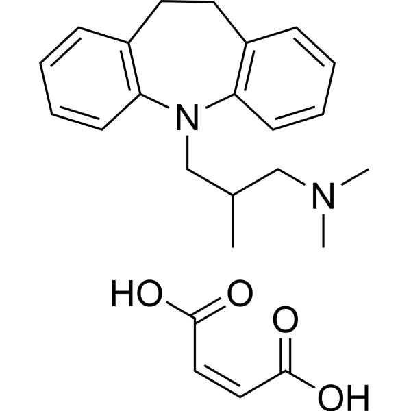
Trimipramine maleate5-HT Receptor Antagonist -
HY-17023R
Esomeprazole sodium (Standard)Proton Pump Inhibitor -

HY-B1213R
Trimipramine maleate (Standard)5-HT Receptor Antagonist
Publications Citing Use of MedChemExpress (MCE) Bacoside A3
More

Biological Activity
Description
In Vitro
Bacoside A3 (0.5-5 μM; 24 h) prevents β-amyloid (10 μM)-mediated growth inhibition of U87MG cells[1].
MedChemExpress (MCE) has not independently confirmed the accuracy of these methods. They are for reference only.
-
Cell Line:U87MG cells
-
Concentration:0.5, 1, 1.5, 2.5 and 5 μM
-
Incubation Time:24 h
-
Result:Inhibited the formation of iNOS mediated by β-amyloid in U87MG cells in a dose-dependent manner and completely alleviated the effect of β-amyloid on iNOS synthesis at a dose of 5 μM.
-
Cell Line:U87MG cells
-
Concentration:0.5, 1, 1.5, 2.5 and 5 μM
-
Incubation Time:24 h
-
Result:Inhibited overexpression of COX-2 mRNA, and the inhibition level of COX-2 level in U87MG cells at a dose of 5 μM is close to that of the control group.


Chemical Information
-
CAS No. 157408-08-7
-
Appearance Solid
-
Molecular Weight 929.10
-
Formula C47H76O18
-
Color White to off-white
-
SMILES
C[C@]12[C@@]34[C@@](CC[C@]1([H])[C@@]5([C@@](C(C)([C@@H](O[C@@]6([H])[C@@H]([C@H]([C@H](O)[C@@H](CO)O6)O[C@]7([H])O[C@@H]([C@@H](O)[C@H](O)[C@H]7O)CO)O[C@]8([H])O[C@@H](CO)[C@H](O)[C@H]8O)CC5)C)([H])CC2)C)([H])[C@]9([H])[C@@](OC4)(O[C@@H](/C=C(C)/C)C[C@@]9(O)C)C3
-
Structure Classification
-
Initial Source
-
Shipping
Room temperature in continental US; may vary elsewhere.
-
Storage
4°C, protect from light
* In solvent : -80°C, 6 months; -20°C, 1 month (protect from light)


Publications (1)
-
Journal Impact Factor
-
Most Recent
-
Int J Mol Sci
From Tradition to Translation: A Critical Appraisal of Bacopa monnieri for Neuroprotection from Preclinical and Clinical Perspectives and Challenges in Utilization. [Abstract]2026 Jun 17;27(12):5488. PMID: 42353204


Solvent & Solubility
In Vitro:
DMSO : 100 mg/mL (107.63 mM; Need ultrasonic; Hygroscopic DMSO has a significant impact on the solubility of product, please use newly opened DMSO)
Please refer to the solubility information to select the appropriate solvent. Once prepared, please aliquot and store the solution to prevent product inactivation from repeated freeze-thaw cycles.
Storage method and period of stock solution: -80°C, 6 months; -20°C, 1 month (protect from light). When stored at -80°C, please use it within 6 months. When stored at -20°C, please use it within 1 month.
Please refer to the solubility information to select the appropriate solvent. Once prepared, please aliquot and store the solution to prevent product inactivation from repeated freeze-thaw cycles.
Storage method and period of stock solution: -80°C, 6 months; -20°C, 1 month (protect from light). When stored at -80°C, please use it within 6 months. When stored at -20°C, please use it within 1 month.
Concentration (start) × Volume (start) = Concentration (final) × Volume (final)


Purity & Documentation
-
Data Sheet (278 KB)
-
SDS (480 KB)
- English - EN (480 KB)
- Français - FR (480 KB)
- Deutsch - DE (480 KB)
- Norwegian - NO (480 KB)
- Español - ES (480 KB)
- Swedish - SV (480 KB)
- Italian - IT (480 KB)
- Korean - KR (480 KB)
- Portuguese - PT (480 KB)
-
Handling Instructions (2659 KB)
References
[1]. Vini C Sekhar, et al. Insights Into the Molecular Aspects of Neuroprotective Bacoside A and Bacopaside I. Curr Neuropharmacol. 2019;17(5):438-446. [Content Brief]
[2]. Bai Q, et al. Isolation and neuronal apoptosis inhibitory property of bacoside‐A3 via downregulation of β‐amyloid induced inflammatory response[J]. Biotechnology and applied biochemistry, 2022, 69(2): 726-734. [Content Brief]


Complete Stock Solution Preparation Table
Please refer to the solubility information to select the appropriate solvent. Once prepared, please aliquot and store the solution to prevent product inactivation from repeated freeze-thaw cycles.
Storage method and period of stock solution: -80°C, 6 months; -20°C, 1 month (protect from light). When stored at -80°C, please use it within 6 months. When stored at -20°C, please use it within 1 month.
| Optional Solvent | Concentration Solvent Mass | 1 mg | 5 mg | 10 mg | 25 mg |
|---|---|---|---|---|---|
| DMSO | 1 mM | 1.0763 mL | 5.3816 mL | 10.7631 mL | 26.9078 mL |
| 5 mM | 0.2153 mL | 1.0763 mL | 2.1526 mL | 5.3816 mL | |
| 10 mM | 0.1076 mL | 0.5382 mL | 1.0763 mL | 2.6908 mL | |
| 15 mM | 0.0718 mL | 0.3588 mL | 0.7175 mL | 1.7939 mL | |
| 20 mM | 0.0538 mL | 0.2691 mL | 0.5382 mL | 1.3454 mL | |
| 25 mM | 0.0431 mL | 0.2153 mL | 0.4305 mL | 1.0763 mL | |
| 30 mM | 0.0359 mL | 0.1794 mL | 0.3588 mL | 0.8969 mL | |
| 40 mM | 0.0269 mL | 0.1345 mL | 0.2691 mL | 0.6727 mL | |
| 50 mM | 0.0215 mL | 0.1076 mL | 0.2153 mL | 0.5382 mL | |
| 60 mM | 0.0179 mL | 0.0897 mL | 0.1794 mL | 0.4485 mL | |
| 80 mM | 0.0135 mL | 0.0673 mL | 0.1345 mL | 0.3363 mL | |
| 100 mM | 0.0108 mL | 0.0538 mL | 0.1076 mL | 0.2691 mL |


Bacoside A3 Related Classifications
HY-N5064 Related Classifications
 Powered by Bioz