Drpitor1a
Based on 1 Customer Validation
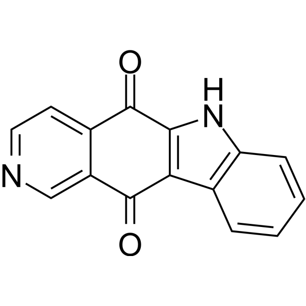
Drpitor1a is an orally active DRP1 GTPase inhibitor. Drpitor1a blocks mitochondrial fission, inhibits DRP1 GTPase activity, and reduces ROS levels. Drpitor1a induces mitochondrial fusion and mitochondria-dependent apoptosis by elevating Bak and Smac/Diablo and inhibiting Bcl-2. Drpitor1a inhibits proliferation in pulmonary arterial hypertension hPASMC and induces necrosis and autophagy mediators. Drpitor1a causes regression of non-small cell lung cancer tumors in mouse xenograft models and exhibits higher accumulation and efficacy in females. Drpitor1a can be used in research related to non-small cell lung cancer, myocardial ischemia-reperfusion injury, and pulmonary arterial hypertension.
For research use only. We do not sell to patients.
- Purity : 96.95%
- CAS No.: 73326-98-4
- Formula: C15H8N2O2
- Molecular Weight:248.24
-
Storage:
4°C, protect from light
* In solvent : -80°C, 6 months; -20°C, 1 month (protect from light)
Biological Activity
Description
IC50 & Target
[4]|
Bcl-2 |
Cellular Effect
|
Cell Line
|
Type | Value | Description | References |
|---|---|---|---|---|
| A549 | IC50 |
0.057 μM
|
Inhibition of mitochondrial fragmentation in human A549 cancer cells.
|
PMC10378656 |
In Vitro
Drpitor1a is an inhibitor of DRP1 GTPase activity, exhibits cardioprotective properties in cardiac I/R injury, and has antiproliferative effects in lung cancer cells[1].
Drpitor1a fused the mitochondrial network in A549 cells expressing wild-type Drp1; moreover, it had no effect on the already fused mitochondrial network in A549 cells expressing the loss-of-function Drp1 K38A mutant[2].
Drpitor1a effectively inhibits mitochondrial fragmentation in A549 cancer cells, with an IC50 of 0.057 μM[2].
Drpitor1a directly inhibits the GTPase activity of recombinant and endogenous Drp1 from PAH hPASMCs[4].
Drpitor1a (0.1 μM; 4-5 h) inhibits mitochondrial fission in PAH hPASMCs but does not alter mitochondrial morphology in normal hPASMCs; it reverses the fragmented mitochondrial network in PAH BOECs but does not change mitochondrial morphology in control human BOECs[4].
Drpitor1a (1 μM; 48 h) inhibits the proliferation of PAH hPASMCs, but has no significant effect on the proliferation of normal hPASMCs[4].
Drpitor1a (5 μM; 48 h) induces apoptosis in PAH hPASMCs through a mitochondria-dependent apoptotic pathway[4].
MedChemExpress (MCE) has not independently confirmed the accuracy of these methods. They are for reference only. Further protocols information, click here.
-
Cell Line:PAH hPASMC, normal hPASMC
-
Concentration:0.1 μM
-
Incubation Time:5 hours
-
Result:Increased the accumulation of endogenous Drp1 on mitochondria in PAH hPASMC.
Did not alter total Drp1 expression in PAH hPASMC.\nIncreased the accumulation of exogenous Drp1 on mitochondria in PAH hPASMC.
-
Cell Line:PAH hPASMC, normal hPASMC
-
Concentration:1 μM
-
Incubation Time:48 hours
-
Result:Reduced cell proliferation in PAH hPASMC without significantly reducing the much lower proliferation rate of control hPASMC.
-
Cell Line:PAH hPASMC
-
Concentration:5 μM
-
Incubation Time:48 hours
-
Result:Increased unstimulated apoptosis in 2 of 3 PAH hPASMC cell lines, with an accompanying significant increase in the expression of the apoptosis mediators Bak, Smac/Diablo, and suppression of the anti-apoptosis mediator, Bcl-2.
Slightly increased the expression of receptor-interacting protein and the LC3B II/I ratio.
Parmacokinetics
| Species | Dose | Route | AUC0-t | AUC0-∞ | MRT | T1/2 (Elimination) | Tmax | Cmax | CLplasma | Vd | Bioavailability |
|---|---|---|---|---|---|---|---|---|---|---|---|
| Rat[4] | 1 mg/kg | i.v. | 552.5 nM·h | 844.3 nM·h | 7.2 h | 6.5 h | 0.08 h | 617.7 nM | 23.9 mL/min | 10.2 L | 26.0 % |
| Rat[4] | 1 mg/kg | i.v. | 1288.2 nM·h | 1433.8 nM·h | 2.4 h | 3.4 h | 0.08 h | 3250.4 nM | 13.1 mL/min | 1.9 L | 12.6 % |
| Rat[4] | 5 mg/kg | i.v. | 2496.9 nM·h | 3211.9 nM·h | 10.4 h | 9.9 h | 0.08 h | 2332.3 nM | 39.8 mL/min | 20.3 L | 21.1 % |
| Rat[4] | 5 mg/kg | i.v. | 5306.6 nM·h | 8577.4 nM·h | 27.5 h | 24.3 h | 0.08 h | 3425.0 nM | 13.7 mL/min | 14.3 L | 19.7 % |
In Vivo
Drpitor1a (1 mg/kg; intravenous injection; once every 48 hours; from day 17 to day 27) regressed Monocrotaline (HY-N0750)-induced PAH in female rats by reversing adverse pulmonary vascular remodeling and RV hypertrophy, improved right heart function, and had no systemic vascular effects or toxicity[4].
Drpitor1a (0.1-1.0 mg/kg; i.v.; every 48 hours) showed a trend toward efficacy in female but not male MCT-PAH rats[4].
MedChemExpress (MCE) has not independently confirmed the accuracy of these methods. They are for reference only.
Chemical Information
-
CAS No. 73326-98-4
-
Appearance Solid
-
Molecular Weight 248.24
-
Formula C15H8N2O2
-
Color Light brown to brown
-
SMILES
O=C1C2=C(C(C=CC=C3)=C3N2)C(C4=CN=CC=C41)=O
-
Shipping
Room temperature in continental US; may vary elsewhere.
-
Storage
4°C, protect from light
* In solvent : -80°C, 6 months; -20°C, 1 month (protect from light)
Solvent & Solubility
In Vitro:
DMSO : 5 mg/mL (20.14 mM; ultrasonic and warming and heat to 60°C; Hygroscopic DMSO has a significant impact on the solubility of product, please use newly opened DMSO)
Please refer to the solubility information to select the appropriate solvent. Once prepared, please aliquot and store the solution to prevent product inactivation from repeated freeze-thaw cycles.
Storage method and period of stock solution: -80°C, 6 months; -20°C, 1 month (protect from light). When stored at -80°C, please use it within 6 months. When stored at -20°C, please use it within 1 month.
Please refer to the solubility information to select the appropriate solvent. Once prepared, please aliquot and store the solution to prevent product inactivation from repeated freeze-thaw cycles.
Storage method and period of stock solution: -80°C, 6 months; -20°C, 1 month (protect from light). When stored at -80°C, please use it within 6 months. When stored at -20°C, please use it within 1 month.
Concentration (start) × Volume (start) = Concentration (final) × Volume (final)
Protocols
-
Annexin V plus membrane-impermeant dye apoptosis staining
Annexin V-based apoptosis assays rely on the detection of phosphatidylserine (PS) externalization from the inner leaflet of the plasma membrane to the outer leaflet, an early biochemical hallmark of apoptosis. Fluorescently labeled Annexin V binds PS in a calcium-dependent manner, enabling identification of early apoptotic cells by flow cytometry or fluorescence microscopy. When combined with a membrane-impermeant DNA-binding dye (e. g. , propidium iodide), this approach allows discrimination between viable (Annexin V−/dye−), early apoptotic (Annexin V+/dye−), and late apoptotic or necrotic (Annexin V+/dye+) cell populations by assessing membrane integrity and PS exposure.
-
ROS/oxidative-stress fluorescent staining
ROS/oxidative-stress fluorescent staining uses cell-permeant fluorogenic probes that become fluorescent after oxidation inside cells or tissues; commonly used examples include DCFH-DA/DCFDA for broad cellular oxidant detection, DHE for superoxide-related signal detection, MitoSOX for mitochondrial superoxide-related signal detection, and CellROX probes for oxidative-stress-associated fluorescence readouts. The assay detects probe oxidation rather than a single ROS species unless the probe and analysis method have been chemically validated for that species. DCFH-DA enters cells, is deacetylated by intracellular esterases to DCFH, and produces fluorescent DCF after oxidation, so the readout is used as an operational measure of total cellular oxidative stress rather than a species-specific ROS measurement. DHE and MitoSOX can report superoxide-related oxidation, but red fluorescence alone can include non-specific ethidium-like oxidation products; HPLC or optimized spectral approaches are
-
Research Protocol for Cardiovascular Diseases
Cardiovascular disease can be modeled as maladaptive cardiac remodeling, where ischemic injury or pressure overload activates inflammatory signaling, fibroblast activation, extracellular-matrix deposition, cardiomyocyte hypertrophy, vascular remodeling, and progressive ventricular dysfunction. The TGF-β/SMAD axis is a central profibrotic pathway after myocardial injury and pressure overload, while innate immune and cytokine pathways regulate leukocyte recruitment, scar formation, and adverse remodeling. Key unresolved questions include which inflammatory signals are reparative versus harmful, when fibrosis is protective versus maladaptive, and whether pathway inhibition improves function without weakening necessary infarct healing or compensatory remodeling.
-
Cell Viability Determination by MTT Colorimetric Assay
The following protocol uses the MTT colorimetric assay as a classic literature-established method for assessing cell viability/metabolic activity in cultured mammalian cells. MTT[3-(4,5-dimethylthiazol-2-yl)-2,5-diphenyltetrazolium bromide] is reduced by metabolically active cells to a colored formazan product; the amount of formazan is quantified spectrophotometrically and provides an indirect measure of metabolically active viable cells. Importantly, MTT reduction reflects cellular oxidoreductase/metabolic activity rather than an absolute direct count of living cells, so changes in cellular metabolism can alter the signal independently of cell number.
-
Apoptosis
Apoptosis, also called programmed cell death, is generally characterized by distinct morphological characteristics.
-
TUNEL staining for apoptotic DNA fragmentation
TUNEL staining detects DNA strand breaks by using terminal deoxynucleotidyl transferase to add labeled nucleotides to exposed 3′-OH DNA termini, generating either microscopic staining in fixed cells or tissue sections, or fluorescence/cytometric signal in cell suspensions. TUNEL positivity reflects DNA fragmentation but should not be interpreted alone as definitive apoptosis, because TUNEL can also label necrotic, autolytic, mechanically damaged, or DNA-repair-associated DNA breaks.
-
Autophagy
Autophagy is a process in which eukaryotic cells use lysosomes to degrade their own cytoplasmic proteins and damaged organelles under the regulation of autophagy related gene (Atg). Microtubule-associated proteins light chain 3 (LC3) is recognized as autophagy marker, which transfers from cytoplasmic LC3 (LC3-I) to membrane type (LC3-II). LC3-II/I ratio could be detected by Western Blot and fluorescence microscopy.
-
Patient-Derived Xenograft (PDX)
Patient-derived xenograft (PDX) models are generated by engrafting primary human tumor tissue directly into immunodeficient mice, allowing in vivo propagation of patient tumor biology without initial in vitro adaptation. These models are used to preserve key histopathological and molecular characteristics of the original tumor and enable assessment of tumor growth dynamics and therapeutic response in a living organism. The biological readout is tumor engraftment and subsequent growth in the murine host, which reflects the ability of human tumor cells to survive, vascularize, and expand in an immunocompromised microenvironment.
-
Apoptosis Solutions
Apoptosis is a regulated, generally non-lytic cell-death pathway that removes unwanted, damaged, infected, or abnormal cells through coordinated morphological changes, caspase activation, DNA fragmentation, and membrane remodeling. The intrinsic apoptosis pathway is controlled mainly by mitochondrial outer membrane permeabilization, BCL-2 family proteins, cytochrome c release, apoptosome formation, caspase-9 activation, and downstream executioner caspase-3/7 activation. The extrinsic apoptosis pathway is initiated by death receptors such as Fas, TNFR, and TRAIL receptors, which recruit adaptor proteins and activate caspase-8 before engaging executioner caspases or mitochondrial amplification through BID cleavage. Apoptosis is linked to many phenotypes, including cancer cell killing, tissue homeostasis, immune regulation, neurodegeneration, infection response, and treatment-induced cytotoxicity; unresolved questions include how apoptosis interacts with necroptosis, pyroptosis, ferroptos
-
Splenic/Portal-Vein Liver Metastasis Xenograft
Splenic and portal-vein liver metastasis xenograft models deliver tumor cells into the portal circulation so that cells reach the liver first and form hepatic metastatic lesions; splenic injection uses the spleen as an access route to the portal system, while direct portal-vein injection introduces cells into the portal vein without requiring splenectomy. The assay detects liver colonization, intrahepatic tumor growth, tumor distribution, treatment response, survival, and liver-metastasis microenvironment changes; readouts include bioluminescence or fluorescence imaging, gross liver nodule counts, liver weight or tumor burden, histology, and survival.
-
Lysosome and acidic-vesicle live-cell staining
Lysosome and acidic-vesicle live-cell staining detects acidic intracellular compartments by using membrane-permeant acidotropic probes that accumulate in low-pH vesicles, including lysosomes, late endosomes, autolysosomes, and acidic phagosomes. LysoTracker staining is commonly used as an intensity-based readout of acidic lysosomal compartment abundance or enlargement, while acridine orange produces green fluorescence in less concentrated compartments and red fluorescence after concentration-dependent accumulation in acidic vesicular organelles. Loss or reduction of acridine-orange red signal can be used as a readout of lysosomal membrane permeabilization or reduced acidic-vesicle integrity. This protocol is designed for live cultured cells and can be adapted for fluorescence microscopy, high-content imaging, plate-reader readout, or flow cytometry when the selected literature supports the readout. Because these dyes report acidotropic accumulation rather than lysosome identity alone,
-
Subcutaneous Cell-Line-Derived Xenograft
Subcutaneous cell-line-derived xenograft (CDX) models are established by implanting cultured human cancer cell lines into immunodeficient mice, where the injected cells form localized tumors that can be monitored in vivo as a measure of tumorigenic potential, growth kinetics, and treatment response. These models are widely used in oncology research because they allow reproducible tumor formation and enable comparative assessment of tumor growth between different cell lines or genetic manipulations in a controlled in vivo microenvironment. Subcutaneous implantation of cancer cells in immunodeficient mice is a standard approach for evaluating tumor growth behavior and therapeutic response across multiple cancer types, including prostate, esophageal, pancreatic, and colon cancer models.
-
Macroautophagy Solutions
Macroautophagy is a conserved lysosome-dependent degradation pathway in which cytoplasmic material is sequestered into double-membrane autophagosomes and delivered to lysosomes for degradation and recycling. The pathway supports cellular homeostasis during nutrient limitation, organelle stress, protein-aggregate accumulation, infection, differentiation, and tissue remodeling by coupling cargo sequestration, autophagosome maturation, lysosomal fusion, and degradation of cargo-derived macromolecules. The core molecular sequence includes initiation by nutrient- and stress-regulated autophagy machinery, autophagosome nucleation, LC3/ATG8-family conjugation to autophagosomal membranes, cargo selection through receptors such as SQSTM1/p62, autophagosome-lysosome fusion, and lysosomal degradation. LC3 was identified as a mammalian homolog of yeast Atg8 that localizes to autophagosomal membranes after processing, and p62/SQSTM1 was shown to connect ubiquitinated cargo with autophagic degradati
-
Orthotopic Cell-Line Xenograft
Orthotopic cell-line xenograft models involve implantation of human cancer cell lines into the anatomically corresponding organ of immunodeficient mice to reproduce tumor growth within a native microenvironment, enabling more clinically relevant tumor behavior compared with subcutaneous models. These models are widely used because orthotopic placement better recapitulates tumor progression, including invasion and metastatic spread, which are often underrepresented in heterotopic implantation systems. Compared with conventional xenografts, orthotopic implantation is described as more technically complex but provides improved simulation of tumor-microenvironment interactions and metastatic behavior, making it particularly valuable for translational oncology research. Surgical orthotopic implantation approaches have been emphasized as enabling faithful reproduction of clinical cancer features, including metastasis and disease progression patterns that align with the tumor’s organ of origi
-
Intraperitoneal/Peritoneal Dissemination Xenograft
Intraperitoneal (IP) or peritoneal dissemination xenograft models are based on the introduction of human cancer cells into the peritoneal cavity of immunodeficient mice, where they attach to peritoneal surfaces, form multicellular aggregates or spheroids, and progressively generate disseminated tumor nodules that mimic advanced peritoneal metastatic disease. These models are widely used to study ovarian cancer progression, tumor-microenvironment interactions, and intraperitoneal therapeutic responses, often incorporating bioluminescence or fluorescence imaging to longitudinally monitor tumor burden in vivo. The biological principle relies on the capacity of tumor cells such as SKOV3 or related ovarian carcinoma lines to survive in suspension, aggregate within ascites-like fluid, adhere to mesothelial surfaces, and invade peritoneal organs, thereby recapitulating human peritoneal carcinomatosis patterns observed in advanced disease.
Purity & Documentation
-
Data Sheet (294 KB)
-
SDS (252 KB)
- English - EN (252 KB)
- Français - FR (252 KB)
- Deutsch - DE (252 KB)
- Norwegian - NO (252 KB)
- Español - ES (252 KB)
- Swedish - SV (252 KB)
- Italian - IT (252 KB)
- Korean - KR (252 KB)
- Portuguese - PT (252 KB)
-
Handling Instructions (2659 KB)
References
Complete Stock Solution Preparation Table
Please refer to the solubility information to select the appropriate solvent. Once prepared, please aliquot and store the solution to prevent product inactivation from repeated freeze-thaw cycles.
Storage method and period of stock solution: -80°C, 6 months; -20°C, 1 month (protect from light). When stored at -80°C, please use it within 6 months. When stored at -20°C, please use it within 1 month.
| Optional Solvent | Concentration Solvent Mass | 1 mg | 5 mg | 10 mg | 25 mg |
|---|---|---|---|---|---|
| DMSO | 1 mM | 4.0284 mL | 20.1418 mL | 40.2836 mL | 100.7090 mL |
| 5 mM | 0.8057 mL | 4.0284 mL | 8.0567 mL | 20.1418 mL | |
| 10 mM | 0.4028 mL | 2.0142 mL | 4.0284 mL | 10.0709 mL | |
| 15 mM | 0.2686 mL | 1.3428 mL | 2.6856 mL | 6.7139 mL | |
| 20 mM | 0.2014 mL | 1.0071 mL | 2.0142 mL | 5.0354 mL |
Keywords
- Drpitor1a
- 73326-98-4
- Dynamin
- Reactive Oxygen Species (ROS)
- Bcl-2 Family
- Apoptosis
- non-small cell lung cancer
- DRP1 GTPase inhibitor
- mitochondrial-dependent apoptosis
- MCT-PAH rats
- mitochondrial fission
- cardiac ischemia/reperfusion injury
- pulmonary arterial hypertension hPASMC
- A549 cells
- reactive oxygen species
- PAH BOEC
- Inhibitor
- inhibitor
- inhibit