



Lysosome Dyes
- 1
- 2

DQ-BSA-Red is a bovine serum albumin labeled with a red fluorescent dye that can be used to detect lysosomal activity. The excitation wavelength and emission wavelength of DQ-BSA-Red are 590 nm and 620 nm, respectively. The BSA molecule in DQ-BSA-Red is labeled with high concentration of red fluorescent dye in multiple sites, which shows high fluorescence self-inhibition. Once DQ-BSA-RED enters the lysosome, DQ-BSA is cleaved by lysosomal proteases, resulting in unquenched and released fluorescent fragments, emitting bright fluorescence. Inactivated lysosomes are unable to degrade the BSA protein and thus have a lower or even no fluorescent signal.
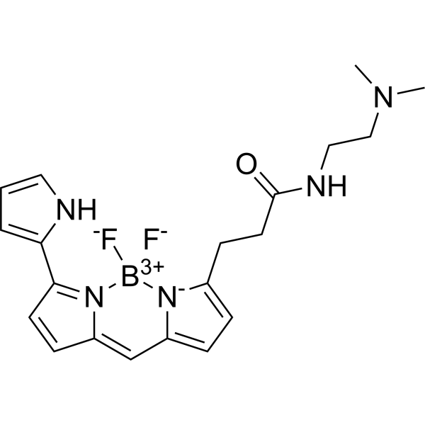
- Formula: C20H24BF2N5O
- Molecular Weight: 399.25
LysoTracker Red is a Red fluorescently labeled lysosomal probe with a maximum excitation/emission wavelength of 577/590 nm. The structure is composed of a fluorescein group and linked weak bases, which can freely cross the cell membrane and gather on spherical organelles. It is suitable for observing the internal biosynthesis and related pathogenesis of lysosomes.
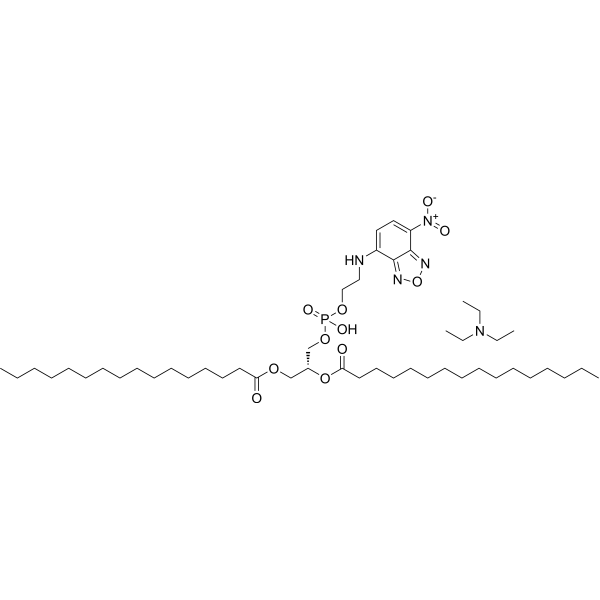
- Formula: C49H90N5O11P
- Molecular Weight: 956.24
NBD-PE is an effective lipid fluorescent probe (Excitation/Emission: 465/535 nm; Color: Green). NBD-PE offers a wide array of applications in membrane and cell biology.
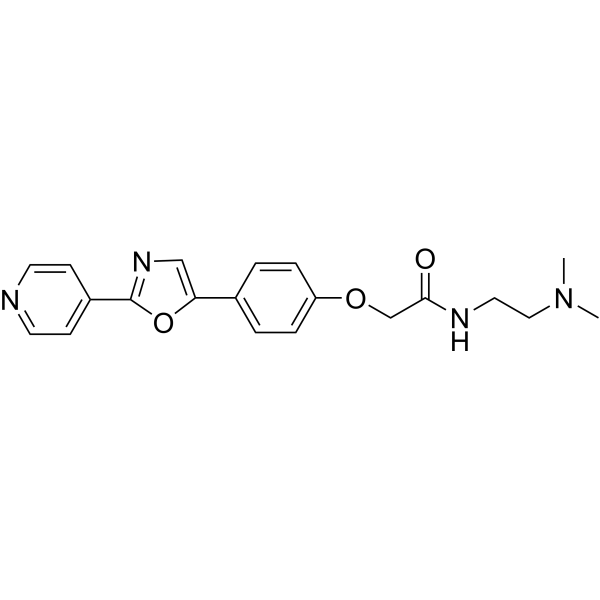
- Formula: C20H22N4O3
- Molecular Weight: 366.41
PDMPO, a lysosome pH indicator, is an excellent fluorescent acidotropic reagent for fluorescence imaging. PDMPO is a potent tool with which to study acidic organelles of live cells. PDMPO exhibits pH-dependent dual-excitation and dual-emission spectral peaks. PDMPO produces a blue fluorescence in weakly acidic organelles and shifts to yellow in more acidic lysosomes. (Abs=329 nm; Em=440/540 nm).
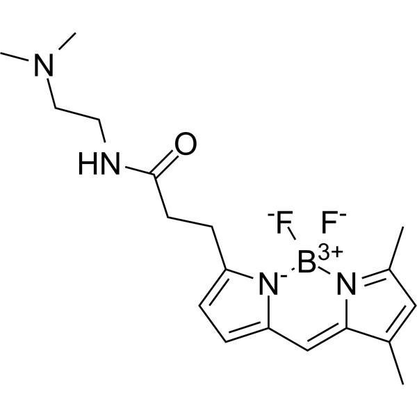
- Formula: C18H25BF2N4O
- Molecular Weight: 362.23
Green DND-26 is a green fluorescently labeled lysosomal probe with a maximum excitation/emission wavelength of 504/511 nm. The structure is composed of a fluorescein group and linked weak bases, which can freely cross the cell membrane and generally gather on spherical organelles. Green DND-26 is suitable for observing the internal biosynthesis and related pathogenesis of lysosomes.

- Formula: C44H68BF2N3O13
- Molecular Weight: 895.83

- Formula: C23H24N2O3S
- Molecular Weight: 408.51
Lyso-VR1 is a fluorescent probe for lysosomal viscosity detection. The detection mechanism of Lyso-VR1 relies on intramolecular rotation restriction. Due to the protonation of its indole moiety in lysosomes, this probe can specifically target lysosomes, thereby localizing to this suborganelle for viscosity monitoring. Its excitation wavelength ranges from 400-500 nm, and its emission peak under excitation at 488 nm is located at ~550 nm to ~560 nm.

- Formula: C25H24N2O5
- Molecular Weight: 432.47
Lyso-OC is a two-photon fluorescent probe that enables real-time monitoring of autophagy through ultrasensitive detection of lysosomal polarity changes. Lyso-OC possesses a coumarin (HY-N0709) solvatochromic group and a morpholine lysosome-targeting group, allowing selective lysosomal localization in living cells. Lyso-OC emits strong fluorescence in low-polarity lysosomes, whereas in high-polarity autolysosomes formed during autophagy, its fluorescence intensity decreases and the emission peak shifts by approximately 30 nm. For two-photon detection, the excitation/emission wavelengths of Lyso-OC are 760/490-550 nm, and its one-photon excitation wavelength is 375 nm. Lyso-OC can be used in cancer-related research.
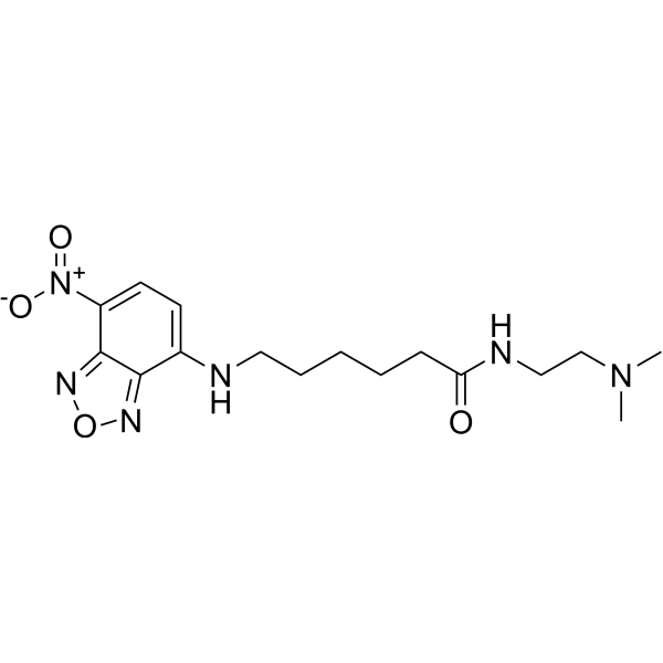
- Formula: C16H24N6O4
- Molecular Weight: 364.40

- Formula: C29H34N4O4
- Molecular Weight: 502.60
NRLyso is a lysosome-targeted solvatochromic fluorescent probe derived from Nile Red (HY-D0718). It can localize to lysosomes and is applicable for imaging lysosomal lipid order and polarity. The excitation wavelength of NRLyso is 520 nm, and its emission maximum varies with the environment. NRLyso partitions well into lipid membranes: it exhibits fluorescence enhancement and emission blue-shift in low-polarity, tightly packed lipid environments, while shows emission red-shift in more polar, disordered lipid environments. NRLyso maintains organelle specificity under oxidative and hyperosmotic stress conditions, and can detect changes in lysosomal lipid order and polarity in response to these stresses, cholesterol extraction, and cholesterol enrichment.

- Formula: C37H36BF2N5O2
- Molecular Weight: 631.52
Lys-VBOD is a Fluorescent probe for lysosomal viscosity monitoring and live cell imaging. Its detection mechanism depends on viscosity: increased viscosity restricts the twisting of the double bond between the indole and BODIPY fluorophore, as well as the rotation of the single bond between quinoline and BODIPY, which reduces non-radiative decay and leads to enhanced fluorescence intensity in high viscosity environments; it also features a morpholine group that provides lysosome-targeting properties, allowing it to localize to lysosomes for viscosity detection. The probe has excitation/emission wavelengths of Ex/Em = 602/637 nm, and its fluorescence intensity shows a good linear relationship with log-transformed viscosity values. Lys-VBOD is stable across a wide pH range of 5.35 to 10.74, is not affected by macromolecules and proteins like BSA, has a fluorescence lifetime that increases with viscosity, and can visualize real-time lysosomal viscosity changes in live cells under Dexamethasone (HY-14648) stimulation[1].

- Formula: C36H34N4O2
- Molecular Weight: 554.68
Lyso-MPCB is a two-photon fluorescent probe used for lysosomal pH detection and autophagy monitoring. Lyso-MPCB integrates a benzimidazole pH-responsive group and a morpholine lysosome-targeting group, enabling specific localization to lysosomes. The fluorescence properties of Lyso-MPCB change with protonation, exhibiting blue fluorescence under alkaline conditions and green fluorescence under acidic conditions.

- Formula: C50H42N2O2
- Molecular Weight: 702.88
LysoAIE2 is a Fluorescent probe for lysosomal viscosity detection and live-cell imaging. Its detection mechanism relies on the aggregation-induced emission effect: it exhibits only weak fluorescence in non-viscous media; in viscous environments, restricted intramolecular motion inhibits non-radiative energy dissipation pathways, thereby significantly enhancing fluorescence intensity. It achieves specific targeting of lysosomes through the proton acceptor property of its indole ring structure, while its hydroxyl group endows it with excellent water solubility. This probe is basically unaffected by microenvironmental polarity and pH within the range of pH 4.0 to pH 8.0, which avoids interference from these factors in viscosity measurement. LysoAIE2 has an emission wavelength of 570 nm. It can be used to monitor lysosomal viscosity changes during processes such as Dexamethasone (HY-14648)-induced lysosomal migration and starvation-induced mitophagy in live cells; at concentrations up to 40 μM, cell viability remains above 80%, demonstrating excellent biocompatibility.

- Formula: C26H28F6N3OP
- Molecular Weight: 543.48

- Formula: C57H64BF2N3O6S4
- Molecular Weight: 1064.20
FLCS1 is a BODIPY-based Fluorescent probe and selective copper (I) ion binder (λex = 630 nm, λemis = 660 nm). Binding of FLCS1 to copper (I) ions induces fluorescence turn-on. Addition of copper (I) to a methanolic solution of FLCS1 causes a slight red shift (4 nm) in the maximum absorption wavelength (λmax) in the UV-Vis spectrum.

- Formula: C31H44BF2N3OS4
- Molecular Weight: 651.77
Ratio-Coppersensor-1 is a selective ratiometric copper ion Fluorescent reporter (excitation wavelength: 480 nm; emission peak central wavelengths at 505 nm and 570 nm, respectively). Ratio-Coppersensor-1 exhibits extremely high selectivity for Cu+ at cellular concentrations, outperforming other competing metal ions, and shows an approximately 20-fold change in fluorescence ratio upon Cu+ binding. Ratio-Coppersensor-1 is capable of reflecting changes in Cu levels.

- Formula: C17H21N5OS
- Molecular Weight: 343.45
DHUCu-1 is a lysosome-targeted NIR divalent copper ion probe with high water solubility. DHUCu-1 serves as a tool for investigating the physiological functions of Cu2+ and related diseases, such as Menke syndrome, Wilson's disease, and Alzheimer's disease.

- Formula: C17H19N7O3
- Molecular Weight: 369.38
Lyso-Cu (II) is a lysosome-targeted divalent copper ion probe. Lyso-Cu (II) is applicable to the research of Wilson's disease, Menkes disease and Alzheimer's disease.

- Formula: C30H30N6O3
- Molecular Weight: 522.60

- Formula: C18H25BF2N4O
- Molecular Weight: 362.23
Green DND-26 (solution) is a green fluorescently labeled lysosomal probe with a maximum excitation/emission wavelength of 504/511 nm. The structure is composed of a fluorescein group and linked weak bases, which can freely cross the cell membrane and generally gather on spherical organelles. Green DND-26 is suitable for observing the internal biosynthesis and related pathogenesis of lysosomes.
Solvent and concentration: DMSO: 2 mM
- 1
- 2



