



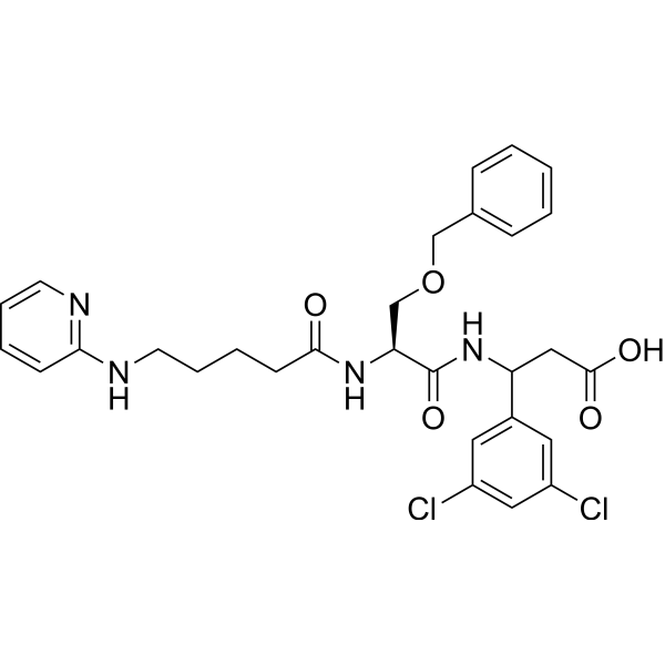
EMD527040
Based on 1 publication(s) in Google Scholar
EMD527040 is a potent and highly selective αvβ6 antagonist with antifibrotic activities. EMD527040 can be used for carcinoma and liver fibrosis research.
Nur für Forschungszwecke. Wir verkaufen nicht an Patienten.
- Reinheit : 99.88%
- CAS. Nr.: 851333-14-7
- Formel: C29H32Cl2N4O5
- Molecular Weight:587.49
-
Speicherung:Powder -20°C, 3 years ; In solvent -80°C, 6 months , -20°C, 1 month

To place orders, for customer services and technical support, please contact: MedChemExpress USA
Tel: 609-228-6898 E-mail: [email protected] [email protected]


-
 Biologische Aktivität
-
Chemical Information
-
Lösungsmittel & Löslichkeit
- Protokoll
- Reinheit & Dokumentation
- Verweise
-
 Help & FAQs


-
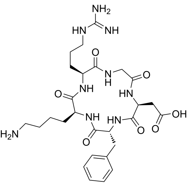
HY-P0023
Cyclo(-RGDfK)αvβ3 Integrin Inhibitor -
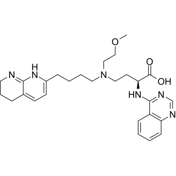
HY-137561
Bexotegrastαvβ6/αvβ1 Integrin Inhibitor -
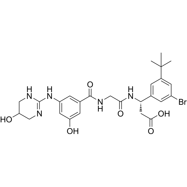
HY-18644
CWHM-12αV Integrins Inhibitor -

HY-Y0320C
Dimethyl sulfoxideAprotic Solvent -
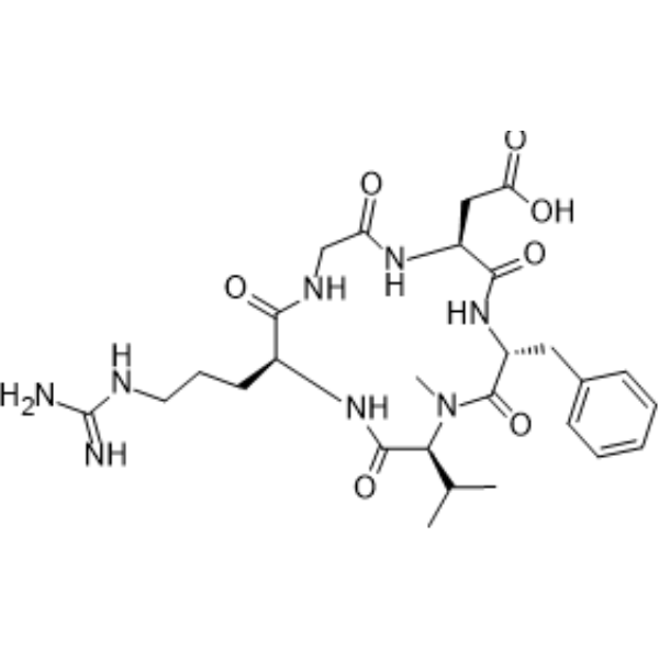
HY-16141
CilengitideIntegrin Inhibitor -
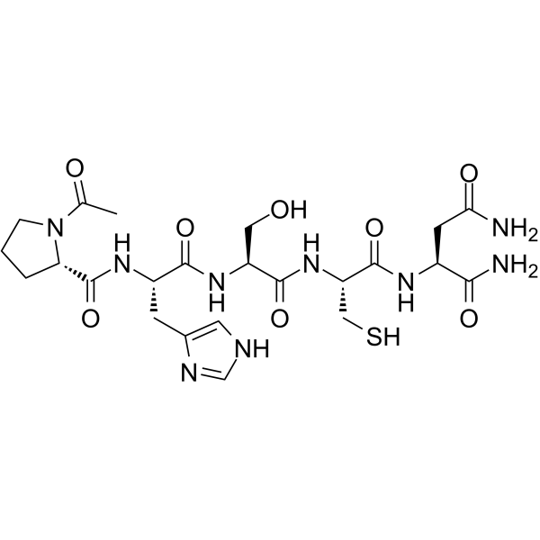
HY-13535
ATN-161α5β1/αvβ3 Integrin Binder -
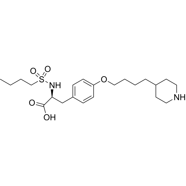
HY-17369B
TirofibanGp IIb/IIIa Receptor Antagonist -
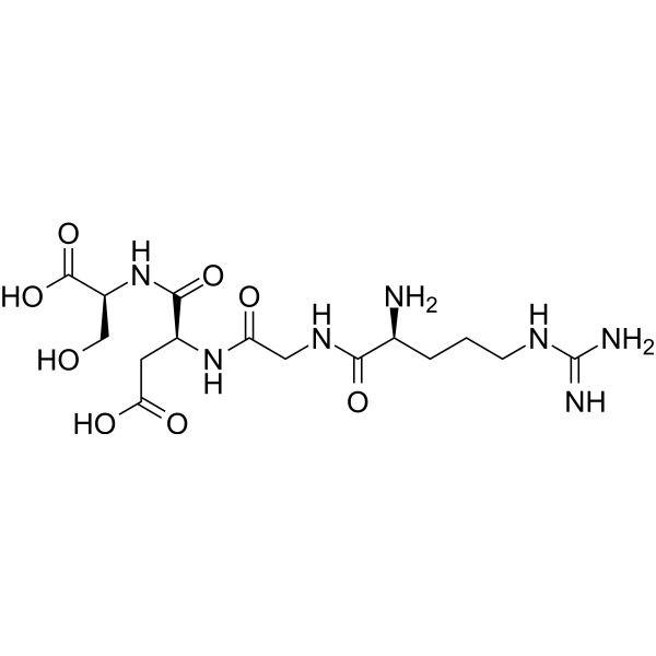
HY-12290
Arg-Gly-Asp-SerIntegrin Inhibitor -
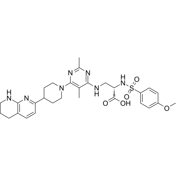
HY-100506
GLPG0187Integrin Receptor Antagonist -
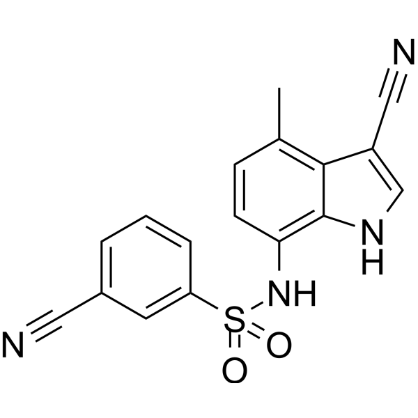
HY-14571
E7820Angiogenesis Inhibitor -
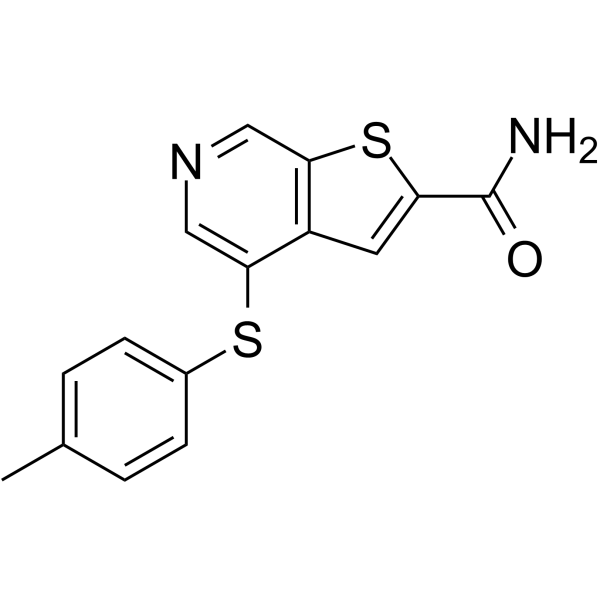
HY-100226
A-205804E-selectin/ICAM-1 Inhibitor -
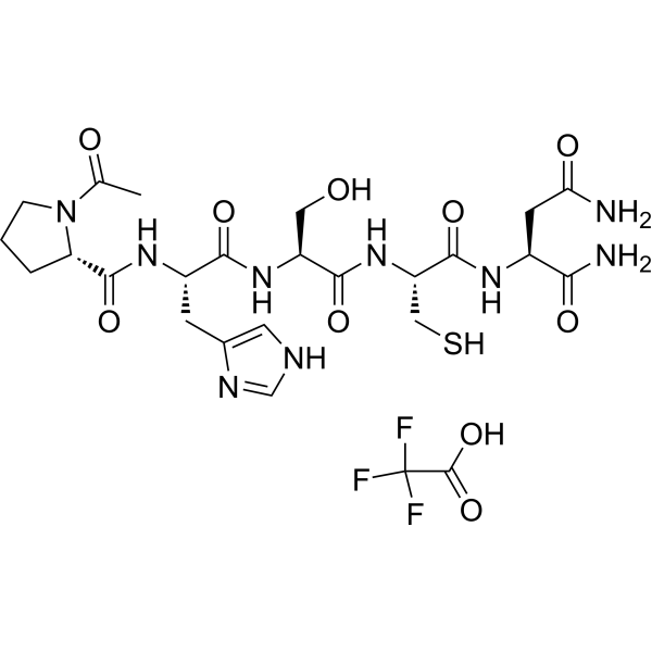
HY-13535A
ATN-161 trifluoroacetate saltIntegrin Antagonist
Publications Citing Use of MedChemExpress (MCE) EMD527040
More

Biologische Aktivität
Beschreibung
IC50 & Target
[1]|
αvβ6 6 nM (IC50) |
In Vitro
EMD527040 inhibits binding of recombinant αvβ6 to fibronectin at 6 nM as compared to >9.5 μM for αvβ3 and αvβ5 integrins (IC5050)[1].EMD527040 inhibits the attachment of αvβ6 expressing cells (UCLAP3 cells) to fibronectin at IC50 of 1.6 μM, as compared to >50 μM for αvβ3 and αvβ5 integrins[1].
MedChemExpress (MCE) has not independently confirmed the accuracy of these methods. They are for reference only.
In Vivo
MedChemExpress (MCE) has not independently confirmed the accuracy of these methods. They are for reference only.
-
Animal Model:Male adult Wistar rats
-
Dosage:20-60 mg/kg
-
Administration:Intraperitoneal injection; 20-60 mg/kg; week 2 to 6 after BDL (bile duct ligation)
-
Result:Ameliorated fibrosis progression in rodents with biliary fibrosis.


Chemical Information
-
CAS. Nr. 851333-14-7
-
Appearance Solid
-
Molecular Weight 587.49
-
Formel C29H32Cl2N4O5
-
Color White to off-white
-
SMILES
O=C(O)CC(NC([C@@H](NC(CCCCNC1=NC=CC=C1)=O)COCC2=CC=CC=C2)=O)C3=CC(Cl)=CC(Cl)=C3
-
Versand
Room temperature in continental US; may vary elsewhere.
-
Speicherung
Powder -20°C 3 years In solvent -80°C 6 months -20°C 1 month


Publications (1)
-
Journal Impact Factor
-
Most Recent


Lösungsmittel & Löslichkeit
In Vitro:
DMSO : 100 mg/mL (170.22 mM; Need ultrasonic; Hygroscopic DMSO has a significant impact on the solubility of product, please use newly opened DMSO)
Please refer to the solubility information to select the appropriate solvent. Once prepared, please aliquot and store the solution to prevent product inactivation from repeated freeze-thaw cycles.
Storage method and period of stock solution: -80°C, 6 months; -20°C, 1 month. When stored at -80°C, please use it within 6 months. When stored at -20°C, please use it within 1 month.
Please refer to the solubility information to select the appropriate solvent. Once prepared, please aliquot and store the solution to prevent product inactivation from repeated freeze-thaw cycles.
Storage method and period of stock solution: -80°C, 6 months; -20°C, 1 month. When stored at -80°C, please use it within 6 months. When stored at -20°C, please use it within 1 month.
Konzentration (Stammlösung) × Volumen (Stammlösung) = Konzentration (Ziellösung) × Volumen (Ziellösung)
In Vivo:
Select the appropriate dissolution method based on your experimental animal and administration route.
- For the following dissolution methods, please ensure to first prepare a clear stock solution using an In Vitro approach and then sequentially add co-solvents:
- To ensure reliable experimental results, the clarified stock solution can be appropriately stored based on storage conditions. As for the working solution for In Vivo experiments, it is recommended to prepare freshly and use it on the same day.
- The percentages shown for the solvents indicate their volumetric ratio in the final prepared solution. If precipitation or phase separation occurs during preparation, heat and/or sonication can be used to aid dissolution.
Add each solvent one by one: 10% DMSO 90% (20% SBE-β-CD in Saline)
Solubility: ≥ 2.5 mg/mL (4.26 mM); Clear solution
This protocol yields a clear solution of ≥ 2.5 mg/mL (saturation unknown).
Taking 1 mL working solution as an example, add 100 μL DMSO stock solution (25.0 mg/mL) to 900 μL 20% SBE-β-CD in Saline, and mix evenly.
Preparation of 20% SBE-β-CD in Saline (4°C, storage for one week): 2 g SBE-β-CD powder is dissolved in 10 mL Saline, completely dissolve until clear.
In Vivo Dissolution Calculator
Please enter the basic information of animal experiments:
-
-
-
-
Recommended: Prepare an additional quantity of animals to account for potential losses during experiments.
Please enter your animal formula composition:
-
%DMSO +
Recommended: Keep the proportion of DMSO in working solution below 2% if your animal is weak.
-
%+
-
+%Tween-80 + +
-
%Saline +
The co-solvents required include: DMSO, . All of co-solvents are available by MedChemExpress (MCE). , Tween 80. All of co-solvents are available by MedChemExpress (MCE).
Working solution concentration: 0.22 mg/mL
Method for preparing stock solution: mg drug dissolved in μL DMSO. Stock solution concentration: mg/mL.
1. Take μL DMSO stock solution;
2. Add μL .
μL , mix evenly;
3. Then add μL Tween 80, mix evenly;
4. Then add μL
Please ensure that the stock solution in the first step is dissolved to a clear state, and add co-solvents in sequence. You can use ultrasonic heating (ultrasonic cleaner, recommended frequency 20-40 kHz), vortexing, etc. to assist dissolution.


Reinheit & Dokumentation
-
Data Sheet (274 KB)
-
SDS (251 KB)
- English - EN (251 KB)
- Français - FR (251 KB)
- Deutsch - DE (251 KB)
- Norwegian - NO (251 KB)
- Español - ES (251 KB)
- Swedish - SV (251 KB)
- Italian - IT (251 KB)
- Korean - KR (251 KB)
- Portuguese - PT (251 KB)
-
Handling Instructions (2659 KB)
Verweise
[1]. Eleonora Patsenker, et al. Inhibition of integrin alphavbeta6 on cholangiocytes blocks transforming growth factor-beta activation and retards biliary fibrosis progression. Gastroenterology. 2008 Aug;135(2):660-70. [Content Brief]
[2]. Yury Popov, et al. Integrin alphavbeta6 is a marker of the progression of biliary and portal liver fibrosis and a novel target for antifibrotic therapies. J Hepatol [Content Brief]


Complete Stock Solution Preparation Table
Please refer to the solubility information to select the appropriate solvent. Once prepared, please aliquot and store the solution to prevent product inactivation from repeated freeze-thaw cycles.
Storage method and period of stock solution: -80°C, 6 months; -20°C, 1 month. When stored at -80°C, please use it within 6 months. When stored at -20°C, please use it within 1 month.
| Optional Solvent | Concentration Solvent Mass | 1 mg | 5 mg | 10 mg | 25 mg |
|---|---|---|---|---|---|
| DMSO | 1 mM | 1.7022 mL | 8.5108 mL | 17.0216 mL | 42.5539 mL |
| 5 mM | 0.3404 mL | 1.7022 mL | 3.4043 mL | 8.5108 mL | |
| 10 mM | 0.1702 mL | 0.8511 mL | 1.7022 mL | 4.2554 mL | |
| 15 mM | 0.1135 mL | 0.5674 mL | 1.1348 mL | 2.8369 mL | |
| 20 mM | 0.0851 mL | 0.4255 mL | 0.8511 mL | 2.1277 mL | |
| 25 mM | 0.0681 mL | 0.3404 mL | 0.6809 mL | 1.7022 mL | |
| 30 mM | 0.0567 mL | 0.2837 mL | 0.5674 mL | 1.4185 mL | |
| 40 mM | 0.0426 mL | 0.2128 mL | 0.4255 mL | 1.0638 mL | |
| 50 mM | 0.0340 mL | 0.1702 mL | 0.3404 mL | 0.8511 mL | |
| 60 mM | 0.0284 mL | 0.1418 mL | 0.2837 mL | 0.7092 mL | |
| 80 mM | 0.0213 mL | 0.1064 mL | 0.2128 mL | 0.5319 mL | |
| 100 mM | 0.0170 mL | 0.0851 mL | 0.1702 mL | 0.4255 mL |


EMD527040 Related Classifications
HY-101473 Related Classifications
 Powered by Bioz