



KU-32
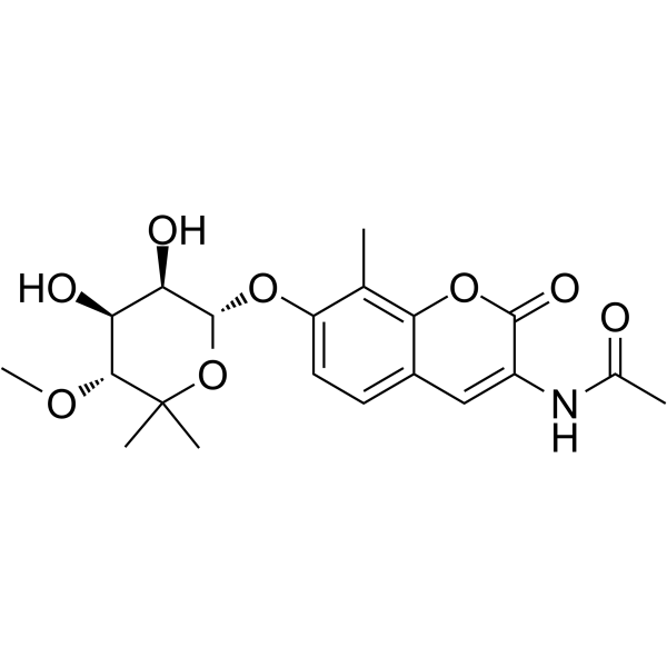
KU-32 is a novel, novobiocin-based Hsp90 inhibitor that can protect against neuronal cell death.
For research use only. We do not sell to patients.
- CAS No.: 956498-70-7
- Formula: C20H25NO8
- Molecular Weight:407.41
-
Storage:
Please store the product under the recommended conditions in the Certificate of Analysis.

To place orders, for customer services and technical support, please contact: MedChemExpress USA
Tel: 609-228-6898 E-mail: [email protected] [email protected]


-
 Biological Activity
-
Chemical Information
- Protocols
- Purity & Documentation
- References
-
 Help & FAQs


-
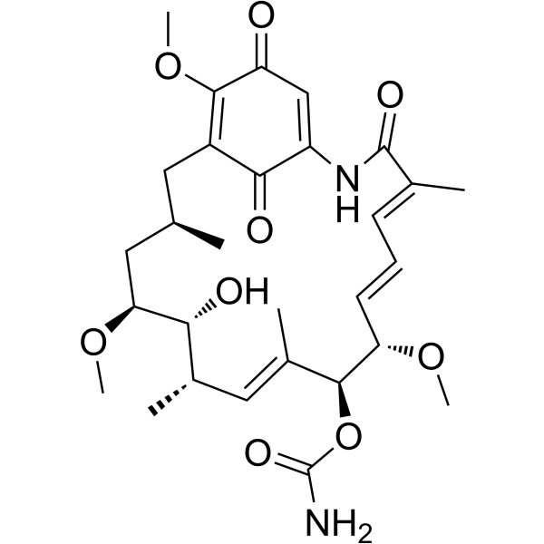
HY-15230
GeldanamycinHsp90 Inhibitor -
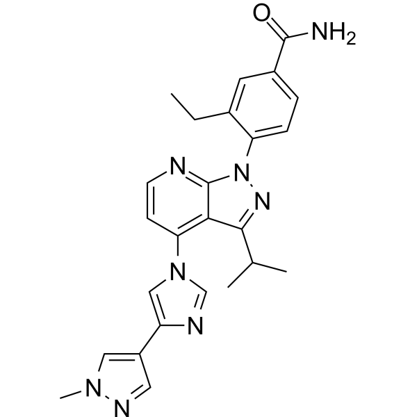
HY-15785
PimitespibHSP90α/HSP90β Inhibitor -
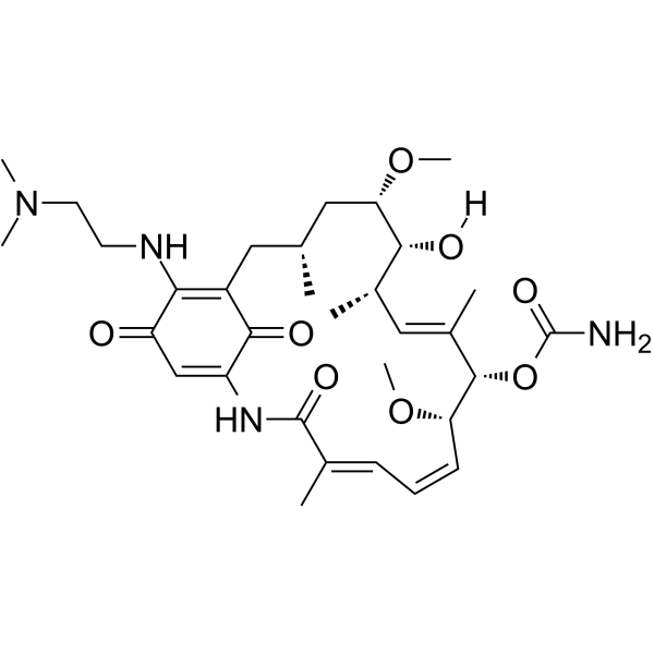
HY-10389
AlvespimycinHsp90 Inhibitor -
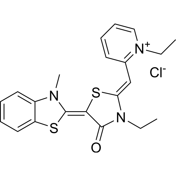
HY-15096
MKT-077Hsp70 Inhibitor -
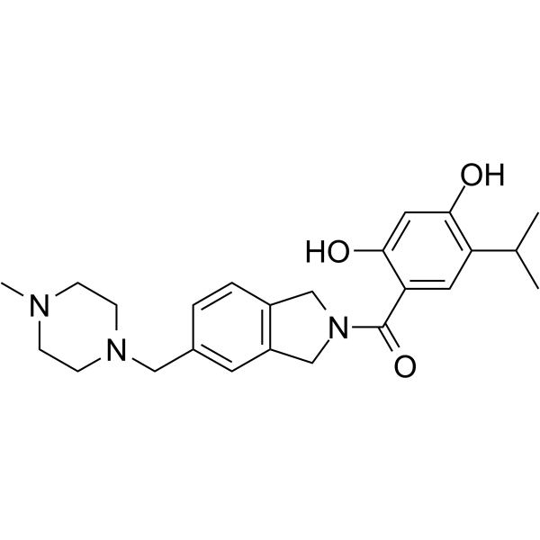
HY-14463
OnalespibHsp90 Inhibitor -
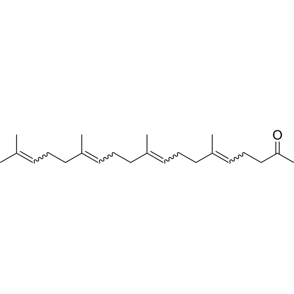
HY-B0779
TeprenoneHSP Inducer -
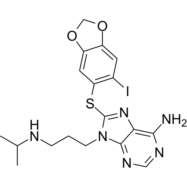
HY-11038
ZelavespibHsp90 Inhibitor -
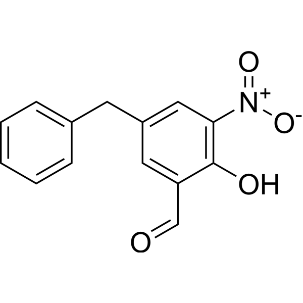
HY-124817
Col003Hsp47 Inhibitor -
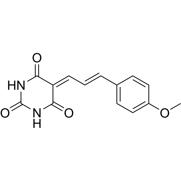
HY-18669
ML346Hsp70 Activator -
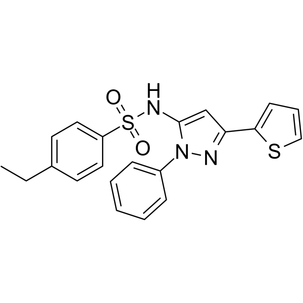
HY-103000
HSF1AHSF1 Activator -
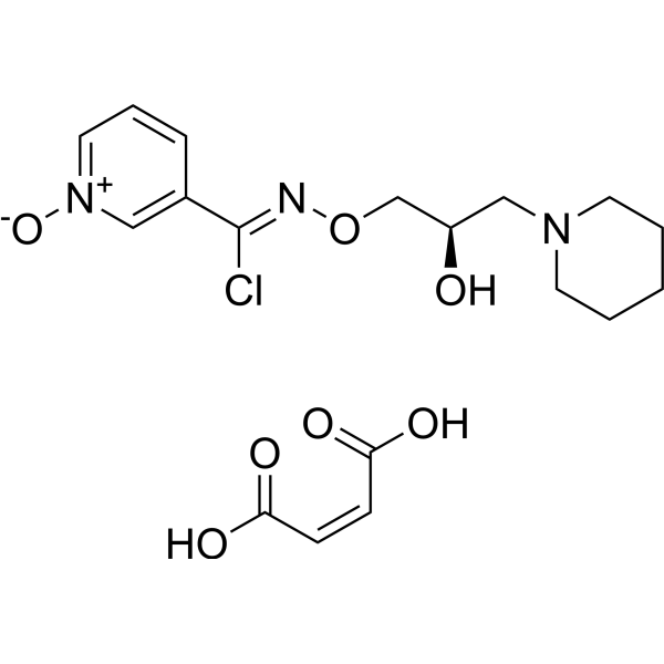
HY-106443A
Arimoclomol maleateHSP Co-Inducer -
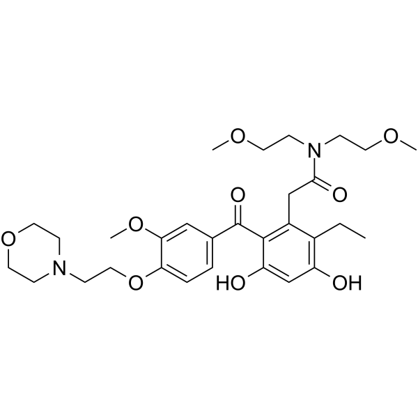
HY-13468
KW-2478


Biological Activity
Description
IC50 & Target
Hsp90[1]
Cellular Effect
|
Cell Line

|
Type | Value | Description | References |
|---|---|---|---|---|
| SH-SY5Y | EC50 |
1.49 nM
Compound: KU32
|
Neuroprotection against beta-amyloid peptide 1-42-induced toxicity in human SH-SY5Y cells assessed as lactate dehydrogenase release
Neuroprotection against beta-amyloid peptide 1-42-induced toxicity in human SH-SY5Y cells assessed as lactate dehydrogenase release
|
[PMID: 19138859] |
| SK-BR-3 | EC50 |
10 nM
Compound: 2, KU-32
|
Antiproliferative activity against human SKBR3 cells
Antiproliferative activity against human SKBR3 cells
|
[PMID: 24953820] |
| SK-BR-3 | IC50 |
>900 μM
Compound: KU32
|
Antiproliferative activity against human SK-BR-3 cells
Antiproliferative activity against human SK-BR-3 cells
|
[PMID: 27563408] |
In Vitro
Treating human islets with KU-32 for 24 hours shows no toxicity. With a minimum of 2-day exposure, KU-32 improves cellular viability by blocking apoptosis. Functionally, isolated human islets release more glucose-stimulated insulin when preincubate in KU-32[1]. KU-32 protects against glucose-induced death of embryonic DRG (dorsal root ganglia) neurons cultured for 3 days in vitro[2].
MedChemExpress (MCE) has not independently confirmed the accuracy of these methods. They are for reference only. Further protocols information, click here.
In Vivo
MedChemExpress (MCE) has not independently confirmed the accuracy of these methods. They are for reference only.


Chemical Information
-
CAS No. 956498-70-7
-
Molecular Weight 407.41
-
Formula C20H25NO8
-
SMILES
CC1=C(O2)C(C=C(NC(C)=O)C2=O)=CC=C1O[C@H]3[C@@H]([C@@H]([C@@H](OC)C(C)(C)O3)O)O
-
Shipping
Room temperature in continental US; may vary elsewhere.
-
Storage
Please store the product under the recommended conditions in the Certificate of Analysis.

Protocols
-
Cell Cytotoxicity Assay
Cytotoxicity assays are usually based on the assessment of cell membrane damage, which can also be indirectly detected by measuring cell viability. Detection methods include MTT assay, CKK-8 assay, LDH assay and ATP assay, etc.
-
Mammalian live/dead viability and cytotoxicity staining
Live/dead viability and cytotoxicity staining assays are based on the simultaneous detection of intracellular esterase activity in metabolically active (viable) cells and membrane integrity loss in non-viable cells. In commonly used dual-staining approaches, membrane-permeant fluorogenic substrates are converted by intracellular esterases into fluorescent products in live cells, while impermeant DNA-binding dyes selectively enter cells with compromised plasma membranes and label nucleic acids in dead or dying cells, enabling discrimination between viable and non-viable populations by fluorescence microscopy or flow cytometry.
-
Apoptosis Solutions
Apoptosis is a regulated, generally non-lytic cell-death pathway that removes unwanted, damaged, infected, or abnormal cells through coordinated morphological changes, caspase activation, DNA fragmentation, and membrane remodeling. The intrinsic apoptosis pathway is controlled mainly by mitochondrial outer membrane permeabilization, BCL-2 family proteins, cytochrome c release, apoptosome formation, caspase-9 activation, and downstream executioner caspase-3/7 activation. The extrinsic apoptosis pathway is initiated by death receptors such as Fas, TNFR, and TRAIL receptors, which recruit adaptor proteins and activate caspase-8 before engaging executioner caspases or mitochondrial amplification through BID cleavage. Apoptosis is linked to many phenotypes, including cancer cell killing, tissue homeostasis, immune regulation, neurodegeneration, infection response, and treatment-induced cytotoxicity; unresolved questions include how apoptosis interacts with necroptosis, pyroptosis, ferroptos


Purity & Documentation
References
[1]. Farmer K, et al. KU-32, a novel drug for diabetic neuropathy, is safe for human islets and improves in vitro insulin secretion and viability. Exp Diabetes Res. 2012;2012:671673. [Content Brief]
[2]. Urban MJ, et al. Inhibiting heat-shock protein 90 reverses sensory hypoalgesia in diabetic mice. ASN Neuro. 2010 Aug 11;2(4):e00040. [Content Brief]

Calculators
Concentration (start) × Volume (start) = Concentration (final) × Volume (final)


KU-32 Related Classifications
HY-108248 Related Classifications
 Powered by Bioz