



ANI-7

Based on 1 Customer Validation
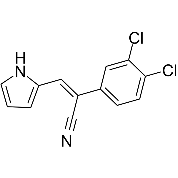
ANI-7 is an activator of aryl hydrocarbon receptor (AhR) pathway. ANI-7 inhibits the growth of multiple cancer cells, and potently and selectively inhibits the growth of MCF-7 breast cancer cells with a GI50 of 0.56 μM. ANI-7 induces CYP1-metabolizing mono-oxygenases by activating AhR pathway, and also induces DNA damage, checkpoint Kinase 2 (Chk2) activation, S-phase cell cycle arrest, and cell death in sensitive breast cancer cell lines.
For research use only. We do not sell to patients.
- Purity : 99.01%
- CAS No.: 931417-26-4
- Formula: C13H8Cl2N2
- Molecular Weight:263.12
-
Storage:Powder -20°C, 3 years , 4°C, 2 years ; In solvent -80°C, 2 years , -20°C, 1 year

To place orders, for customer services and technical support, please contact: MedChemExpress USA
Tel: 609-228-6898 E-mail: [email protected] [email protected]


-
 Biological Activity
-
Chemical Information
-
Solvent & Solubility
- Protocols
- Purity & Documentation
- References
-
 Help & FAQs


-
HY-14715B
CCT241533 hydrochlorideCHK2 Inhibitor -
HY-109044
TapinarofAhR Agonist -
HY-13946
BML-277 -
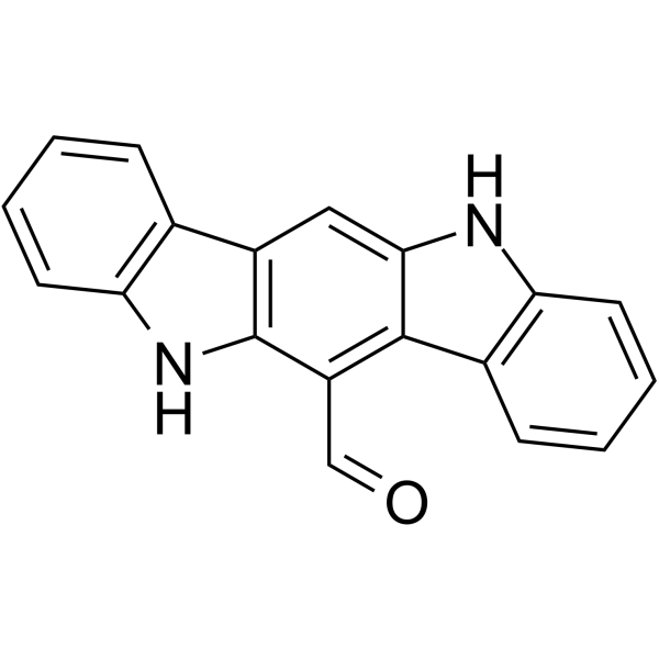
HY-12451
FICZAhR Agonist -
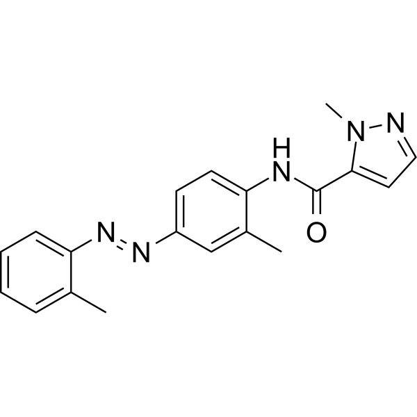
HY-12684
CH-223191AhR Antagonist -
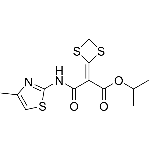
HY-100242
MivotilateAryl Hydrocarbon Receptor Activator -
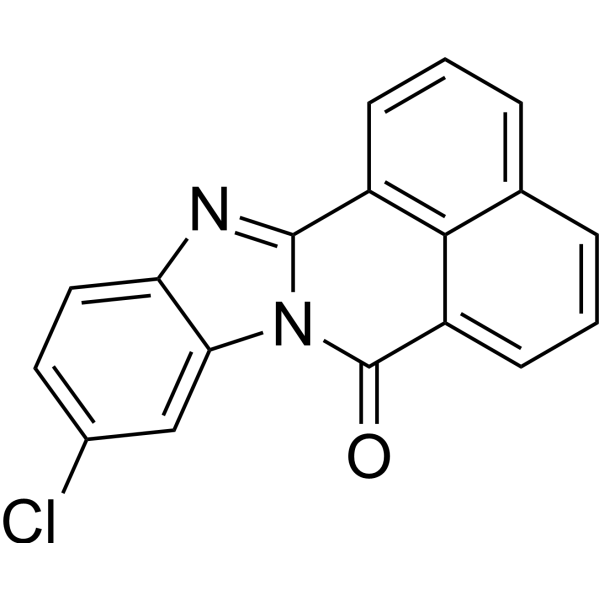
HY-118438
10-Cl-BBQAhR Ligand -
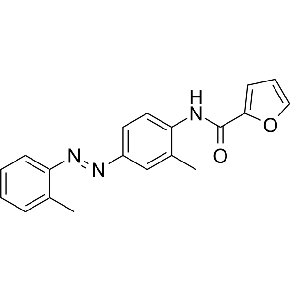
HY-118780
CHD-5AhR antagonist -
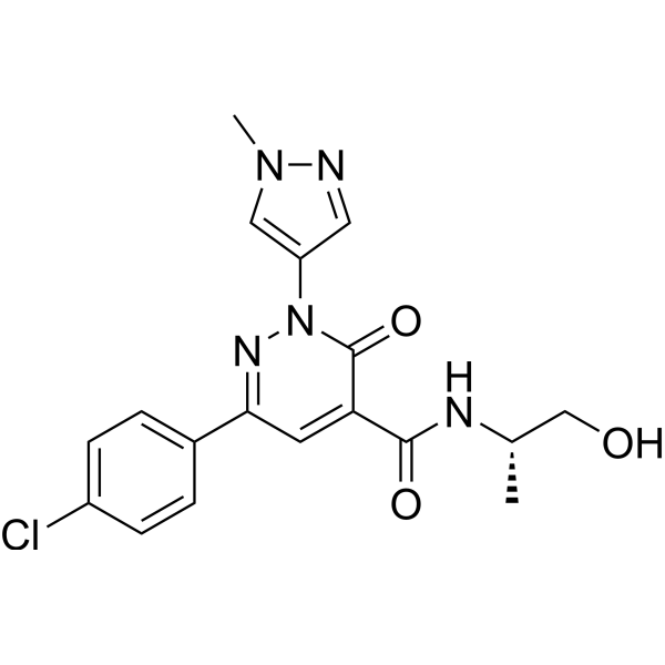
HY-135829
BAY 2416964AHR Antagonist -
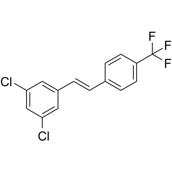
HY-112627
CAY 10465Aryl Hydrocarbon Receptor Agonist -
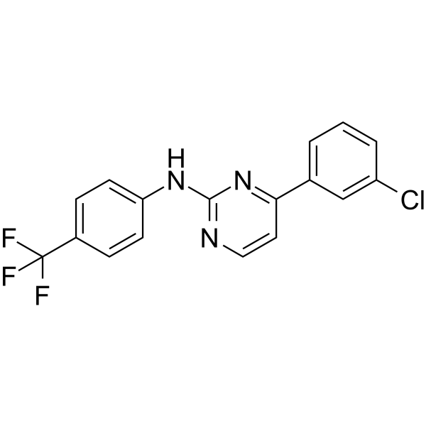
HY-135750
VAF347AhR Agonist -
HY-18174
PrexasertibCHK1 Inhibitor


Biological Activity
Description
IC50 & Target
[1]|
Aryl Hydrocarbon Receptor |
Chk2 |
Cellular Effect
|
Cell Line

|
Type | Value | Description | References |
|---|---|---|---|---|
| A2780 | GI50 |
16 μM
Compound: 5
|
Cytotoxicity against human A2780 cells after 72 hrs by MTT assay
Cytotoxicity against human A2780 cells after 72 hrs by MTT assay
|
10.1039/C0MD00147C |
| A-431 | GI50 |
3.2 μM
Compound: 5
|
Cytotoxicity against human A431 cells after 72 hrs by MTT assay
Cytotoxicity against human A431 cells after 72 hrs by MTT assay
|
10.1039/C0MD00147C |
| BE(2)-C | GI50 |
25 μM
Compound: 5
|
Cytotoxicity against human BE(2)-C cells after 72 hrs by MTT assay
Cytotoxicity against human BE(2)-C cells after 72 hrs by MTT assay
|
10.1039/C0MD00147C |
| DU-145 | GI50 |
41 μM
Compound: 5
|
Cytotoxicity against human DU145 cells after 72 hrs by MTT assay
Cytotoxicity against human DU145 cells after 72 hrs by MTT assay
|
10.1039/C0MD00147C |
| HT-29 | GI50 |
15 μM
Compound: 5
|
Cytotoxicity against human HT-29 cells after 72 hrs by MTT assay
Cytotoxicity against human HT-29 cells after 72 hrs by MTT assay
|
10.1039/C0MD00147C |
| MCF-10A | GI50 |
28 μM
Compound: 5
|
Cytotoxicity against human MCF10A cells after 72 hrs by MTT assay
Cytotoxicity against human MCF10A cells after 72 hrs by MTT assay
|
10.1039/C0MD00147C |
| MCF7 | GI50 |
0.56 μM
Compound: 5
|
Cytotoxicity against human MCF7 cells after 72 hrs by MTT assay
Cytotoxicity against human MCF7 cells after 72 hrs by MTT assay
|
10.1039/C0MD00147C |
| MDA-MB-231 | GI50 |
46 μM
Compound: 5
|
Cytotoxicity against human MDA-MB-231 cells after 72 hrs by MTT assay
Cytotoxicity against human MDA-MB-231 cells after 72 hrs by MTT assay
|
10.1039/C0MD00147C |
| NCI-H460 | GI50 |
5.7 μM
Compound: 5
|
Cytotoxicity against human H460 cells after 72 hrs by MTT assay
Cytotoxicity against human H460 cells after 72 hrs by MTT assay
|
10.1039/C0MD00147C |
| SW480 | GI50 |
23 μM
Compound: 5
|
Cytotoxicity against human SW480 cells after 72 hrs by MTT assay
Cytotoxicity against human SW480 cells after 72 hrs by MTT assay
|
10.1039/C0MD00147C |
In Vitro
ANI-7 (2.5 μM; 24 hours; MCF10A and MDA-MB-468 cells) treatment induces significant S-phase and G2 + M-phase cell cycle arrest within 24 hours of treatment in MDA-MB-468 cells, and negligible effect in normal breast MCF10A cells[1].
ANI-7 (2 μM; 12-24 hours; MDA-MB-468 cells) treatment results in a significant increase in the content and phosphorylation of CHK2, and induces a significant increase in H2AX in MDA-MB-468 cells, indicative of DNA double-strand damage[1].
Inhibition of the AhR pathway ameliorates the effects of ANI-7. ANI-7 activates XRE activity and expression of the AhR and CYP1 members[1].
Comparisons of the GI50 values show that ANI-7 produces a GI50 value of 0.38 μM in MCF-7 cells, whereas values of 3.0-42 μM are observed in cell lines from lung, colon, ovary, neuronal, glial, prostate, and pancreas. The only other tumor type that shows appreciable growth inhibition by ANI-7 is the A431 vulva cell line (GI50 of 0.51μM)[1][1].
ANI-7 potently inhibits the growth of T47D, ZR-75-1, MCF-7, SKBR3, and MDA-MB-468 breast cancer cells (GI50 range of 0.16-0.38 μM), moderately inhibits the growth of BT20 and BT474 cells (GI50 range of 1-2 μM), and essentially fails to inhibit the growth of MDA-MB-231 and MCF10A cells (GI50 range of 17-26 μM). Moreover, ANI-7 maintained its ability to inhibit the growth of drug-resistant cells (MCF-7/VP16: GI50 of 0.21 μM)[1].
MedChemExpress (MCE) has not independently confirmed the accuracy of these methods. They are for reference only.
-
Cell Line:MCF10A and MDA-MB-468 cells
-
Concentration:2.5 μM
-
Incubation Time:24 hours
-
Result:Induced significant S-phase and G2 + M-phase cell cycle arrest in MDA-MB-468 cells, and negligible effect in normal breast MCF10A cells.
-
Cell Line:MDA-MB-468 cells
-
Concentration:2 μM
-
Incubation Time:12 hours, 24 hours
-
Result:Resulted in a significant increase in the content and phosphorylation of CHK2 (25-fold increase),and induced a significant increase in H2AXɣ (3.5-fold increase).


Chemical Information
-
CAS No. 931417-26-4
-
Appearance Solid
-
Molecular Weight 263.12
-
Formula C13H8Cl2N2
-
Color Light yellow to yellow
-
SMILES
ClC1=CC(/C(C#N)=C/C2=CC=CN2)=CC=C1Cl
-
Shipping
Room temperature in continental US; may vary elsewhere.
-
Storage
Powder -20°C 3 years 4°C 2 years In solvent -80°C 2 years -20°C 1 year


Solvent & Solubility
In Vitro:
DMSO : 20.83 mg/mL (79.17 mM; Need ultrasonic; Hygroscopic DMSO has a significant impact on the solubility of product, please use newly opened DMSO)
Please refer to the solubility information to select the appropriate solvent. Once prepared, please aliquot and store the solution to prevent product inactivation from repeated freeze-thaw cycles.
Storage method and period of stock solution: -80°C, 2 years; -20°C, 1 year. When stored at -80°C, please use it within 2 years. When stored at -20°C, please use it within 1 year.
Please refer to the solubility information to select the appropriate solvent. Once prepared, please aliquot and store the solution to prevent product inactivation from repeated freeze-thaw cycles.
Storage method and period of stock solution: -80°C, 2 years; -20°C, 1 year. When stored at -80°C, please use it within 2 years. When stored at -20°C, please use it within 1 year.
Concentration (start) × Volume (start) = Concentration (final) × Volume (final)
In Vivo:
Select the appropriate dissolution method based on your experimental animal and administration route.
- For the following dissolution methods, please ensure to first prepare a clear stock solution using an In Vitro approach and then sequentially add co-solvents:
- To ensure reliable experimental results, the clarified stock solution can be appropriately stored based on storage conditions. As for the working solution for In Vivo experiments, it is recommended to prepare freshly and use it on the same day.
- The percentages shown for the solvents indicate their volumetric ratio in the final prepared solution. If precipitation or phase separation occurs during preparation, heat and/or sonication can be used to aid dissolution.
Add each solvent one by one: 10% DMSO 40% PEG300 5% Tween-80 45% Saline
Solubility: ≥ 2.08 mg/mL (7.91 mM); Clear solution
This protocol yields a clear solution of ≥ 2.08 mg/mL (saturation unknown).
Taking 1 mL working solution as an example, add 100 μL DMSO stock solution (20.8 mg/mL) to 400 μL PEG300, and mix evenly; then add 50 μL Tween-80 and mix evenly; then add 450 μL Saline to adjust the volume to 1 mL.
Preparation of Saline: Dissolve 0.9 g sodium chloride in ddH₂O and dilute to 100 mL to obtain a clear Saline solution.
In Vivo Dissolution Calculator
Please enter the basic information of animal experiments:
-
-
-
-
Recommended: Prepare an additional quantity of animals to account for potential losses during experiments.
Please enter your animal formula composition:
-
%DMSO +
Recommended: Keep the proportion of DMSO in working solution below 2% if your animal is weak.
-
%+
-
+%Tween-80 + +
-
%Saline +
The co-solvents required include: DMSO, . All of co-solvents are available by MedChemExpress (MCE). , Tween 80. All of co-solvents are available by MedChemExpress (MCE).
Working solution concentration: 0.22 mg/mL
Method for preparing stock solution: mg drug dissolved in μL DMSO. Stock solution concentration: mg/mL.
1. Take μL DMSO stock solution;
2. Add μL .
μL , mix evenly;
3. Then add μL Tween 80, mix evenly;
4. Then add μL
Please ensure that the stock solution in the first step is dissolved to a clear state, and add co-solvents in sequence. You can use ultrasonic heating (ultrasonic cleaner, recommended frequency 20-40 kHz), vortexing, etc. to assist dissolution.


Purity & Documentation
-
Data Sheet (278 KB)
-
SDS (251 KB)
- English - EN (251 KB)
- Français - FR (251 KB)
- Deutsch - DE (251 KB)
- Norwegian - NO (251 KB)
- Español - ES (251 KB)
- Swedish - SV (251 KB)
- Italian - IT (251 KB)
- Korean - KR (251 KB)
- Portuguese - PT (251 KB)
-
Handling Instructions (2659 KB)
References
[1]. ilbert J, et al. (Z)-2-(3,4-Dichlorophenyl)-3-(1H-Pyrrol-2-yl)Acrylonitrile Exhibits Selective Antitumor Activity in Breast Cancer Cell Lines via the Aryl Hydrocarbon Receptor Pathway. Mol Pharmacol. 2018 Feb;93(2):168-177. [Content Brief]
[2]. Baker JR, et al. Dichlorophenylacrylonitriles as AhR Ligands That Display Selective Breast Cancer Cytotoxicity in vitro. ChemMedChem. 2018 Jul 18;13(14):1447-1458. [Content Brief]


Complete Stock Solution Preparation Table
Please refer to the solubility information to select the appropriate solvent. Once prepared, please aliquot and store the solution to prevent product inactivation from repeated freeze-thaw cycles.
Storage method and period of stock solution: -80°C, 2 years; -20°C, 1 year. When stored at -80°C, please use it within 2 years. When stored at -20°C, please use it within 1 year.
| Optional Solvent | Concentration Solvent Mass | 1 mg | 5 mg | 10 mg | 25 mg |
|---|---|---|---|---|---|
| DMSO | 1 mM | 3.8005 mL | 19.0027 mL | 38.0055 mL | 95.0137 mL |
| 5 mM | 0.7601 mL | 3.8005 mL | 7.6011 mL | 19.0027 mL | |
| 10 mM | 0.3801 mL | 1.9003 mL | 3.8005 mL | 9.5014 mL | |
| 15 mM | 0.2534 mL | 1.2668 mL | 2.5337 mL | 6.3342 mL | |
| 20 mM | 0.1900 mL | 0.9501 mL | 1.9003 mL | 4.7507 mL | |
| 25 mM | 0.1520 mL | 0.7601 mL | 1.5202 mL | 3.8005 mL | |
| 30 mM | 0.1267 mL | 0.6334 mL | 1.2668 mL | 3.1671 mL | |
| 40 mM | 0.0950 mL | 0.4751 mL | 0.9501 mL | 2.3753 mL | |
| 50 mM | 0.0760 mL | 0.3801 mL | 0.7601 mL | 1.9003 mL | |
| 60 mM | 0.0633 mL | 0.3167 mL | 0.6334 mL | 1.5836 mL |


ANI-7 Related Classifications
HY-117102 Related Classifications
 Powered by Bioz