



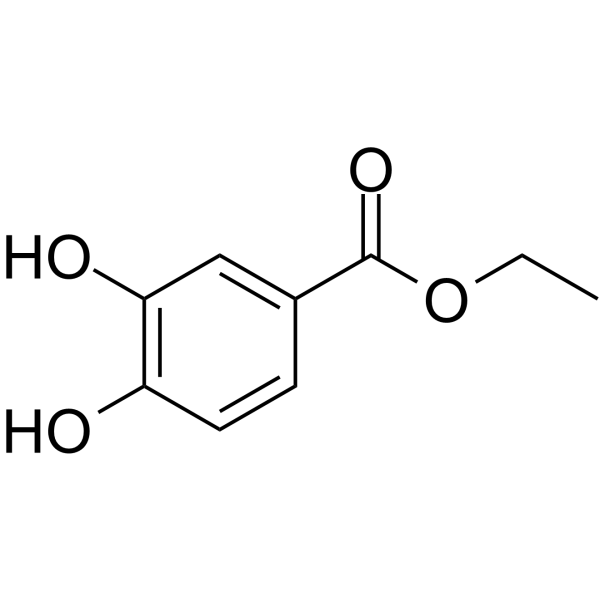
Ethyl 3,4-dihydroxybenzoate
Based on 1 publication(s) in Google Scholar
Ethyl 3,4-dihydroxybenzoate (Protocatechuic acid ethyl ester) is an orally effective, blood-brain barrier-permeable, competitive prolyl hydroxylase (PHD) inhibitor that inhibits the hydroxylation modification of hypoxia-inducible factor (HIF) by PHD. Ethyl 3,4-dihydroxybenzoate stabilizes HIF-1α by inhibiting PHD, activates downstream pathways to induce autophagy and apoptosis of tumor cells, and regulates inflammatory responses, inhibits the NF-κB pathway, improves vascular permeability, and promotes osteoblast differentiation. Ethyl 3,4-dihydroxybenzoate has anti-tumor, anti-hypoxic injury, and bone metabolism regulation effects. It can also be used in the research of cardiovascular protection (such as reducing myocardial ischemic damage), bone tissue engineering (promoting osteogenesis/inhibiting osteoclast differentiation), and prevention and treatment of high-altitude cerebral edema.
For research use only. We do not sell to patients.
- Purity : 99.96%
- CAS No.: 3943-89-3
- Formula: C9H10O4
- Molecular Weight:182.18
-
Storage:
4°C, stored under nitrogen
* In solvent : -80°C, 6 months; -20°C, 1 month (stored under nitrogen)

To place orders, for customer services and technical support, please contact: MedChemExpress USA
Tel: 609-228-6898 E-mail: [email protected] [email protected]


-
 Biological Activity
-
Chemical Information
-
Solvent & Solubility
- Protocols
- Purity & Documentation
- References
-
 Help & FAQs


-
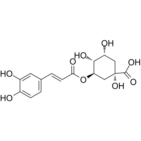
HY-N0055
Chlorogenic acidAntioxidant -
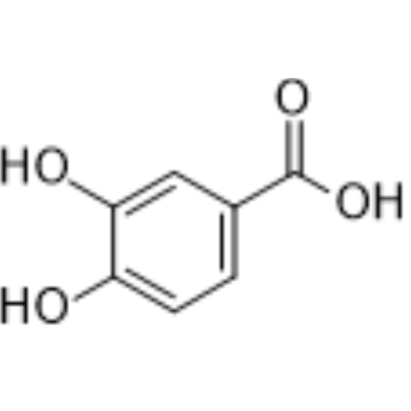
HY-N0294
Protocatechuic acidEndogenous Metabolite -
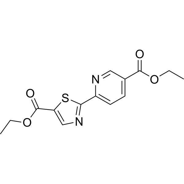
HY-103068
Diethyl-pythiDCCP4H Inhibitor -
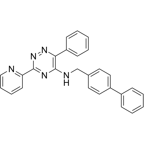
HY-12754
ML228HIF Activator -

HY-W016409R
Ethyl 3,4-dihydroxybenzoate (Standard)Prolyl-hydroxylase Inhibitor -

HY-N0055R
Chlorogenic acid (Standard)Antioxidant -
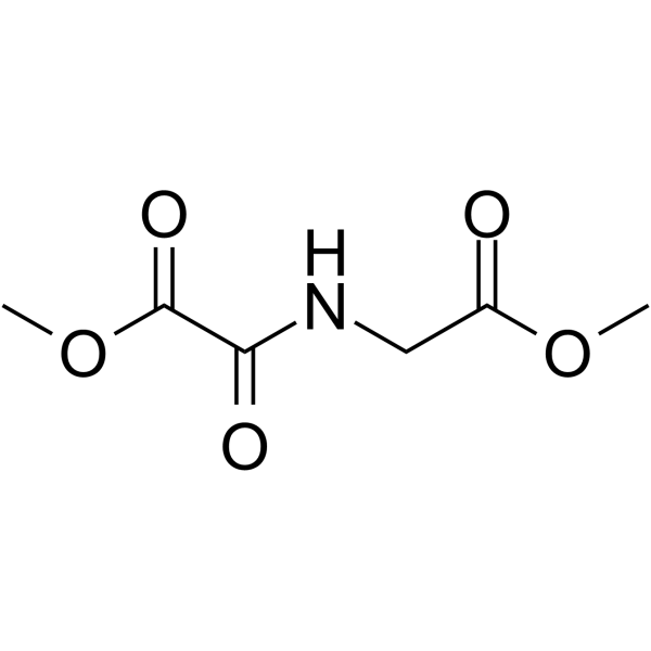
HY-15893
DMOGHIF-PH Inhibitor -
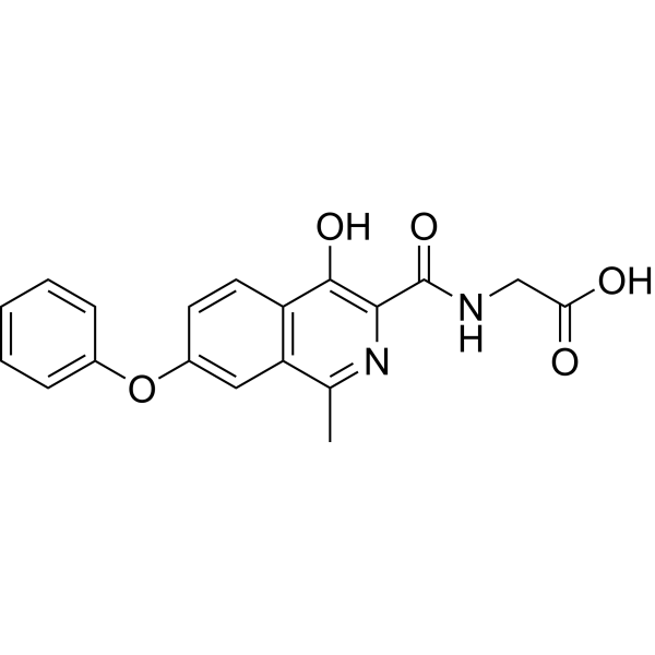
HY-13426
RoxadustatHIF-PH Inhibitor -
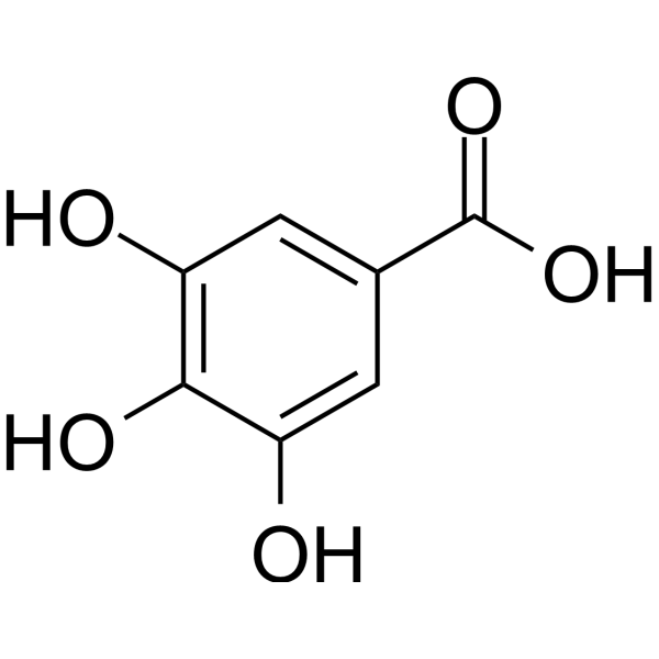
HY-N0523
Gallic acidBioactive Polyhydroxyphenolic Compound -
HY-W001179
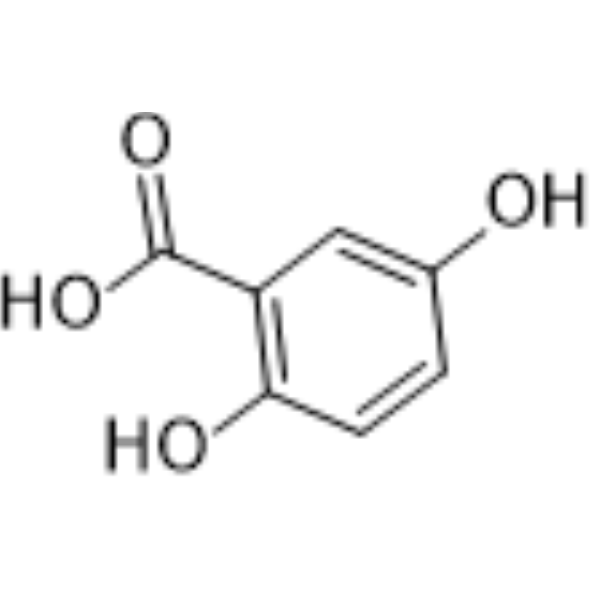
2,5-Dihydroxybenzoic acid -
HY-10231
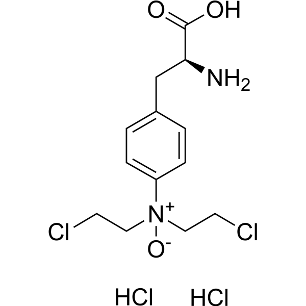
PX-478HIF-1α Inhibitor -
HY-N0524
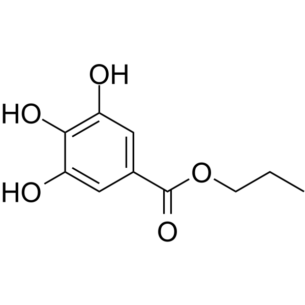
Propyl gallateAntioxidant
Publications Citing Use of MedChemExpress (MCE) Ethyl 3,4-dihydroxybenzoate
More

Biological Activity
Description
Cellular Effect
|
Cell Line

|
Type | Value | Description | References |
|---|---|---|---|---|
| BV-2 | IC50 |
>10 μM
Compound: 25
|
Antiinflammatory activity in C57BL6/J mouse BV2 cells assessed as inhibition of LPS-induced nitric oxide production after 24 hrs by Griess assay
Antiinflammatory activity in C57BL6/J mouse BV2 cells assessed as inhibition of LPS-induced nitric oxide production after 24 hrs by Griess assay
|
[PMID: 26110443] |
| MDCK | CC50 |
>169.99 μM
Compound: 3d
|
Cytotoxicity against MDCK cells after 72 hrs by MTT assay
Cytotoxicity against MDCK cells after 72 hrs by MTT assay
|
[PMID: 24803363] |
| MDCK | IC50 |
>35 μM
Compound: 25
|
Antiviral activity against Influenza A virus (A/FM/1/47(H1N1)) infected in MDCK cells assessed as inhibition of virus-induced cytopathic effect
Antiviral activity against Influenza A virus (A/FM/1/47(H1N1)) infected in MDCK cells assessed as inhibition of virus-induced cytopathic effect
|
[PMID: 26110443] |
| MDCK | IC50 |
16.11 μM
Compound: 3d
|
Antiviral activity against Influenza A virus (A/Vietnam/1194/2004(H5N1)) infected in MDCK cells assessed as inhibition of viral replication after 2 to 3 days by crystal violet staining-based plaque reduction assay
Antiviral activity against Influenza A virus (A/Vietnam/1194/2004(H5N1)) infected in MDCK cells assessed as inhibition of viral replication after 2 to 3 days by crystal violet staining-based plaque reduction assay
|
[PMID: 24803363] |
| Vero | IC50 |
11.3 μM
Compound: 25
|
Antiviral activity against Coxsackievirus B3 infected in African green monkey Vero cells assessed as inhibition of virus-induced cytopathic effect
Antiviral activity against Coxsackievirus B3 infected in African green monkey Vero cells assessed as inhibition of virus-induced cytopathic effect
|
[PMID: 26110443] |
In Vitro
1. Apoptosis experiment of esophageal cancer cells:
Ethyl 3,4-dihydroxybenzoate (50 μg/mL; 24-72 h) induces S phase cell cycle arrest, loss of mitochondrial membrane permeability and caspase-dependent apoptosis in KYSE 170 and EC109 esophageal squamous cell carcinoma cells, and upregulates the expression of NDRG1, BNIP3, Beclin and other proteins[1].
2. Myocardial protection experiment:
Ethyl 3,4-dihydroxybenzoate (1 mM; 15 min, pretreatment) can activate NOS in isolated cardiomyocytes through KATP channels, increase mitochondrial ROS levels, and exert myocardial protection[2].
3. Osteocyte differentiation assay:
Ethyl 3,4-dihydroxybenzoate (5 mg/mL; 3-14 d) significantly increases alkaline phosphatase (ALP) activity, type I collagen deposition and mineralizes nodule formation in MC3T3-E1 osteoblast precursor cells, and inhibits TRAP-positive multinucleate cell differentiation and TRAP enzyme activity in RAW264.7 osteoclasts in a dose-dependent manner (2-4 mg/mL; 6 d)[3].
MedChemExpress (MCE) has not independently confirmed the accuracy of these methods. They are for reference only. Further protocols information, click here.
-
Cell Line:KYSE 170 and EC109 esophageal squamous cell carcinoma cells
-
Concentration:50 μg/mL
-
Incubation Time:24 h, 36 h, 72 h
-
Result:Induced S-phase cell cycle arrest, mitochondrial membrane permeabilization, and caspase-dependent apoptosis in KYSE 170 and EC109 cells.
Upregulated autophagy- and apoptosis-related proteins, including NDRG1, BNIP3, and Beclin, with significant reduction in cell viability by 50-70% compared to controls.
-
Cell Line:MC3T3-E1 preosteoblasts and RAW264.7 monocyte cells
-
Concentration:5 mg/mL (MC3T3-E1); 1-4 mg/mL (RAW264.7)
-
Incubation Time:3-14 days (MC3T3-E1); 6 days (RAW264.7)
-
Result:In MC3T3-E1 cells, significantly increased alkaline phosphatase (ALP) activity (2-fold at day 10), collagen type I deposition (1.5-fold at day 14), and mineralized nodule formation (34% increase at week 5).
In RAW264.7 cells, at 2-4 mg/mL dose-dependently reduced TRAP-positive multinucleated osteoclast formation by 54-77% and decreased TRAP enzyme activity, indicating inhibition of osteoclast differentiation.
In Vivo
Ethyl 3,4-dihydroxybenzoate (5 mg/bead, 50 mg/bead; subcutaneous injection after Alginate gel embedding; single dose; 5-8 weeks) promotes the osteogenic differentiation of human mesenchymal stem cells, increases the expression of type I collagen and osteocalcin, and induces the formation of new bone tissue in the nude mouse subcutaneous transplantation model[3].
2. High-altitude cerebral edema prevention and treatment experiment:
Ethyl 3,4-dihydroxybenzoate (50-100 mg/kg; intraperitoneal injection; once daily; 3 days) significantly reduces brain water content and vascular permeability in the rat acute hypobaric hypoxia model, down-regulates pro-inflammatory factors such as TNF-α and IL-6, and up-regulates antioxidant proteins such as HIF-1α and HO-1[4].
3. Myocardial ischemia protection experiment:
Ethyl 3,4-dihydroxybenzoate (75 mg/kg; intraperitoneal injection; once daily; 3 days) reduces infarct area and inhibited NF-κB-mediated inflammatory response in the rat myocardial ischemia-reperfusion model by activating the NOS-NO pathway and mitochondrial KATP channel[4].
MedChemExpress (MCE) has not independently confirmed the accuracy of these methods. They are for reference only.
-
Animal Model:Subcutaneous bone tissue engineering model (Balb/c nude mice) (6 weeks old), subcutaneous transplantation of alginate-encapsulated hMSCs[3]
-
Dosage:5 mg/bead or 50 mg/bead, incorporated into alginate gel
-
Administration:Single subcutaneous injection into the back, observed for 5-8 weeks
-
Result:Promoted osteogenic differentiation of human mesenchymal stem cells (hMSCs), as shown by von Kossa and Alizarin Red staining for calcium deposition.
Increased expression of collagen type I and osteocalcin in the transplanted tissue, with dose-dependent enhancement (50 mg/bead showing stronger effects).
Resulted organized new bone formation with reduced fibrous tissue compared to control groups.
-
Animal Model:Myocardial ischemia-reperfusion injury model (Sprague-Dawley rats) (180±20 g), myocardial ischemia-reperfusion injury model[4]
-
Dosage:75 mg/kg, dissolved in DMSO/saline
-
Administration:Intraperitoneal injection, daily for 3 days, followed by 30-min coronary artery occlusion and 120-min reperfusion
-
Result:Significantly reduced infarct size by 68% compared to controls, associated with increased nitric oxide (NO) production, activation of mitochondrial ATP-sensitive potassium (mitoKATP) channels, and downregulation of nuclear factor-κB (NF-κB)-dependent pro-inflammatory cytokines (TNF-α, IL-6). Western blot analysis showed upregulation of hypoxia-inducible factor-1α (HIF-1α) and heme oxygenase-1 (HO-1), while immunohistochemistry revealed reduced neutrophil infiltration in the ischemic myocardium.
-
Animal Model:Acute hypobaric hypoxia model (Sprague-Dawley rats) (180±20 g), acute hypobaric hypoxia at 9144 m for 5 h[4]
-
Dosage:75 mg/kg, dissolved in DMSO/saline
-
Administration:Intraperitoneal injection, daily for 3 days prior to hypoxia exposure
-
Result:Reduced brain water content (edema index) by 10% and vascular permeability (sodium fluorescein leakage) by 49% compared to hypoxic controls.
Decreased levels of pro-inflammatory cytokines (TNF-α, IL-6, IFN-γ) and increased anti-inflammatory IL-10.
Upregulated HIF-1α, HO-1, and metallothionein-1 (MT-1) expression, while NF-κB nuclear translocation and vascular cell adhesion molecule-1 (VCAM-1) expression were significantly attenuated.


Chemical Information
-
CAS No. 3943-89-3
-
Appearance Solid
-
Molecular Weight 182.18
-
Formula C9H10O4
-
Color White to off-white
-
SMILES
O=C(OCC)C1=CC=C(O)C(O)=C1
-
Synonyms
Protocatechuic acid ethyl ester
-
Structure Classification
-
Initial Source
-
Shipping
Room temperature in continental US; may vary elsewhere.
-
Storage
4°C, stored under nitrogen
* In solvent : -80°C, 6 months; -20°C, 1 month (stored under nitrogen)


Publications (1)
-
Journal Impact Factor
-
Most Recent


Solvent & Solubility
In Vitro:
DMSO : 100 mg/mL (548.92 mM; Need ultrasonic; Hygroscopic DMSO has a significant impact on the solubility of product, please use newly opened DMSO)
Please refer to the solubility information to select the appropriate solvent. Once prepared, please aliquot and store the solution to prevent product inactivation from repeated freeze-thaw cycles.
Storage method and period of stock solution: -80°C, 6 months; -20°C, 1 month (stored under nitrogen). When stored at -80°C, please use it within 6 months. When stored at -20°C, please use it within 1 month.
Please refer to the solubility information to select the appropriate solvent. Once prepared, please aliquot and store the solution to prevent product inactivation from repeated freeze-thaw cycles.
Storage method and period of stock solution: -80°C, 6 months; -20°C, 1 month (stored under nitrogen). When stored at -80°C, please use it within 6 months. When stored at -20°C, please use it within 1 month.
Concentration (start) × Volume (start) = Concentration (final) × Volume (final)
In Vivo:
Select the appropriate dissolution method based on your experimental animal and administration route.
- For the following dissolution methods, please ensure to first prepare a clear stock solution using an In Vitro approach and then sequentially add co-solvents:
- To ensure reliable experimental results, the clarified stock solution can be appropriately stored based on storage conditions. As for the working solution for In Vivo experiments, it is recommended to prepare freshly and use it on the same day.
- The percentages shown for the solvents indicate their volumetric ratio in the final prepared solution. If precipitation or phase separation occurs during preparation, heat and/or sonication can be used to aid dissolution.
Add each solvent one by one: 10% DMSO 40% PEG300 5% Tween-80 45% Saline
Solubility: ≥ 2.5 mg/mL (13.72 mM); Clear solution
This protocol yields a clear solution of ≥ 2.5 mg/mL (saturation unknown).
Taking 1 mL working solution as an example, add 100 μL DMSO stock solution (25.0 mg/mL) to 400 μL PEG300, and mix evenly; then add 50 μL Tween-80 and mix evenly; then add 450 μL Saline to adjust the volume to 1 mL.
Preparation of Saline: Dissolve 0.9 g sodium chloride in ddH₂O and dilute to 100 mL to obtain a clear Saline solution.
Add each solvent one by one: 10% DMSO 90% (20% SBE-β-CD in Saline)
Solubility: ≥ 2.5 mg/mL (13.72 mM); Clear solution
This protocol yields a clear solution of ≥ 2.5 mg/mL (saturation unknown).
Taking 1 mL working solution as an example, add 100 μL DMSO stock solution (25.0 mg/mL) to 900 μL 20% SBE-β-CD in Saline, and mix evenly.
Preparation of 20% SBE-β-CD in Saline (4°C, storage for one week): 2 g SBE-β-CD powder is dissolved in 10 mL Saline, completely dissolve until clear.
In Vivo Dissolution Calculator
Please enter the basic information of animal experiments:
-
-
-
-
Recommended: Prepare an additional quantity of animals to account for potential losses during experiments.
Please enter your animal formula composition:
-
%DMSO +
Recommended: Keep the proportion of DMSO in working solution below 2% if your animal is weak.
-
%+
-
+%Tween-80 + +
-
%Saline +
The co-solvents required include: DMSO, . All of co-solvents are available by MedChemExpress (MCE). , Tween 80. All of co-solvents are available by MedChemExpress (MCE).
Working solution concentration: 0.22 mg/mL
Method for preparing stock solution: mg drug dissolved in μL DMSO. Stock solution concentration: mg/mL. * In solvent : -80°C, 6 months; -20°C, 1 month (stored under nitrogen)
1. Take μL DMSO stock solution;
2. Add μL .
μL , mix evenly;
3. Then add μL Tween 80, mix evenly;
4. Then add μL
Please ensure that the stock solution in the first step is dissolved to a clear state, and add co-solvents in sequence. You can use ultrasonic heating (ultrasonic cleaner, recommended frequency 20-40 kHz), vortexing, etc. to assist dissolution.

Protocols
-
3D Hydrogel Synthetic Scaffold Culture
3D hydrogel synthetic scaffold culture embeds cells, spheroids, organoids, or tissue fragments inside a hydrated crosslinked polymer network so that cells receive matrix and cell-cell cues in three dimensions rather than from a flat plastic surface. A literature-supported model protocol is PEG-4MAL hydrogel culture, in which four-arm maleimide-terminated PEG is functionalized with cysteine-containing adhesive peptides such as RGD and crosslinked with protease-degradable peptides such as GPQ-W; this creates a defined, modular scaffold that supports human organoid generation and culture. The readouts are scaffold-supported growth, morphology, lumen formation, budding, viability, proliferation, lineage-marker expression, and matrix-dependent expansion or differentiation; reported assays include transmitted-light imaging, immunofluorescence, in situ hybridization, qRT-PCR, and rheological characterization.
-
Apoptosis
Apoptosis, also called programmed cell death, is generally characterized by distinct morphological characteristics.
-
TUNEL staining for apoptotic DNA fragmentation
TUNEL staining detects DNA strand breaks by using terminal deoxynucleotidyl transferase to add labeled nucleotides to exposed 3′-OH DNA termini, generating either microscopic staining in fixed cells or tissue sections, or fluorescence/cytometric signal in cell suspensions. TUNEL positivity reflects DNA fragmentation but should not be interpreted alone as definitive apoptosis, because TUNEL can also label necrotic, autolytic, mechanically damaged, or DNA-repair-associated DNA breaks.
-
Annexin V plus membrane-impermeant dye apoptosis staining
Annexin V-based apoptosis assays rely on the detection of phosphatidylserine (PS) externalization from the inner leaflet of the plasma membrane to the outer leaflet, an early biochemical hallmark of apoptosis. Fluorescently labeled Annexin V binds PS in a calcium-dependent manner, enabling identification of early apoptotic cells by flow cytometry or fluorescence microscopy. When combined with a membrane-impermeant DNA-binding dye (e. g. , propidium iodide), this approach allows discrimination between viable (Annexin V−/dye−), early apoptotic (Annexin V+/dye−), and late apoptotic or necrotic (Annexin V+/dye+) cell populations by assessing membrane integrity and PS exposure.
-
Mesenchymal stromal/stem cell osteogenic differentiation
Mesenchymal stromal/stem cells can be induced toward an osteoblast-like lineage in vitro by culture in osteogenic medium containing dexamethasone, ascorbic acid or ascorbate-2-phosphate, and β-glycerophosphate; the differentiation process is commonly evaluated by alkaline phosphatase activity, osteogenic marker expression, collagenous matrix formation, and calcium-rich matrix mineralization. The main readouts are alkaline phosphatase activity as an early osteogenic marker and Alizarin Red S staining as a calcium-deposit readout for mineralized extracellular matrix; Alizarin Red S can be inspected microscopically or extracted and measured colorimetrically at 405 nm.
-
Autophagy
Autophagy is a process in which eukaryotic cells use lysosomes to degrade their own cytoplasmic proteins and damaged organelles under the regulation of autophagy related gene (Atg). Microtubule-associated proteins light chain 3 (LC3) is recognized as autophagy marker, which transfers from cytoplasmic LC3 (LC3-I) to membrane type (LC3-II). LC3-II/I ratio could be detected by Western Blot and fluorescence microscopy.
-
Apoptosis Solutions
Apoptosis is a regulated, generally non-lytic cell-death pathway that removes unwanted, damaged, infected, or abnormal cells through coordinated morphological changes, caspase activation, DNA fragmentation, and membrane remodeling. The intrinsic apoptosis pathway is controlled mainly by mitochondrial outer membrane permeabilization, BCL-2 family proteins, cytochrome c release, apoptosome formation, caspase-9 activation, and downstream executioner caspase-3/7 activation. The extrinsic apoptosis pathway is initiated by death receptors such as Fas, TNFR, and TRAIL receptors, which recruit adaptor proteins and activate caspase-8 before engaging executioner caspases or mitochondrial amplification through BID cleavage. Apoptosis is linked to many phenotypes, including cancer cell killing, tissue homeostasis, immune regulation, neurodegeneration, infection response, and treatment-induced cytotoxicity; unresolved questions include how apoptosis interacts with necroptosis, pyroptosis, ferroptos
-
Transepithelial/transendothelial electrical resistance assay
TEER measures electrical resistance across epithelial or endothelial monolayers cultured on permeable supports, and the readout reflects ionic conductance through the cell barrier, especially the paracellular pathway regulated by junctional integrity. TEER can be measured without destroying the monolayer and is commonly used before or during transport, permeability, barrier-disruption, and barrier-maturation experiments. TEER values are influenced by biological maturation and technical conditions; reported factors include temperature, medium formulation, passage number, electrode geometry, membrane properties, and junctional length during early monolayer maturation. Therefore, TEER should be interpreted with blank-insert subtraction, area normalization, repeated readings, and, when possible, orthogonal barrier readouts such as FITC-dextran flux or tight-junction staining.
-
Lysosome and acidic-vesicle live-cell staining
Lysosome and acidic-vesicle live-cell staining detects acidic intracellular compartments by using membrane-permeant acidotropic probes that accumulate in low-pH vesicles, including lysosomes, late endosomes, autolysosomes, and acidic phagosomes. LysoTracker staining is commonly used as an intensity-based readout of acidic lysosomal compartment abundance or enlargement, while acridine orange produces green fluorescence in less concentrated compartments and red fluorescence after concentration-dependent accumulation in acidic vesicular organelles. Loss or reduction of acridine-orange red signal can be used as a readout of lysosomal membrane permeabilization or reduced acidic-vesicle integrity. This protocol is designed for live cultured cells and can be adapted for fluorescence microscopy, high-content imaging, plate-reader readout, or flow cytometry when the selected literature supports the readout. Because these dyes report acidotropic accumulation rather than lysosome identity alone,
-
Research Protocol for Inflammation-related Diseases
The NLRP3 inflammasome is a cytosolic innate immune signaling platform that integrates priming signals and danger-signal activation to promote caspase-1 activation, maturation of IL-1β and IL-18, and gasdermin D-mediated pyroptotic cell death. The core experimental logic is to determine whether inflammatory disease phenotypes are driven by increased NLRP3 expression, ASC-containing inflammasome assembly, caspase-1 cleavage, GSDMD cleavage, and extracellular release of IL-1β/IL-18 rather than by nonspecific cell injury alone. The pathway is strongly linked to inflammation-related disease phenotypes because monosodium urate crystals activate NALP3/NLRP3 inflammasome signaling in gout-like crystal inflammation, cholesterol crystals activate NLRP3 inflammasomes in atherogenesis models, and DSS-induced intestinal inflammation has been reported to involve NLRP3 inflammasome activity. However, experimental colitis studies also show context-dependent protective effects of NLRP3 inflammasome co
-
Macroautophagy Solutions
Macroautophagy is a conserved lysosome-dependent degradation pathway in which cytoplasmic material is sequestered into double-membrane autophagosomes and delivered to lysosomes for degradation and recycling. The pathway supports cellular homeostasis during nutrient limitation, organelle stress, protein-aggregate accumulation, infection, differentiation, and tissue remodeling by coupling cargo sequestration, autophagosome maturation, lysosomal fusion, and degradation of cargo-derived macromolecules. The core molecular sequence includes initiation by nutrient- and stress-regulated autophagy machinery, autophagosome nucleation, LC3/ATG8-family conjugation to autophagosomal membranes, cargo selection through receptors such as SQSTM1/p62, autophagosome-lysosome fusion, and lysosomal degradation. LC3 was identified as a mammalian homolog of yeast Atg8 that localizes to autophagosomal membranes after processing, and p62/SQSTM1 was shown to connect ubiquitinated cargo with autophagic degradati
-
Osteoclast differentiation from monocyte/macrophage precursors
Osteoclast differentiation is an in vitro induction assay in which monocyte/macrophage-lineage precursors are exposed to macrophage colony-stimulating factor (M-CSF) and receptor activator of NF-κB ligand (RANKL), generating multinucleated osteoclasts that are commonly identified by tartrate-resistant acid phosphatase (TRAP) staining and functionally confirmed by resorption pits on dentin, bone, or mineralized substrates. M-CSF supports survival and expansion of osteoclast precursors, while RANKL binding to RANK drives osteoclast commitment, fusion, maturation, and resorptive function; osteoprotegerin inhibits this pathway by binding RANKL and preventing RANK activation. The main readouts are the number of TRAP-positive multinucleated cells, formation of F-actin rings, and resorbed surface area; TRAP-positive multinucleated cells indicate osteoclast differentiation, whereas pit formation on dentin, bone, or mineralized coating indicates functional bone-resorbing activity.


Purity & Documentation
-
Data Sheet (284 KB)
-
SDS (393 KB)
- English - EN (393 KB)
- Français - FR (393 KB)
- Deutsch - DE (393 KB)
- Norwegian - NO (393 KB)
- Español - ES (393 KB)
- Swedish - SV (393 KB)
- Italian - IT (393 KB)
- Korean - KR (393 KB)
- Portuguese - PT (393 KB)
-
Handling Instructions (2659 KB)
References
[1]. Bo Han, et al. A prolyl-hydroxylase inhibitor, ethyl-3,4-dihydroxybenzoate, induces cell autophagy and apoptosis in esophageal squamous cell carcinoma cells via up-regulation of BNIP3 and N-myc downstream-regulated gene-1. PLoS One. 2014 Sep 18;9(9):e107204. [Content Brief]
[2]. Sebastian Philipp, et al. Desferoxamine and ethyl-3,4-dihydroxybenzoate protect myocardium by activating NOS and generating mitochondrial ROS. Am J Physiol Heart Circ Physiol. 2006 Jan;290(1):H450-7. [Content Brief]
[3]. Byeong-Ju Kwon, et al. Ethyl-3,4-dihydroxybenzoate with a dual function of induction of osteogenic differentiation and inhibition of osteoclast differentiation for bone tissue engineering. Tissue Eng Part A. 2014 Nov;20(21-22):2975-84. [Content Brief]
[4]. Singh DP, et al. Ethyl 3,4-dihydroxybenzoate (EDHB): a prolyl hydroxylase inhibitor attenuates acute hypobaric hypoxia mediated vascular leakage in brain. J Physiol Sci. 2016 Jul;66(4):315-26. [Content Brief]


Complete Stock Solution Preparation Table
Please refer to the solubility information to select the appropriate solvent. Once prepared, please aliquot and store the solution to prevent product inactivation from repeated freeze-thaw cycles.
Storage method and period of stock solution: -80°C, 6 months; -20°C, 1 month (stored under nitrogen). When stored at -80°C, please use it within 6 months. When stored at -20°C, please use it within 1 month.
| Optional Solvent | Concentration Solvent Mass | 1 mg | 5 mg | 10 mg | 25 mg |
|---|---|---|---|---|---|
| DMSO | 1 mM | 5.4892 mL | 27.4461 mL | 54.8923 mL | 137.2307 mL |
| 5 mM | 1.0978 mL | 5.4892 mL | 10.9785 mL | 27.4461 mL | |
| 10 mM | 0.5489 mL | 2.7446 mL | 5.4892 mL | 13.7231 mL | |
| 15 mM | 0.3659 mL | 1.8297 mL | 3.6595 mL | 9.1487 mL | |
| 20 mM | 0.2745 mL | 1.3723 mL | 2.7446 mL | 6.8615 mL | |
| 25 mM | 0.2196 mL | 1.0978 mL | 2.1957 mL | 5.4892 mL | |
| 30 mM | 0.1830 mL | 0.9149 mL | 1.8297 mL | 4.5744 mL | |
| 40 mM | 0.1372 mL | 0.6862 mL | 1.3723 mL | 3.4308 mL | |
| 50 mM | 0.1098 mL | 0.5489 mL | 1.0978 mL | 2.7446 mL | |
| 60 mM | 0.0915 mL | 0.4574 mL | 0.9149 mL | 2.2872 mL | |
| 80 mM | 0.0686 mL | 0.3431 mL | 0.6862 mL | 1.7154 mL | |
| 100 mM | 0.0549 mL | 0.2745 mL | 0.5489 mL | 1.3723 mL |


Ethyl 3,4-dihydroxybenzoate Related Classifications
HY-W016409 Related Classifications
 Powered by Bioz