



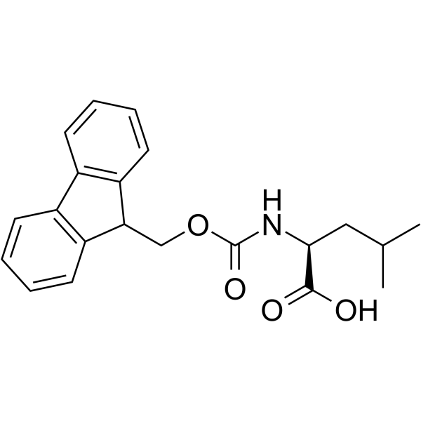
Fmoc-leucine

Based on 1 Customer Validation
Fmoc-Leucine (N-FMOC-leucine) is an anti-inflammatory agent that not only promotes extracellular Ca2+ influx but also facilitates intracellular Ca2+ release. Fmoc-Leucine is a selective ligand for PPARγ (Ki = 15 μM), exhibiting insulin-sensitizing effects but with weak fatogenic activity. Fmoc-Leucine exhibits unique self-assembly properties and can form transient gels, stable gels, or crystals/2D sheets through different pathways. Fmoc-Leucine can be used in the research of diabetes, colitis, and bladder cancer.
For research use only. We do not sell to patients.
- Purity : 99.9%
- CAS No.: 35661-60-0
- Formula: C21H23NO4
- Molecular Weight:353.42
-
Storage:Powder -20°C, 3 years , 4°C, 2 years ; In solvent -80°C, 6 months , -20°C, 1 month

To place orders, for customer services and technical support, please contact: MedChemExpress USA
Tel: 609-228-6898 E-mail: [email protected] [email protected]


-
 Biological Activity
-
Chemical Information
-
Solvent & Solubility
- Protocols
- Purity & Documentation
- References
-
 Help & FAQs


-
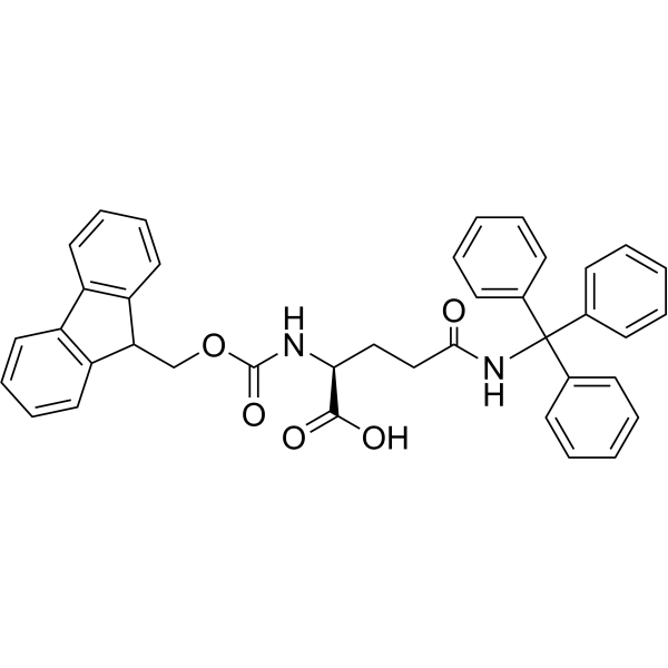
HY-W013081
Fmoc-Gln(Trt)-OHGlutamine Derivative -
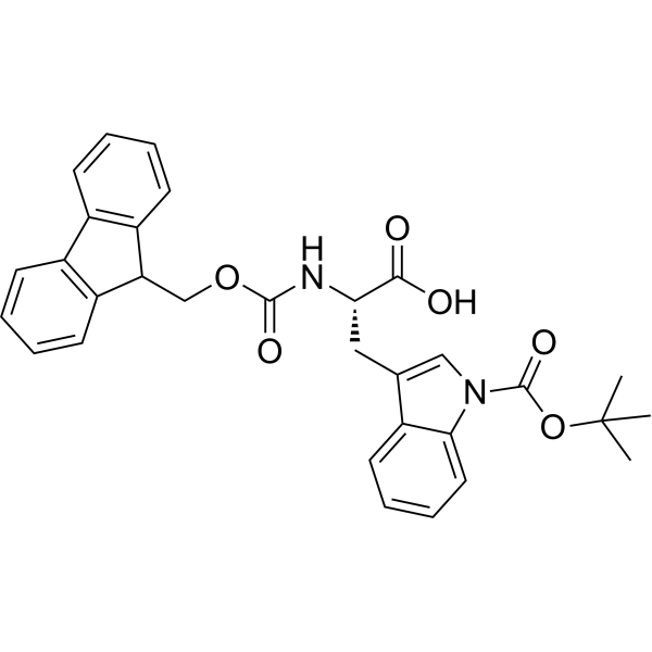
HY-W008034
Fmoc-Trp(Boc)-OHAmino Acid Derivative -
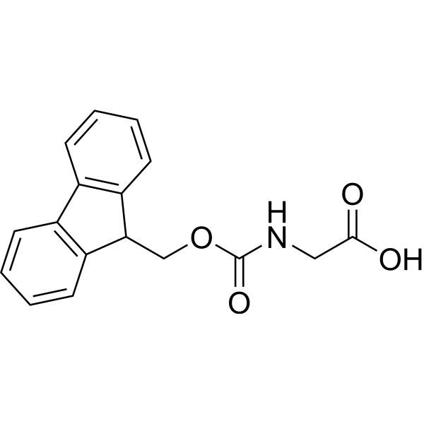
HY-Y1250
Fmoc-Gly-OHGlycine Derivative -
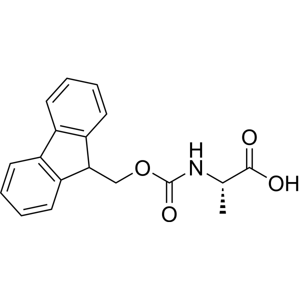
HY-W009204
Fmoc-Ala-OHAmino Acid Derivative -
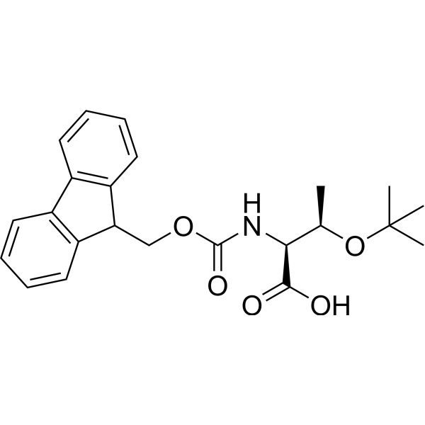
HY-W007706
Fmoc-Thr(tBu)-OHAmino Acid Derivative -
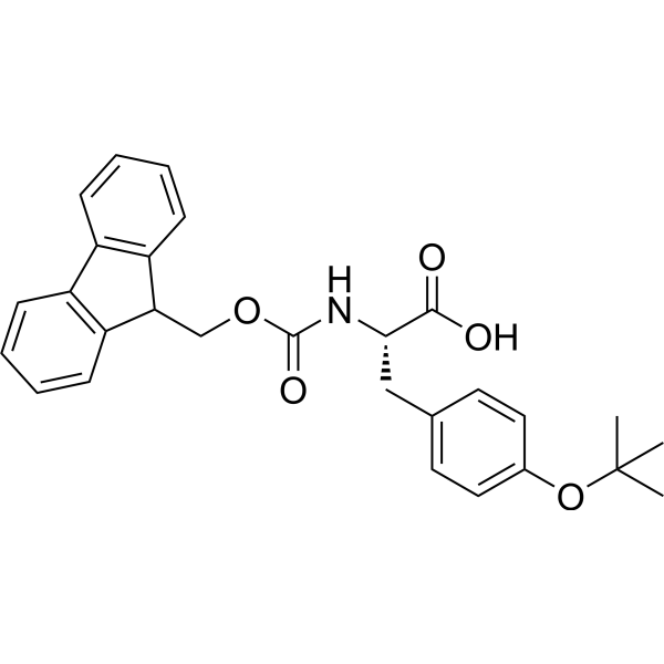
HY-W008016
Fmoc-Tyr(tBu)-OHAmino Acid Derivative -
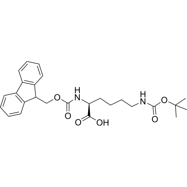
HY-79128
Fmoc-L-Lys (Boc)-OHAmino Acid Derivative -

HY-Y1169
Fmoc-Asp(OtBu)-OHAspartate Derivative -
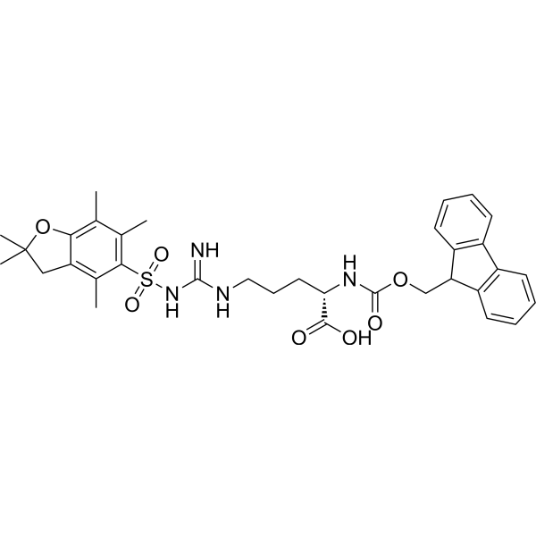
HY-Y1636
Fmoc-Arg(Pbf)-OHArginine Derivative -
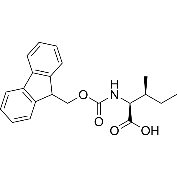
HY-19821
Fmoc-Ile-OHAmino Acid Derivative -
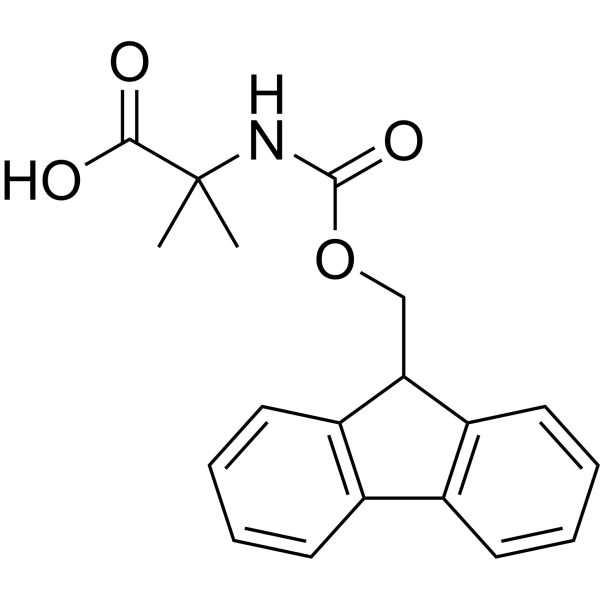
HY-79648
Fmoc-Aib-OHAmino Acid Derivative -
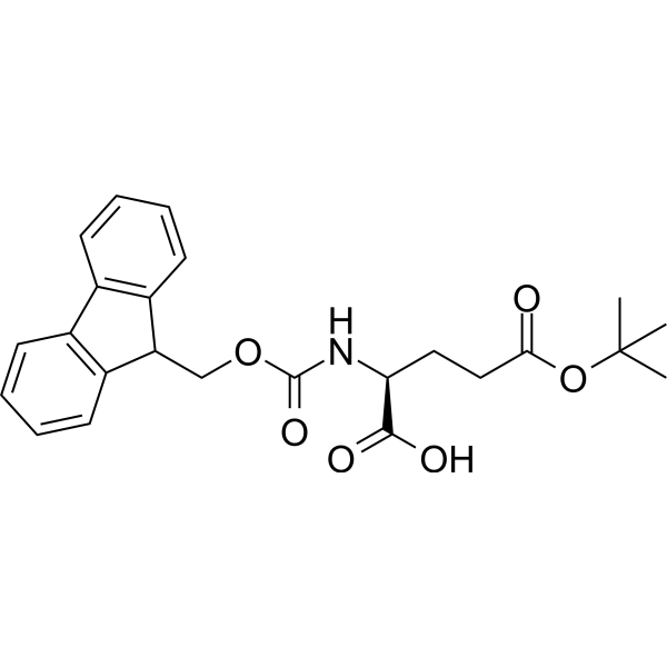
HY-Y0134
Fmoc-Glu(OtBu)-OHAmino Acid Derivative
All Calcium Channel Isoforms
More

Biological Activity
Description
IC50 & Target
|
PPARγ |
Cellular Effect
|
Cell Line

|
Type | Value | Description | References |
|---|---|---|---|---|
| HEK293 | IC50 |
23.3 μM
Compound: 13
|
Inhibition of human recombinant-Sialin expressed in HEK293 cells assessed as reduction in [3H]Neu5Ac uptake at 30 to 300 uM incubated for 15 mins by liquid scintillation counting method
Inhibition of human recombinant-Sialin expressed in HEK293 cells assessed as reduction in [3H]Neu5Ac uptake at 30 to 300 uM incubated for 15 mins by liquid scintillation counting method
|
[PMID: 32608236] |
In Vitro
Fmoc-Leucine (1 mM, 24 h) significantly enhances the interaction between PPARγ and SRC-1 in RK13 cells[1].
Fmoc-Leucine (10 μM, 0-6 d) induces the expression of LPL and aP2 in 3T3-L1 cells, but its ability to accumulate lipids is significantly lower than that of Rosiglitazone (HY-17386)[1].
Fmoc-Leucine (0.1-2 mM) rapidly and continuously increases [Ca²⁺]i in MDCK and BFTC cells in a concentration-dependent manner with an EC50 in MDCK cells of 0.5 mM[3][4].
Fmoc-Leucine induces the release of Ca²⁺ from the endoplasmic reticulum and activated the influx of Ca²⁺ in BFTC bladder cancer cells[4].
MedChemExpress (MCE) has not independently confirmed the accuracy of these methods. They are for reference only. Further protocols information, click here.
-
Cell Line:3T3-L1 cells
-
Concentration:10 μM
-
Incubation Time:0, 2, 3, 4, 6 d
-
Result:Induced the expression of LPL and aP2.
In Vivo
Fmoc-Leucine (50 mg/kg, i.p., once daily for 4 days) reduces the severity of TNBS-induced colitis in the mice[1].
MedChemExpress (MCE) has not independently confirmed the accuracy of these methods. They are for reference only.
-
Animal Model:Normal mice, diet-induced insulin resistance model and diabetic db/db mice model established in C57BL/6J mice[1]
-
Dosage:10, 30 and 60 mg/kg
-
Administration:Intraperitoneal injection (i.p.), once daily for 7 or 14 days
-
Result:Significantly improved glucose clearance rate, and reduce the plasma glucose and insulin levels 1 hour after the meal.
Induced the expression of aP2 and LPL mRNA in adipose tissue.
Did not cause weight gain.
-
Animal Model:TNBS-induced colitis model established in C57BL/6J mice[1]
-
Dosage:50 mg/kg
-
Administration:Intraperitoneal injection (i.p.), once daily for 4 days
-
Result:Reduced the severity of colitis.


Chemical Information
-
CAS No. 35661-60-0
-
Appearance Solid
-
Molecular Weight 353.42
-
Formula C21H23NO4
-
Color White to off-white
-
SMILES
CC(C)C[C@@H](C(O)=O)NC(OCC1C2=C(C3=C1C=CC=C3)C=CC=C2)=O
-
Synonyms
N-FMOC-leucine; NPC 15199; NSC 334290
-
Shipping
Room temperature in continental US; may vary elsewhere.
-
Storage
Powder -20°C 3 years 4°C 2 years In solvent -80°C 6 months -20°C 1 month


Solvent & Solubility
In Vitro:
DMSO : 50 mg/mL (141.47 mM; Need ultrasonic; Hygroscopic DMSO has a significant impact on the solubility of product, please use newly opened DMSO)
Please refer to the solubility information to select the appropriate solvent. Once prepared, please aliquot and store the solution to prevent product inactivation from repeated freeze-thaw cycles.
Storage method and period of stock solution: -80°C, 6 months; -20°C, 1 month. When stored at -80°C, please use it within 6 months. When stored at -20°C, please use it within 1 month.
Please refer to the solubility information to select the appropriate solvent. Once prepared, please aliquot and store the solution to prevent product inactivation from repeated freeze-thaw cycles.
Storage method and period of stock solution: -80°C, 6 months; -20°C, 1 month. When stored at -80°C, please use it within 6 months. When stored at -20°C, please use it within 1 month.
Concentration (start) × Volume (start) = Concentration (final) × Volume (final)
In Vivo:
Select the appropriate dissolution method based on your experimental animal and administration route.
- For the following dissolution methods, please ensure to first prepare a clear stock solution using an In Vitro approach and then sequentially add co-solvents:
- To ensure reliable experimental results, the clarified stock solution can be appropriately stored based on storage conditions. As for the working solution for In Vivo experiments, it is recommended to prepare freshly and use it on the same day.
- The percentages shown for the solvents indicate their volumetric ratio in the final prepared solution. If precipitation or phase separation occurs during preparation, heat and/or sonication can be used to aid dissolution.
Add each solvent one by one: 10% DMSO 40% PEG300 5% Tween-80 45% Saline
Solubility: ≥ 2.5 mg/mL (7.07 mM); Clear solution
This protocol yields a clear solution of ≥ 2.5 mg/mL (saturation unknown).
Taking 1 mL working solution as an example, add 100 μL DMSO stock solution (25.0 mg/mL) to 400 μL PEG300, and mix evenly; then add 50 μL Tween-80 and mix evenly; then add 450 μL Saline to adjust the volume to 1 mL.
Preparation of Saline: Dissolve 0.9 g sodium chloride in ddH₂O and dilute to 100 mL to obtain a clear Saline solution.
Add each solvent one by one: 10% DMSO 90% (20% SBE-β-CD in Saline)
Solubility: ≥ 2.5 mg/mL (7.07 mM); Clear solution
This protocol yields a clear solution of ≥ 2.5 mg/mL (saturation unknown).
Taking 1 mL working solution as an example, add 100 μL DMSO stock solution (25.0 mg/mL) to 900 μL 20% SBE-β-CD in Saline, and mix evenly.
Preparation of 20% SBE-β-CD in Saline (4°C, storage for one week): 2 g SBE-β-CD powder is dissolved in 10 mL Saline, completely dissolve until clear.
In Vivo Dissolution Calculator
Please enter the basic information of animal experiments:
-
-
-
-
Recommended: Prepare an additional quantity of animals to account for potential losses during experiments.
Please enter your animal formula composition:
-
%DMSO +
Recommended: Keep the proportion of DMSO in working solution below 2% if your animal is weak.
-
%+
-
+%Tween-80 + +
-
%Saline +
The co-solvents required include: DMSO, . All of co-solvents are available by MedChemExpress (MCE). , Tween 80. All of co-solvents are available by MedChemExpress (MCE).
Working solution concentration: 0.22 mg/mL
Method for preparing stock solution: mg drug dissolved in μL DMSO. Stock solution concentration: mg/mL.
1. Take μL DMSO stock solution;
2. Add μL .
μL , mix evenly;
3. Then add μL Tween 80, mix evenly;
4. Then add μL
Please ensure that the stock solution in the first step is dissolved to a clear state, and add co-solvents in sequence. You can use ultrasonic heating (ultrasonic cleaner, recommended frequency 20-40 kHz), vortexing, etc. to assist dissolution.

Protocols
-
Research Protocol for Endocrine Diseases
Endocrine diseases often arise from disrupted hormone production, hormone signaling, or target-tissue responsiveness; for diabetes-focused endocrine disease models, insulin signaling regulates glucose uptake, hepatic glucose output, lipid metabolism, and β-cell compensation. Type 2 diabetes develops through interacting defects in insulin resistance, β-cell dysfunction, adipose inflammation, hepatic glucose overproduction, altered incretin signaling, and ectopic lipid metabolism. A major unresolved question is whether endocrine dysfunction is driven primarily by target-tissue insulin resistance, intrinsic β-cell failure, immune/inflammatory stress, or combined multi-organ failure that differs by disease stage.
-
DSS-Induced Colitis
Dextran sulfate sodium (DSS)-induced colitis is generated by administering DSS in mouse drinking water, producing epithelial injury, barrier disruption, weight loss, diarrhea, fecal blood, colon shortening, histologic mucosal damage, and inflammatory mediator changes; the model is mainly used to study acute or chronic intestinal inflammation resembling selected features of ulcerative colitis. DSS injury is interpreted through clinical and tissue readouts rather than a single molecular endpoint: daily body weight, stool consistency, and bleeding are combined into a disease activity index, while colon length, histology, cytokines, myeloperoxidase activity, intestinal permeability, and tight-junction markers provide complementary measures of inflammation and barrier damage.
-
TNBS-Induced Colitis
TNBS-induced colitis is produced by intrarectal delivery of 2,4,6-trinitrobenzene sulfonic acid in ethanol, where ethanol disrupts the mucosal barrier and TNBS haptenates colonic proteins, generating immune-mediated colonic inflammation with weight loss, diarrhea, ulceration, transmural injury, inflammatory-cell infiltration, and cytokine responses. The model is used as an experimental intestinal inflammation model with Crohn’s disease–like features, especially when Th1-type responses, IL-12–dependent inflammation, chronic relapsing inflammation, or fibrosis-related endpoints are studied.
-
Research Protocol for Inflammation-related Diseases
The NLRP3 inflammasome is a cytosolic innate immune signaling platform that integrates priming signals and danger-signal activation to promote caspase-1 activation, maturation of IL-1β and IL-18, and gasdermin D-mediated pyroptotic cell death. The core experimental logic is to determine whether inflammatory disease phenotypes are driven by increased NLRP3 expression, ASC-containing inflammasome assembly, caspase-1 cleavage, GSDMD cleavage, and extracellular release of IL-1β/IL-18 rather than by nonspecific cell injury alone. The pathway is strongly linked to inflammation-related disease phenotypes because monosodium urate crystals activate NALP3/NLRP3 inflammasome signaling in gout-like crystal inflammation, cholesterol crystals activate NLRP3 inflammasomes in atherogenesis models, and DSS-induced intestinal inflammation has been reported to involve NLRP3 inflammasome activity. However, experimental colitis studies also show context-dependent protective effects of NLRP3 inflammasome co
-
Ca2+ Staining Technique
Ca2+ staining is an experimental technique that utilizes specific fluorescent probes (such as Fluo-4 AM, Fura-2, etc.) to qualitatively or quantitatively detect dynamic changes in intracellular Ca2+ concentrations; this is achieved by monitoring the changes in fluorescent signals generated when these probes bind to free intracellular calcium ions. The underlying principle relies primarily on the presence of chelating groups within the probe's molecular structure that possess high affinity for calcium ions.
-
Research Protocol for Metabolic Diseases
AMP-activated protein kinase, AMPK, is a conserved cellular energy sensor that responds to reduced cellular energy status and coordinates metabolism by increasing ATP-generating catabolic pathways while suppressing ATP-consuming anabolic processes. In metabolic disease research, the AMPK pathway is experimentally relevant because it regulates hepatic lipid synthesis, fatty acid oxidation, glucose production, skeletal-muscle glucose disposal, mTORC1-linked biosynthesis, autophagy, mitochondrial homeostasis, and whole-body energy balance. The central pathway logic is that energy stress, metformin, exercise-like stimulation, or direct AMPK activators increase AMPKα Thr172 phosphorylation and downstream substrate phosphorylation, including ACC and RAPTOR. Phosphorylation of ACC suppresses lipogenesis and supports fatty acid oxidation, whereas phosphorylation of RAPTOR suppresses mTORC1 signaling and links cellular energy status to growth and protein synthesis control. The pathway is linked


Purity & Documentation
-
Data Sheet (281 KB)
-
SDS (393 KB)
- English - EN (393 KB)
- Français - FR (393 KB)
- Deutsch - DE (393 KB)
- Norwegian - NO (393 KB)
- Español - ES (393 KB)
- Swedish - SV (393 KB)
- Italian - IT (393 KB)
- Korean - KR (393 KB)
- Portuguese - PT (393 KB)
-
Handling Instructions (2659 KB)
References
[1]. Rocchi S, et al. A unique PPARgamma ligand with potent insulin-sensitizing yet weak adipogenic activity. Mol Cell. 2001 Oct;8(4):737-47. [Content Brief]
[2]. Paul S, et al. Complex Pathways Drive Pluripotent Fmoc-Leucine Self-Assemblies. Angew Chem Int Ed Engl. 2024 Sep 9;63(37):e202406220. [Content Brief]
[3]. Jan CR, Yu CC, Huang JK. NPC-15199, a novel anti-inflammatory agent, mobilizes intracellular Ca2+ in bladder female transitional carcinoma (BFTC) cells. Chin J Physiol. 2000 Mar 31;43(1):29-33. [Content Brief]
[4]. Jan CR, et al. Effect of NPC-15199 on Ca2+ levels in renal tubular cells. Chin J Physiol. 2002 Sep 30;45(3):117-22. Erratum in: Chin J Physiol. 2003 Dec 31;45(4):200. [Content Brief]


Complete Stock Solution Preparation Table
Please refer to the solubility information to select the appropriate solvent. Once prepared, please aliquot and store the solution to prevent product inactivation from repeated freeze-thaw cycles.
Storage method and period of stock solution: -80°C, 6 months; -20°C, 1 month. When stored at -80°C, please use it within 6 months. When stored at -20°C, please use it within 1 month.
| Optional Solvent | Concentration Solvent Mass | 1 mg | 5 mg | 10 mg | 25 mg |
|---|---|---|---|---|---|
| DMSO | 1 mM | 2.8295 mL | 14.1475 mL | 28.2949 mL | 70.7374 mL |
| 5 mM | 0.5659 mL | 2.8295 mL | 5.6590 mL | 14.1475 mL | |
| 10 mM | 0.2829 mL | 1.4147 mL | 2.8295 mL | 7.0737 mL | |
| 15 mM | 0.1886 mL | 0.9432 mL | 1.8863 mL | 4.7158 mL | |
| 20 mM | 0.1415 mL | 0.7074 mL | 1.4147 mL | 3.5369 mL | |
| 25 mM | 0.1132 mL | 0.5659 mL | 1.1318 mL | 2.8295 mL | |
| 30 mM | 0.0943 mL | 0.4716 mL | 0.9432 mL | 2.3579 mL | |
| 40 mM | 0.0707 mL | 0.3537 mL | 0.7074 mL | 1.7684 mL | |
| 50 mM | 0.0566 mL | 0.2829 mL | 0.5659 mL | 1.4147 mL | |
| 60 mM | 0.0472 mL | 0.2358 mL | 0.4716 mL | 1.1790 mL | |
| 80 mM | 0.0354 mL | 0.1768 mL | 0.3537 mL | 0.8842 mL | |
| 100 mM | 0.0283 mL | 0.1415 mL | 0.2829 mL | 0.7074 mL |


Fmoc-leucine Related Classifications
HY-101064 Related Classifications
 Powered by Bioz